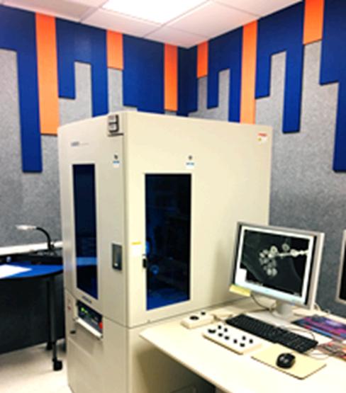
✅ SAXS/WAXS Under Low Vacuum – Ideal for nanostructure analysis
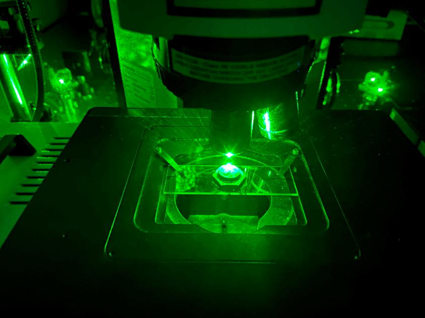
Raman Imaging MicroscopeUnlock Chemistry of Materials
Building
✅ Raman Imaging & Spectroscopy
✅ Analyze chemical composition, identify phases, and assess crystallinity at the microscale.
✅Multi-Laser Excitation Options
�� Blue (473 nm)
�� Green (532 nm)
�� Red (785 nm)
✅Automated X-Y Raman Mapping
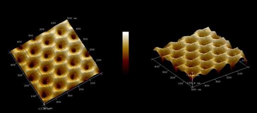
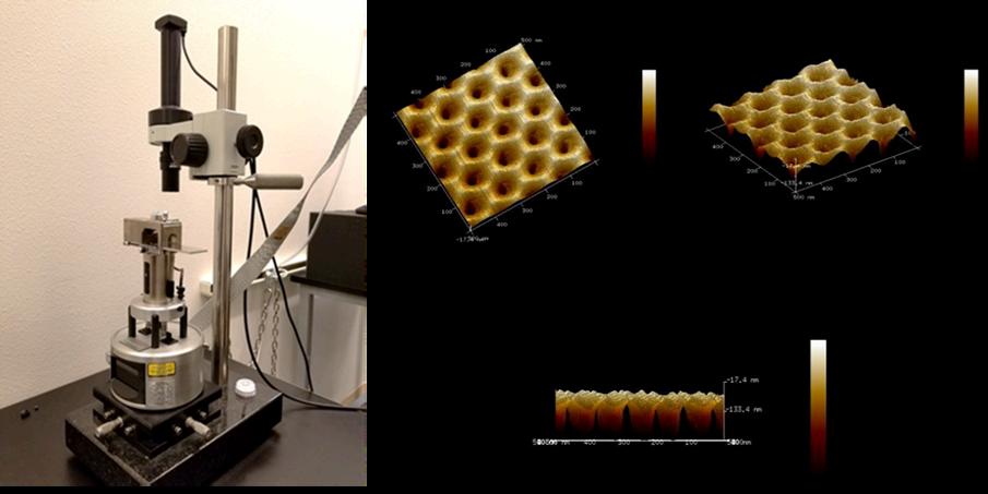
Atomic Force MicroscopeSurface Characterization
✅ Nanoscale materials and coatings characterization
✅ Biological and polymer material studies in liquid or ambient environments
✅ Surface roughness, adhesion, and mechanical analysis
N B u i l d i n g
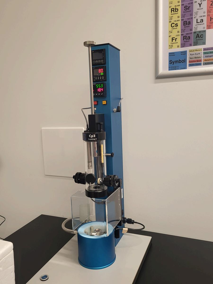
Cryoplunge
SAMPLE PREPARATION
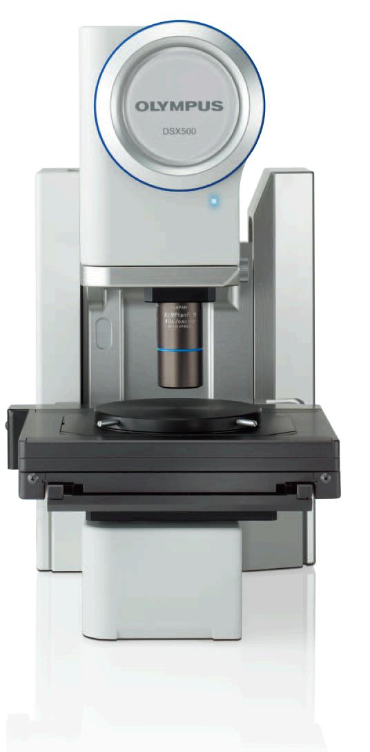
3D Imaging & Versatile Optical Analysis
Surface Inspection
Quality Control & Defect Analysis
Microstructure Imaging
3D imaging geological samples
3D imaging biological tissues.
Rapidly freezes hydrated samples to preserve their native state for cryo-electron microscopy
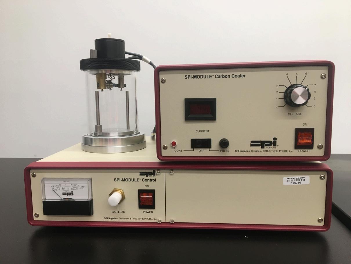
Carbon Coater Gold Sputter Coater
Deposits a thin carbon film to samples, improving conductivity and image quality in SEM
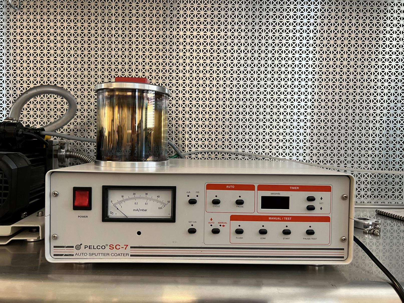
Deposits a thin layer of gold (Au) onto nonconductive samples to enhance imaging quality in SEM
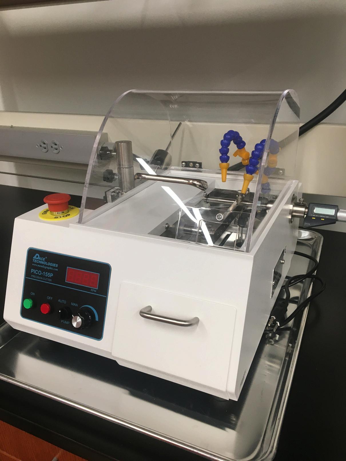
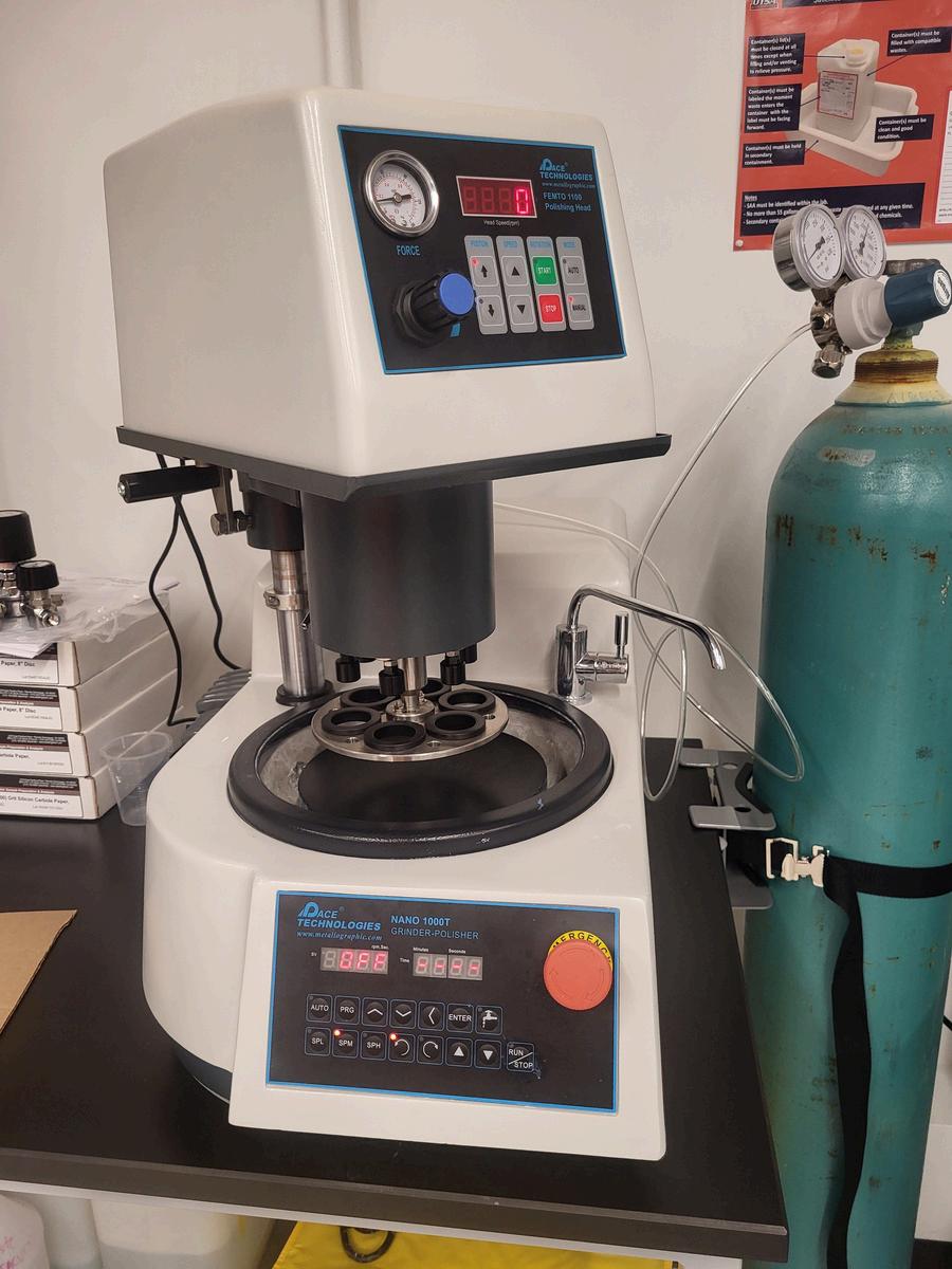
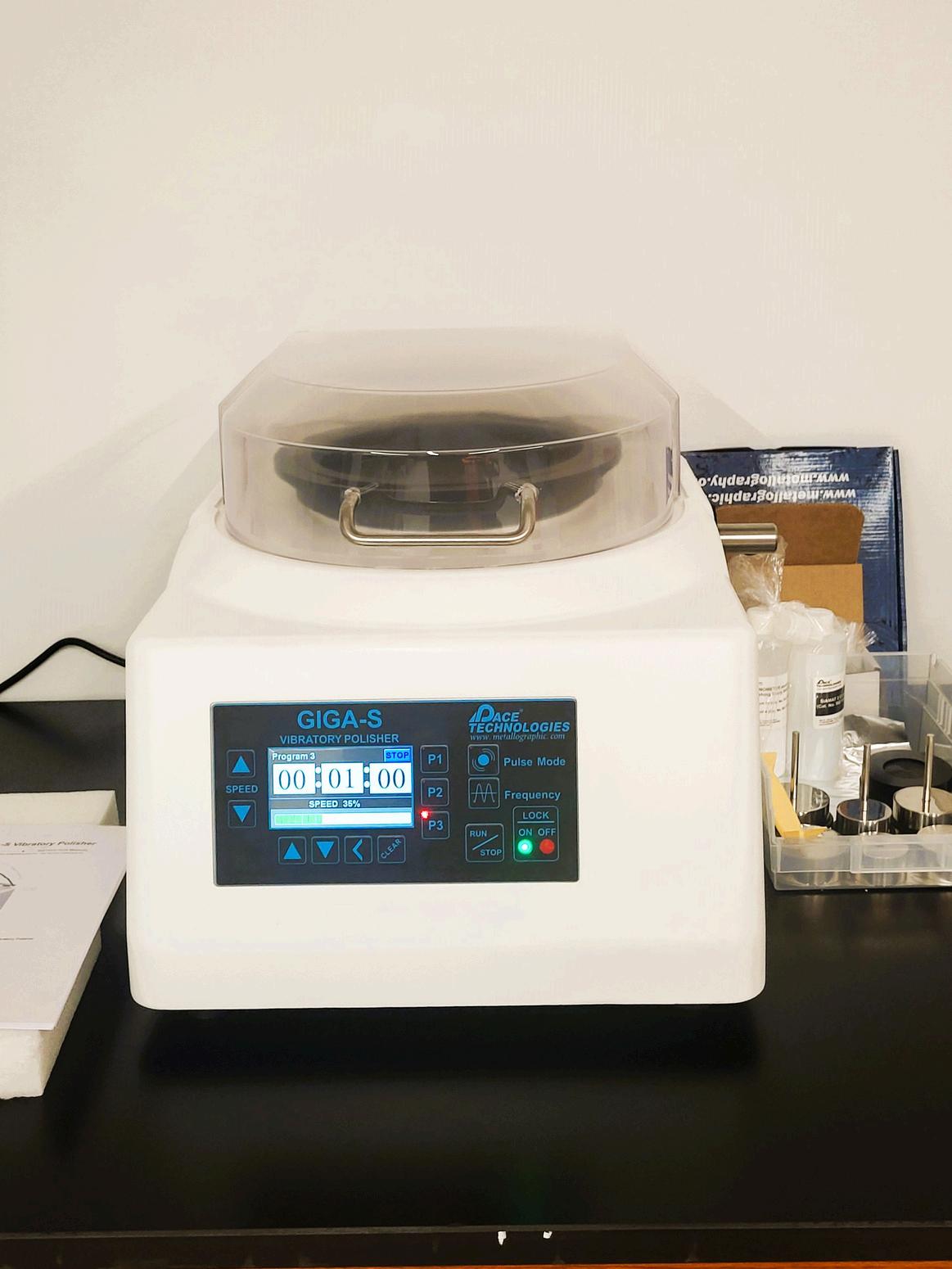
Vibrating Polisher
Grinding and polishing of metallographic specimens
Ultra-fine polishing for EBSD analysis
For preparing biological samples for SEM, as it preserves surface structure
John Jay High School & UTSA Day & NIH Estimeed Hands-On STEM Outreach
High School Student operating SEM
High School Student operating SEM
High School hands-on TEM
High School hands-on TEM
John Jay High School Visit John Jay High School Visit
Dr Stevanovic comes to UTSA after previous stints as a postdoctoral fellow at Harvard University and the National Institute of Standards and Technology Her expertise involves insitu TEM/SEM characterization of materials under externally applied stimuli: temperature and gas.
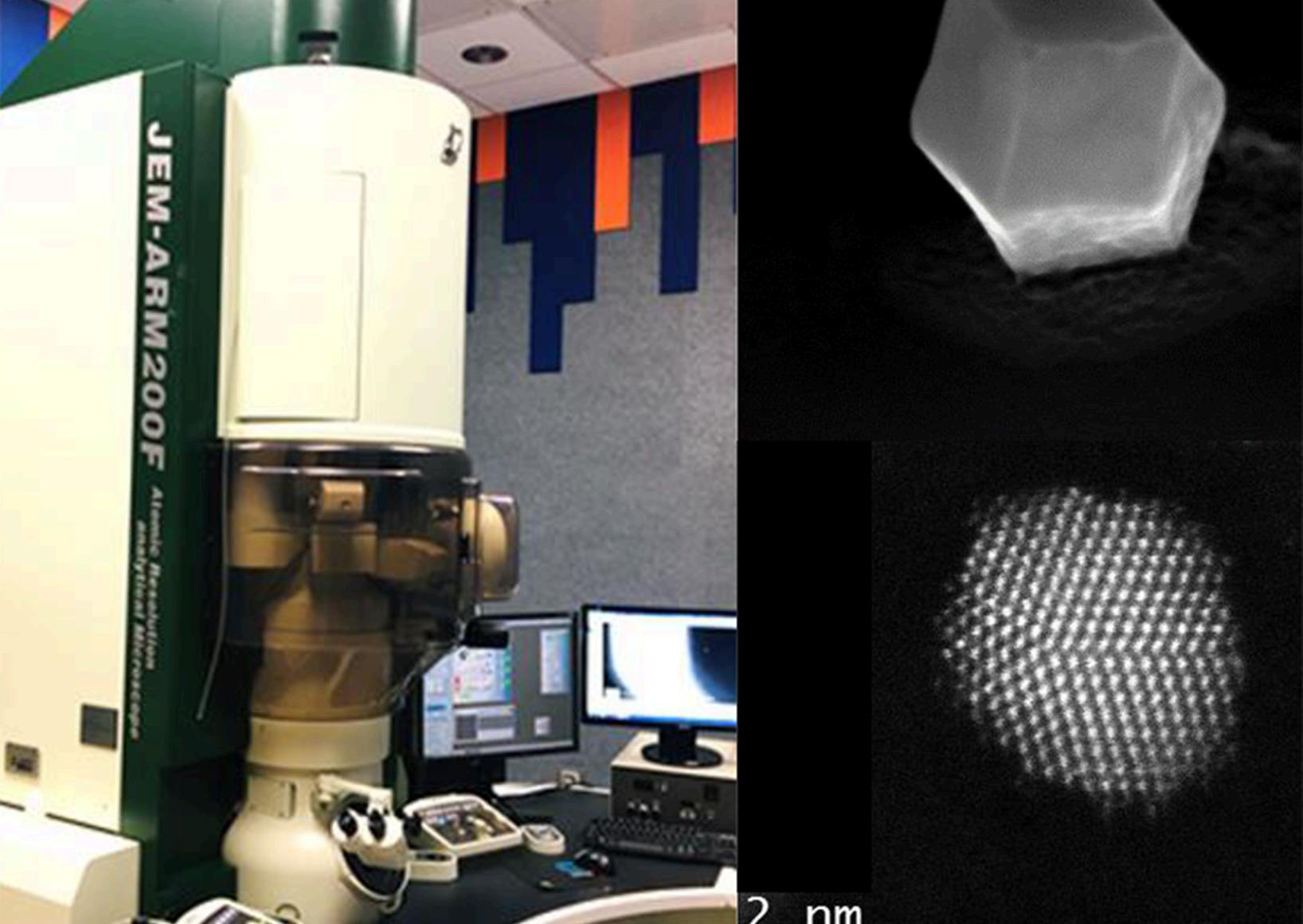
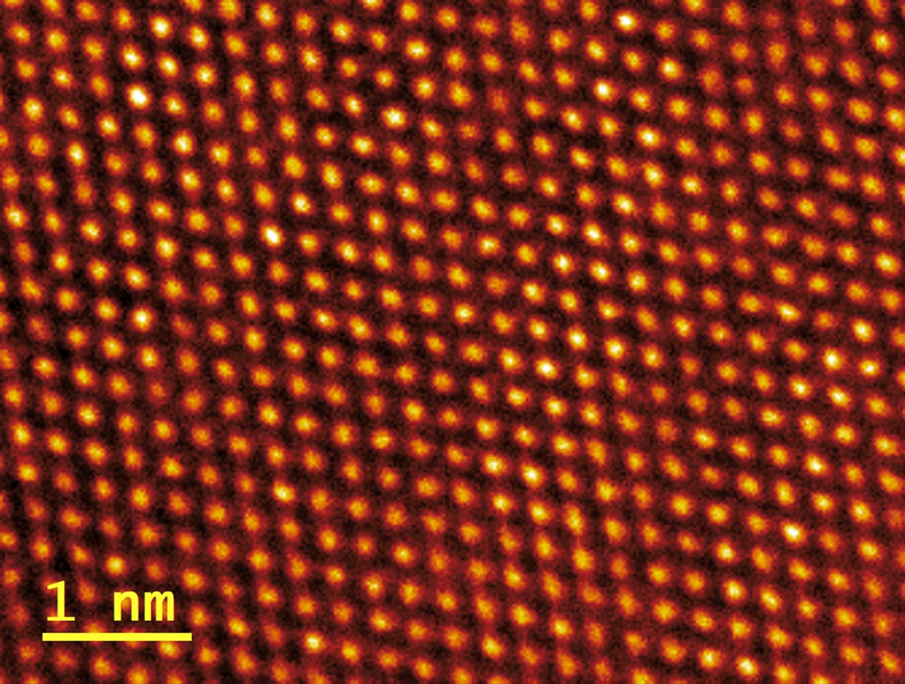
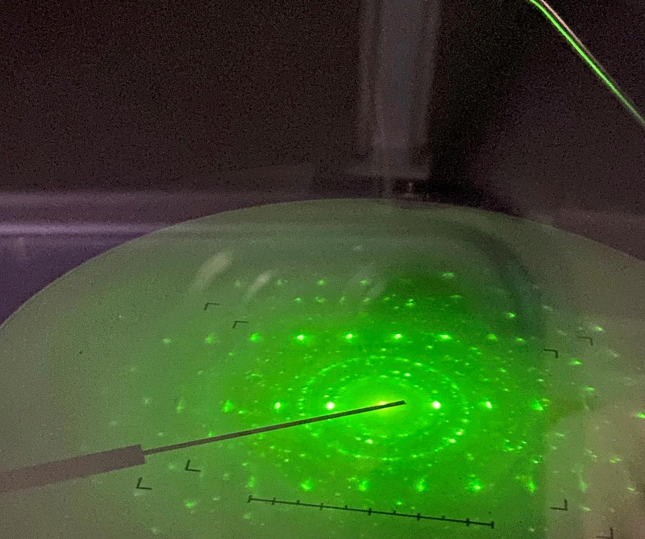
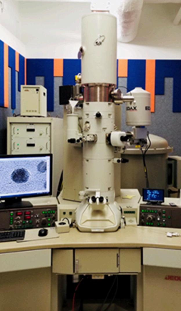
Dr Morales earned his Ph D in Physics and Astronomy from The University of Texas at San Antonio (UTSA) He provides user support at KAMC for the Panalytical Empyrean X-ray Diffractometer, JEOL JEM-2010F TEM, ARM 200F TEM, and the Focused Ion Beam system.
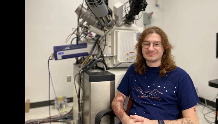
Adis is a Ph.D. student in the Department of Physics and Astronomy at The University of Texas at San Antonio (UTSA). He supports KAMC’s daily operations by assisting users with instruments such as the SEM, Raman Spectroscope, Panalytical X-ray Diffractometer, and Atomic Force Microscope.