The International Handbook of Social Enterprise Law 1st edition by Henry Peter, Carlos Vargas Vasserot, Jaime Alcalde Silva ISBN 3031142161 9783031142161
This work is subject to copyright. All rights are reserved by the Publisher, whether the whole or part of the material is concerned, specifically the rights of translation, reprinting, reuse of illustrations, recitation, broadcasting, reproduction on microfilms or in any other physical way, and transmission or information storage and retrieval, electronic adaptation, computer software, or by similar or dissimilar methodology now known or hereafter developed.
The use of general descriptive names, registered names, trademarks, service marks, etc. in this publication does not imply, even in the absence of a specific statement, that such names are exempt from the relevant protective laws and regulations and therefore free for general use. The publisher, the authors and the editors are safe to assume that the advice and information in this book are believed to be true and accurate at the date of publication. Neither the publisher nor the authors or the editors give a warranty, expressed or implied, with respect to the material contained herein or for any errors or omissions that may have been made. The publisher remains neutral with regard to jurisdictional claims in published maps and institutional affiliations.
This Springer imprint is published by the registered company Springer Nature Switzerland AG The registered company address is: Gewerbestrasse 11, 6330 Cham, Switzerland
To my Masters, and to S., my Muse
– Carlos Francisco Silva
Preface
Healthcare delivery is experiencing a great transition in terms of “value-based healthcare” or “value-based radiology”. The idea behind this transformation is that the providers are paid based on patient’s health outcomes and not for the amount of service they delivered. Care for a medical condition usually involves different specialties and a number of interventions, and the value for the patient can only be created by combined efforts of all stakeholders on the entire cycle of care. In this regard, the specialty of radiology is one of the important outcomes influencing players in the whole healthcare cycle, whether contributing to diagnosis, or by minimally invasive interventional procedures, radiation therapy or therapy monitoring. Consequently, radiology departments are facing many challenges to improve operational efficiency, performance and quality to keep pace with this rapid transition in the healthcare delivery. The duty and workload of the radiologist has changed rapidly in the last decades; times when radiologists have analysed “just a film” are long gone. Today, radiologists face an ever increasing workload and yet have to provide the most possible value to the patients, in an adverse context of shortage of imaging specialists and lack of time spent for interpreting and communicating the imaging exams with patients and referring clinicians.
The following issues are inevitable for creating value and contributing to patient outcome in radiology departments.
• well-organized utilization plans for patient scheduling and consequently shorter waiting times for patients;
• guideline compliance, adherence to appropriateness criteria and identification of redundancy;
• accurate and timely exam reporting with adherence to incidental finding reporting criteria, use of standardized lexicon and structured reports;
• proper communication of reports with referring physicians and with patients;
• continuous research for better imaging, intervention and therapy.
The main goal is to achieve a sustainable and affordable care, creating value, better outcomes and satisfaction to both patients and all players in the health cycle.
This book offers a cutting-edge guide to value-based radiology and provides readers with the latest and comprehensive information on all aspects of
value-based radiology. All topics are discussed by prominent experts in the field in a clearly organized and illustrated form, which will help readers gain the most from each chapter. Accordingly, the book offers a valuable resource for radiologists and healthcare managers working in public or private institutions, as well as a quick reference guide for all other physicians interested in the topic.
Heidelberg, Baden-Württemberg, Germany Carlos Francisco Silva Heidelberg, Baden-Württemberg, Germany Oyunbileg von Stackelberg Heidelberg, Baden-Württemberg, Germany Hans-Ulrich Kauczor
Part I
Michael Fuchsjäger, Lorenzo Derchi, and Adrian Brady
Carlos Francisco Silva, Kheng L. Lim, Teresa Guerra, Gianluca Ficarra, and Ricarda von Krüchten
Carlos Francisco Silva, Claus Peter Heussel, and Eduardo Mortani Barbosa Jr. Value-Based
Sabine Weckbach and Oyunbileg von Stackelberg
Ramandeep Singh, Fatemeh Homayounieh, Rachel Vining, Subba R. Digumarthy, and Mannudeep K. Kalra
The Value in 3D
Namkug Kim, Sangwook Lee, Eunseo Gwon, and Joon Beom Seo
Incentivizing
Florian Hofer, Carlos Francisco Silva, and Tom Stargardt
Part II Practical Applications in Specific Areas of Radiology
Value-Based Radiology in Neuro/Head and Neck Imaging
David Rodrigues
Value-Based Radiology in Thoracic Imaging
Carlos Francisco Silva and Hans-Ulrich Kauczor
Value-Based Radiology in Abdominal and Pelvic Imaging
Kheng L. Lim
Value-Based Radiology in MSK Imaging
Catarina Ruivo and Diogo Roriz
Value-Based Radiology in Breast Imaging
Inês Leite and Elisa Melo Abreu
Value-Based Radiology in Pediatric Imaging 143
Daniela Pinto and Sílvia Costa Dias
Value-Based Radiology in Cardiovascular Imaging 159
Carlos Francisco Silva
Part I
Theoretical Basis and General Concepts
Value-Based Radiology: A New Era Begins
M. Fuchsjäger (*)
Department of Radiology, Medical University of Graz, Graz, Austria e-mail: michael.fuchsjaeger@medunigraz.at
L. Derchi
San Martino University of Genoa, Genoa, Italy
A. Brady
Mercy University Hospital, Cork, Ireland
Med Radiol Diagn Imaging (2019) https://doi.org/10.1007/174_2019_220,
Published Online: 08 August 2019
Abstract
This introduction chapter is written by Prof. Michael Fuchsjäger, Chair of the European Society of Radiology (ESR)’s Value-Based Radiology Subcommittee, Prof. Lorenzo Derchi, Chair of the ESR Board of Directors, and Dr. Adrian Brady, Chair of the ESR Quality, Safety and Standards Committee. Prof. Derchi and Dr. Brady are also ex officio members of the ValueBased Radiology Subcommittee. The ValueBased Radiology Subcommittee was established with the aim of supporting radiologists in fulfilling their central role in healthcare while assisting them in the development of “appropriate metrics, which capture their true contribution and added value to patient care” (European Society of Radiology (ESR), https://www.myesr.org/ about/organisation/executive-council# paragraph_grid_16643, Accessed 28 May 2019, 2019). The Subcommittee takes an active role in promoting value-based radiology to patients, through patient groups, and to healthcare professionals, through various publications and other activities, including dedicated sessions and lectures at the European Congress of Radiology (ECR) and stakeholder events such as the ECCO (European CanCer Organisation) summit 2018 and COCIR (European Coordination Committee of the Radiological, Electromedical and Healthcare IT Industry) General Assembly 2018. Furthermore, the Subcommittee collaborates with other radiological societies around the world on various initiatives.
M. Fuchsjäger et
1 Introduction
In medical terms, radiology is a young specialty with a relatively short history, beginning with the discovery of X-rays by Wilhelm Conrad Röntgen in 1895. Despite its youth, it has had a transformative impact on the practice of medicine, introducing increasingly complex equipment, capabilities and modalities, and interrupting the direct diagnostic link between physician and patient that had previously existed for millenia (van Gelderen 2004). As a matter of fact, the radiologist’s work is now in the middle between the patient and his/her primary care physician, making the patient’s body visible and understandable. As radiology has become increasingly technologically sophisticated in recent decades, the distance between patients and radiologists has grown, despite the concomitant growth in the influence of radiology on patient care. Work practices have led to radiologists, with the exception of a few subspecialties (e.g. interventional radiology, breast imaging), retreating to reading rooms, with limited direct patient contact. The rise of teleradiology makes this distance increasingly spatial as well as personal. Consequently, a significant degree exists of ignorance amongst patients about the actual role of the radiologist, as revealed by a 2008 survey conducted by the American College of Radiology (ACR): approximately half of respondents were unable to tell whether radiologists administer or interpret scans nor whether radiologists were licensed physicians or technicians (Glazer and RuizWibbelsmann 2011).
The increasing digitalisation of radiology has required radiologists to take on an ever-increasing workload: quicker scans and higher patient throughput have resulted in a greater number of examinations and an increased number of images per examination (especially for CT and MRI). Yet the increase in productivity facilitated by improvements in technology has, arguably, only contributed to the increasing commoditisation of radiology as a profession, its work being measured predominantly in terms of volume (Brady 2011a, b). As a matter of fact, it is usually considered that the examinations we perform are fully standardised (as a commodity) and our results are
measured predominantly in terms of volume of studies performed. In addition, the current financial difficulties encountered by all healthcare systems and the consequent focus on efficiency make the perception of radiology as nothing more than a numbers game increasingly problematic. Indeed, the question of whether the increasing use of technology such as artificial intelligence (AI) solutions in radiology could make radiologists obsolete has even been raised by some (Ridly 2019; Goedert 2019). The need for radiologists to demonstrate the value that they add to the healthcare value-chain every day has never been more acute.
Value-based healthcare is a conceptualisation of healthcare centred on quality, rather than quantity. It is a response to the increasing costs of healthcare provision, particularly in developed countries. Traditionally, healthcare has focused primarily on responding to acute and emergency episodes. This focus meant there were few incentives for healthcare providers to invest in “prevention, longitudinal chronic disease management, [or] population health” (Philips Position Paper 2019). Furthermore, timeconsuming activities such as direct patient consultation were actually dis-incentivised. Value-based healthcare seeks to invert this, placing patients at the centre of the care model.
Value-based healthcare, as a framework, originated in the seminal work of Harvard economist Michael Porter (2010). Its goal is to simultaneously improve health outcomes and reduce costs. By placing patients’ outcomes at the centre of the model, value-based healthcare seeks to incentivise improved outcomes for patients instead of merely an increase in workload (including not just potentially unnecessary procedures, but potentially harmful ones) (Kimpen 2019). Specifically, value is defined by Porter as patient health outcome divided by money spent. This formula would suggest two ways of increasing value to patients: either reducing costs for the same outcome, or increasing outcomes relative to costs. This is, of course, not a universally accepted definition of value, the Utah Value in Health Care Survey providing just one example of alternative ways in which value can be assessed (Albo et al. 2018). The European
Commission has convened an expert panel on health, which has produced a draft opinion on value-based healthcare. Their conclusion that value may be measured according to four metrics, personal value (“appropriate care to achieve patients’ personal goals” (Expert Panel on Effective Ways of Investigating in Health 2019)), technical value (“achievement of best possible outcomes with available resources” (Expert Panel on Effective Ways of Investigating in Health 2019)), allocative value (“equitable resource distribution across all patient groups” (Expert Panel on Effective Ways of Investigating in Health 2019)), and societal value (“contribution of healthcare to social participation and connectedness” (Expert Panel on Effective Ways of Investigating in Health 2019)), is due to be discussed in June 2019, providing stakeholders, such as radiologists, an opportunity to offer their perspectives.
In the context of the Utah Value in Health Care Survey amongst patients, physicians and employers, University of Utah Health defines value as the “product of the quality of care plus the patient experience at a given cost” (Albo et al. 2018). Thus, University of Utah Health added a subjective element to Porter’s value equation by including the service aspect in order to reflect the patient’s assessment of value. Adapted to employers, the equation considers employee productivity resulting from better health combined with employee satisfaction, divided by the cost of providing health benefits. One of the main findings of the survey was that patients, physicians, and employers who pay for medical benefits have different opinions of what is most valuable in healthcare. The study’s authors therefore conclude that mutual understanding of all stakeholders’ positions is a first step towards a value-based healthcare system.
Porter explicitly states that the “proper unit for measuring value should encompass all services or activities that jointly determine success in meeting a set of patient needs” (emphasis added) (Porter 2010). Yet, under both the Porter and Utah frameworks, radiology’s place in the value chain has, to a large extent, been overlooked thus far. The combined effect of the dislocation of radiologists from patients, a phenomenon Glazer
and Ruiz-Wibbelsmann (2011) describe as leading to the ‘invisibility’ of radiologists, and the commoditisation of the service they provide has been that diagnosis is not seen as part of the patient health outcome, and radiology is subsequently either absent from the value chain or viewed only as a cost: ‘radiology is widely viewed as a contributor to health care costs without an adequate understanding of its contribution to downstream cost savings or improvement in patient outcomes’ (Sarwar et al. 2015).
Swift and, above all, accurate diagnosis is irrefutably integral to determining the success of meeting patient needs. Porter himself acknowledges this: “Delays in diagnosis or formulation of treatment plans can cause unnecessary anxiety” (Porter 2010), a factor that would certainly adversely affect value under the subjective element of the University of Utah Health framework. Aside from patient anxiety, it should go without saying that erroneous diagnosis can lead to worse health outcomes, both through failure to optimally treat disease and the performance of unnecessary procedures.
In recent years, radiologists have sought to increase their visibility, for example, through initiatives such as the International Day of Radiology (IDOR) (2019), inaugurated in 2012 by the ESR in association with the Radiological Society of North America (RSNA) and the ACR. IDOR has since become an annual event held with the aim of building greater awareness of the value that radiology contributes to safe patient care, and improving understanding of the vital role radiologists play in the healthcare continuum. IDOR is now celebrated by more than 170 societies all over the world with special publications, social media activities, courses and charity events.
The ESR was amongst the first medical scientific societies to create a patient group (the Patient Advisory Group—PAG) within the society structure, with the specific goal of bringing together “patients, the public and imaging professionals in order to positively influence advances in the field of medical imaging to the benefit of patients in Europe” (European Society of Radiology (ESR) Patient Advisory Group 2019). The ESR-PAG thus serves as a role model for others as it works towards the improvement of radiologist-patient Value-Based Radiology:
dialogue. The ESR’s Value-Based Radiology Subcommittee purposefully included a Patient Advisory Group (PAG) representative with the aim of working with them to boost the visibility of the concept of value-based radiology amongst patients and to consider their perspective.
Other examples of ways radiologists have sought to raise the profile of radiology and build closer connections to patients include informative websites, such as the RSNA and ACR’s radiologyinfo.org (Radiology Info 2019), and making increased efforts to routinely speak to patients; Glazer and Ruiz-Wibbelsmann give the examples of personally explaining mammographic results, communicating paediatric imaging results to parents, and utilising online portals to provide enhanced contact with patients (Glazer and RuizWibbelsmann 2011).
Yet, despite these attempts to make radiology more ‘visible’, particularly to patients, the precise position of radiology in the value chain remains uncertain as the healthcare industry begins its shift towards value-based metrics (Brandt-Zawadski and Kerlan 2009). As future planning and resource allocation will, more than likely, depend upon such models, it is vital to ensure that radiology’s position is recognised. As such, the discussion and perspectives presented in this volume are most welcome, although it should, of course, be noted that the discussion of value-based healthcare and its application to radiology has taken slightly different perspectives on different sides of the Atlantic, largely due to differing models of funding, governance, and payment for healthcare in Europe and the USA (Kimpen 2019), and within different branches of radiology.
2 Where Is the Value in Radiology Delivered?
Impact on patients’ outcome, and therefore ‘value’, is delivered in all aspects of radiology, ranging from screening and disease prevention to detection, diagnosis, image-guided biopsy, staging of disease, evaluation of patient progress during treatment, the provision of high-level subspecialist interpretation, reassurance and confirmation of resolution of disease, clinical decision support,
M. Fuchsjäger et al.
imaging biomarkers, radiation protection, interventional radiology and teleradiology. It is generated by justified indications, appropriate criteria and appropriate dose, personalised patient protocols, structured reporting, reporting of incidental findings, therapeutic decisions based on radiological diagnoses and improved patient outcome. The added value that radiology provides to the healthcare value chain has been documented in various longitudinal studies (Alberle et al. 2013; Mehanna et al. 2016; The SCOT-HEART Investigators 2018). Furthermore, Sarwar et al. (2015) provide a clear illustration of how radiology can deliver value at each step of the imaging chain. Every step of this whole chain can be broken down to several processes, from decision support tools and proper scheduling at the front end, to appropriate communication and follow-up recommendation at the back end. In addition, every process can be measured by specific indicators to help improve practice.
The ESR’s 2017 concept paper on value-based radiology (European Society of Radiology (ESR) 2017) adds new aspects to the value chain by identifying five key factors that relate to the quality of the diagnosis and, similar to the University of Utah Health model, focus particularly on the human aspect of the value chain, including the patient’s well-being and relations with patients and referring physicians. The first key factor concerns the appropriateness of an imaging request. Clinical decision support systems developed by the radiological community for referring physicians are designed to enhance appropriateness. The ESR’s solution is the ESR iGuide (2019), a system for making imaging referral guidelines available to referring physicians at the point of care, providing evidence-based information and decision support. The value of this step consists in ensuring the appropriate use of radiation, avoiding unnecessary exposure and related risks, and contributing to correct protocolling of exams.
Appropriately prioritising patients enables treatment of urgent cases at an early stage, thus reducing patient burden and costs that would be incurred by diagnosis and treatment at a more advanced stage. This enables value to be added during the processes associated with Sarwar et al.’s first step in the value chain (Sarwar et al. 2015).
The second key factor is attention to radiation protection measures. Major radiological societies and organisations have launched radiation protection initiatives, such as ESR EuroSafe Imaging and, following its lead, AFROSAFE, Arab Safe, CanadaSafe, Image Gently, Image Wisely, Japan Safe Imaging, and LATINSAFE. EuroSafe Imaging strives to support and strengthen medical radiation protection across Europe following a holistic, inclusive approach (EuroSafe Imaging 2019), focusing on optimisation, justification, quality and safety, education, research and regulatory compliance. A number of metrics concerning radiation protection should ideally be put in place in every department, for example: the presence of diagnostic protocols which entail the choice of non-ionising examinations whenever possible; the presence of low-dose protocols in all CT equipment; a framework for reporting the percentage of use of such protocols; a requirement to report all exposures to a radiation dose index registry; and training programmes on radiation protection. Visser notes dose monitoring— comparing dosages with diagnostic reference levels (DRLs)—as another step towards ensuring maximum value is provided to patients in terms of safety. Patient preparation, including the choice and administration of contrast media, is another factor that may generate value (Sarwar et al. 2015; Visser 2019).
The third key factor concerns reporting, specifically the characteristics of the radiology report: it should be correct, concise, complete, clearly structured and easily comprehensible to the referring physician (Brady 2018). Following such rules provides value to the referring physician by supplying them with all the information they need to make decisions optimally. Structured reporting will be particularly helpful in the future as it allows the use of decision support tools which can guide the radiologist (Visser 2019). Every radiology report should use standardised terminology, provide specific recommendations about further imaging or treatment, give full contact information, and, ideally, should be made available to the patient via an online portal.
The fourth key factor for adding value is the relationship between patients and radiology personnel. The availability of detailed instructions for
different examinations, the distribution of patient satisfaction questionnaires (developed together with PAGs), followed by audits, as well as formal relationships between radiology departments and patient organisations are factors and possible metrics of the radiologist’s availability and thus visibility to patients. The importance of this factor to patients was underlined by a survey conducted by the ESR’s Value-Based Radiology Subcommittee in 2019 (European Society of Radiology (ESR) 2019b) in which preliminary results indicated that patients in various countries expressed a degree of dissatisfaction with the availability of radiologists for personal consultation, and, to a lesser extent, with both the way their results were communicated to them and the information provided following diagnosis by radiology staff [unpublished]. This is an area in which radiologists may provide significant improvements in perceived value at relatively little expense (assuming sufficient workforce availability).
The fifth key factor according to the ESR concept paper is continuous professional education, research, and innovation. Again, the ESR’s ValueBased Radiology Subcommittee’s patient survey revealed that a key element that patients regarded as providing value was their confidence in their radiologist’s qualifications and expertise. While it is obvious that staying abreast of new developments and using state-of-the-art technology increases value, this factor is particularly difficult to measure. Regarding continuous professional education, compliance with national regulations on continuous medical education (CME) could serve as metrics.
With so many factors through which radiology may contribute to enhancing value for the patient, the referring physician, and health policy makers, it is high time that radiology’s place within valuebased healthcare models be fully recognised.
3 What Is the Status of ValueBased Radiology in Other Parts of the World?
The ESR dedicated its International Forum 2018 to the topic of value-based radiology in an attempt to gain global perspectives on the current status
of value-based radiology in different regions and contexts, as well as what efforts are being made to promote value-based radiology. The International Forum is convened annually by the ESR during the ECR and offers the ESR’s partner and member societies from outside Europe the opportunity to present the situation regarding a particular topic in their respective country or region. A report on the ESR International Forum 2018 was published in Insights into Imaging in 2019 (European Society of Radiology (ESR) 2019c) and can be summarised as follows:
North America
In 2017, the Conference Board of Canada, Canada’s largest non-partisan, not-forprofit, evidence-based research organisation published a primer document ‘The value of radiology in Canada’, demonstrating to lawmakers and policymakers that radiology adds value to the health system (The Value of Radiology in Canada 2016). This primer provides three examples: breast cancer screening, teleradiology, and interventional neuroradiology. The Canadian Association of Radiologists (CAR) has been very active in promoting the role of radiology and radiologists through various initiatives designed to raise awareness of who radiologists are, what role they perform, and demonstrate the ways in which radiologists help patients, or improve patients’ care in general, e.g. through advocacy activities, such as meetings with stakeholders, or patient care initiatives, such as practice guidelines and various patient resources (European Society of Radiology (ESR) 2019c).
In the United States, the RSNA provides material to enable patients to properly inform themselves about radiology procedures as well as its RadLex and Structured Reporting initiatives to help encourage radiologists to adopt structured and stan-
dardised terminology for drafting their reports (Radiological Society of North America 2019). The RSNA takes the perspective that, radiologists can demonstrate the value they add to the patient by taking full responsibility for managing their imaging, thereby assuring the patient that they are fully engaged in their diagnosis/treatment (European Society of Radiology (ESR) 2019c).
The ACR offers its Imaging 3.0 initiative as a roadmap towards value-based imaging, which should be achieved with the help of clinical decision support (CDS), structured reporting, data mining, and other information technology tools (American College of Radiology 2019). Unlike in traditional radiological care, radiologists have to actively take responsibility for all aspects of imaging care, thereby enhancing patients’ experience and relevance to the clinical team (European Society of Radiology (ESR) 2019c).
Latin America
Latin America suffers from considerable disparities in both health and socioeconomic terms between urban and rural areas. Technological developments and valuebased radiology initiatives are largely limited to private hospitals. While, overall, efforts still focus on improving access to and coverage of health services rather than on fee for value, the Inter-American College of Radiology (CIR), as well as the major national radiological societies, such as those in Brazil, Colombia, and Mexico, are making efforts to move towards a valuebased approach. For example, LATINSAFE is mentioned as an initiative dedicated to education in radiation protection (European Society of Radiology (ESR) 2019c).
M. Fuchsjäger
Asia
In India, like in Latin America, the situation is highly heterogeneous, ranging from modern hospitals with state-of-the-art facilities to villages without any access to imaging whatsoever. According to the Indian Radiological and Imaging Association (IRIA), radiologists should be perceived as clinicians interacting with their patients. In Korea, the government has increased the budget for assessing and increasing medical quality, and the Korean Society of Radiology (KSR) embraces the value-based healthcare system. The Japan Radiological Society (JRS) launched Japan Safe Radiology, a government-supported project to promote safety, standardisation and optimisation of imaging, and plans to add value-based radiology to the project’s safety and efficiency related targets (European Society of Radiology (ESR) 2019c).
Asia-Oceania is yet another region where the practice of value-based radiology is very diverse. In most countries, radiology departments are seen primarily as service providers, with turnaround times of reports still viewed as the key indicator. However, some moves are being made towards value-based metrics: Choosing Wisely Australia is a clinician-led global initiative aiming to improve safety and quality in healthcare by avoiding unnecessary examinations, treatments and procedures (Choosing Wisely Australia 2019). With InsideRadiology, the Royal Australian and New Zealand College of Radiologists (RANZCR) offers patients and referring physicians information on clinical radiology tests, treatments and procedures (Inside Radiology 2019). Furthermore, RANZCR offers educational modules to promote appropriateness of referrals (European Society of Radiology (ESR) 2019c). Value-Based Radiology: A New
To summarise, although the extent to which value-based radiology has been adopted still varies between countries and within countries, the world’s major radiological societies agree that the value-based approach is the concept to follow in the future.
4 Perspective
Radiology has finally begun to appreciate that the quality and the value it provides is more important than the mere volume, previously the main driver of and unit used for measuring productivity and efficacy. As in healthcare as a whole, radiology will in future be measured according to patient outcome, which will certainly be better with the improving quality and safety of the entire imaging chain: decision to image, performance of procedure, interpretation of study, reporting of study, and the highly important last step of communication of results to our patients and referring physicians.
Change is inevitable in healthcare, especially in specialities which rely heavily on technology, such as radiology. Radiologists must continue to show themselves to be adaptable and willing to change: it is the only way to survive evolutionary processes, and emerge stronger and better. The evolution which this new era of value-based radiology ushers in is an opportunity to enhance the ability of radiologists to provide the best possible care for patients and secure their position at the heart of ensuring optimum outcomes.
To accomplish all this in the near future the role and—very importantly—the self-image of the radiologist will have to change considerably: from that of the traditional image interpreter to that of the leader of the whole imaging process, and perhaps even of integrated diagnostics in the more distant future. Accepting this new role entails accepting heightened responsibility as a large number of processes—many of which have been managed separately by radiology for a long time—have to be integrated into one cohesive body around the framework of value-based radiology.
The more active role of radiologists in creating value will necessarily include a better understanding for the needs of the referral base through active engagement with referring physicians, for example, having daily consultations with subspecialties within the department or embedding reading rooms in specialty clinics of referring physicians and, obviously, improving radiology reports themselves with regard to structure and standardisation.
Artificial Intelligence (AI) will undoubtedly play a role in this. Currently, AI seems to show most promise in certain specific fields or niches, e.g. helping with repetitive tasks like lesion detection and feature description; it has also offered potential as a decision support tool (Savadjiev et al. 2019). This could be of vital importance in the future as AI frees time for interpretation and communication and/or makes coping with the ever-increasing workload possible, especially in regions where radiologists are scarce (teleradiology could also play a vital role here). In short, AI offers radiologists further potential to generate increased value. Rather than seeing AI as an existential threat (Ridly 2019; Goedert 2019), radiologists should embrace AI as an additional means through which they can enhance value to patients (e.g. by using deep learning to lower dosage for CT scans) (Visser 2019).
5 Conclusion
At the end of the day, each radiologist has to provide the best possible care for his/her patients; therefore, any definition of the “value” provided by our work should rightly be focused on patient outcome. For all patients, radiology can have impact in different moments of each episode of care, thus continuously providing value and contributing to patient outcome. Furthermore, such contributions are extremely broad and involve well-managed imaging utilisation plans, shortening of waiting times for imaging exams, improved appropriateness, attention to radiation protection, use of structured reporting, using the ‘drivers seat’ position in introducing technological
M. Fuchsjäger et al.
innovation including AI tools and solutions to improve diagnostic imaging, interventional radiology and image-guide therapy. However, the two most crucial aspects through which we add value to patient outcome remain close collaboration with our referring physicians and communication with our patients. As regards the first, liaising with colleagues and working together as a team, both informally and in regular multidisciplinary meetings, is the basis of appropriate use of imaging as well as of correct therapeutic choices based on the resulting images. As regards the latter, communication with patients not only makes radiologists ‘visible’, but contributes to giving radiology the prominence its importance to value-based healthcare deserves.
Acknowledgements The authors acknowledge and thank the following individuals for their particular contributions: Jonathan Clark, Martina Szucsich, and Monika Hierath (ESR Department of European & International Affairs).
References
Alberle DR, DeMello S, Berg CD et al (2013) Results of the two incidence screenings in national lung screening trial. N Engl J Med 369(10):920–931
Albo A, Bracken S, Orlandi R et al (2018) Bringing value into focus: the state of value in U.S. Health Care. University of Utah Health, Salt Lake City, UT American College of Radiology (2019) Imaging 3.0. https://www.acr.org/Practice-Management-QualityInformatics/Imaging-3. Accessed 6 Jun 2019
Brady AP (2011a) Measuring radiologist workload: how to do it, and why it matters. Eur Radiol 21(11):2315–2317
Brady AP (2011b) Measuring consultant radiologist workload: method and results from a national survey. Insights Imaging 2:247–260
Brady AP (2018) Radiology reporting—from Hemingway to HAL? Insights Imaging 9:237–246
Brandt-Zawadski M, Kerlan RK (2009) Patient-centered radiology: use it or lose it! Acad Radiol 16:521–523
Choosing Wisely Australia (2019). http://www.choosingwisely.org.au/home. Accessed 6 Jun 2019
ESR iGuide (2019). https://www.myesr.org/esriguide Accessed 3 Jun 2019
European Society of Radiology (ESR) (2017) ESR concept paper on value-based radiology. Insights Imaging 8:447–454
European Society of Radiology (ESR) (2019a). https:// www.myesr.org/about/organisation/executive-
council#paragraph_grid_16643. Accessed 28 May 2019
European Society of Radiology (ESR) (2019b) Valuebased radiology subcommittee patient survey. https:// www.myesr.org/esr-patient-survey-value-based-radiology. Accessed 20 May 2019
European Society of Radiology (ESR) (2019c) Summary of the proceedings of the international forum 2018: “value-based radiology”. Insights Imaging 10:34
European Society of Radiology (ESR) Patient Advisory Group (2019). https://www.myesr.org/sites/default/ files/ESR-PAG-leaflet-web.pdf. Accessed 3 Jun 2019
EuroSafe Imaging (2019). http://www.eurosafeimaging. org/about. Accessed 21 May 2019
Expert Panel on Effective Ways of Investigating in Health (2019) Opinion on defining value in ‘value-based healthcare’. https://ec.europa.eu/health/expert_panel/ sites/expertpanel/files/024_valuebasedhealthcare_ en.pdf. Accessed 3 Jun 2019
Glazer GM, Ruiz-Wibbelsmann JA (2011) The invisible radiologist. Radiology 258:18–22
Goedert J (2019) Are radiologists becoming obsolete? https://www.healthdatamanagement.com/news/areradiologists-becoming-obsolete. Accessed 23 May 2019
Inside Radiology (2019). https://www.insideradiology. com.au/. Accessed 6 Jun 2019
International Day of Radiology (2019). https://www.internationaldayofradiology.com/. Accessed 24 May 2019
Kimpen J (2019) How health care informatics supports increased productivity and better patient experiences. https://www.politico.eu/sponsored-content/how-healthcare-informatics-supports-increased-productivity-andbetter-patient-experiences/. Accessed 21 May 2019
Mehanna H, Wong W-L, McConkey CC et al (2016) PET-CT surveillance versus neck dissection in advanced head and neck cancer. N Engl J Med 374:1444–1454
Philips Position Paper (2019) Value-based care: turning healthcare theory into a dynamic and patient-focused reality. https://www.philips.com/a-w/about/news/ archive/blogs/innovation-matters/20190212-howinformatics-supports-increased-productivity-betteroutcomes-and-improved-experiences-in-healthcare. html. Accessed 20 May 2019
Porter EM (2010) What is value in healthcare? N Engl J Med 363:26
Radiological Society of North America (2019) RadLex radiology lexicon. https://www.rsna.org/en/practicetools/data-tools-and-standards/radlex-radiology-lexicon. Accessed 6 Jun 2019
Radiology Info (2019). https://www.radiologyinfo.org/ Accessed 3 Jun 2019
Ridly EL (2019) Will AI soon put radiologists out of a job? https://www.auntminnie.com/index.aspx?sec=su p&sub=aic&pag=dis&ItemID=114604. Accessed 22 May 2019
Sarwar A, Boland G, Monks A et al (2015) Metrics for radiologists in the era of value-based health care delivery. Radiographics 35:866–878
Savadjiev P, Chong J, Dohan A et al (2019) Demystification of AI-driven medical image interpretation: past, present and future. Eur Radiol 29:1616–1624
The SCOT-HEART Investigators (2018) Coronary CT angiography and 5-year risk of myocardial infarction. N Engl J Med 379:924–933
The Value of Radiology in Canada (2016) The Conference Board of Canada. https://www.conferenceboard.ca/temp/9a7de99e-3869-4676-addd823dedcb3968/8532_ValueofRadiology_BR_.pdf. Accessed 6 Jun 2019
van Gelderen F (2004) Understanding X-rays. Springer, Berlin
Visser JJ (2019) EuSoMII Webinar Series 2019 ‘Value based Imaging’. https://www.eusomii.org/6496-2/ Accessed 22 May 2019
Patient-Centered Care
Carlos Francisco Silva, Kheng L. Lim, Teresa Guerra, Gianluca Ficarra, and Ricarda von Krüchten
C. F. Silva (*) · R. von Krüchten
Department of Diagnostic and Interventional Radiology, University Hospital Heidelberg, Heidelberg, Germany
e-mail: Carlos.dasilva@med.uni-heidelberg.de
K. L. Lim
Department of Radiology, Pennsylvania Hospital, University of Pennsylvania Health System, Philadelphia, PA, USA
T. Guerra
IMA—Imagens Médicas Associadas, Setúbal, Portugal
G. Ficarra
Department of Diagnostic and Interventional Radiology, University Hospital Heidelberg, Heidelberg, Germany
Department of Diagnostic and Interventional Radiology, University of Genoa Hospital, Genoa, Italy
In this chapter we focus on the topics of patient-centered care, or more broadly speaking patient- and family-centered care. The various aspects of improving patient experience in healthcare are discussed. These include patient comfort in a healthcare facility, surveying patients of the care they receive, patient education and providing compassion in delivering bad news, and involvement of patient social circles, among others. A five-step approach in communicating bad news is discussed. In addition, we highlight the importance of promoting the well-being of healthcare providers and its impact on improving patient health outcomes.
1 Introduction
Patient-centered care, or more inclusively patient- and family-centered care (PFCC), is generating lots of discussion and gaining momentum in the medical community, especially in the last 5 years. In this model, healthcare delivery revolves around the patient with the emphasis on generating a more pleasant experience from the patient perspective (Itri 2015 ). In radiology, this includes but is not
Med Radiol Diagn Imaging (2019) https://doi.org/10.1007/174_2019_209,
Published Online: 24 May 2019
limited to timely scheduling of exams, efficient registration, compassionate and knowledgeable staff, peaceful and comfortable environment, radiologist expertise, timely reports, radiologist availability for consultation with the patients and referring physicians, and transparent billing with easy accessibility when questions arise. As patient experience gains traction in influencing reimbursement for health services, it is more important than ever that physicians adopt PFCC paradigm. Radiology consult is discussed separately on the next chapter. Here we discuss the access and waiting times (patient comfort), the involvement of family and friends, the patient education (fear and anxiety alleviation), and finally the PFCC model coexistence with the triple/quadruple aim.
2 Access and Waiting Times: Patient Comfort
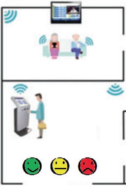
In a recent study published by Boos et al., it was found that cleanliness, waiting time, patient-staff communication, and especially courtesy of the receptionist were the most important factors for patient satisfaction (Boos et al. 2017). In their tertiary-care academic radiology department, they analyzed patient satisfaction surveys obtained either via online or via electronic kiosks. Interestingly, electronic kiosks generated higher patient response rates than online surveys (92.4% vs. 7.6%; p < 0.001), and the location of the electronic kiosks (Fig. 1) also influenced the patient response rates which were found to be lower in changing and waiting areas compared to those next to elevators (63.8% vs. 77.8%; p < 0.0001) (Boos et al. 2017).
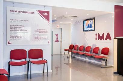
The importance of a good design in the radiology department environment was demonstrated by Holbrook et al. in their study showing that patients underestimated waiting times when the environment was specifically designed to optimize the patient experience (Holbrook et al. 2016). Their outpatient waiting room was set very well with the ultimate patient experience in mind: ample reading materials, multiple large-
WAITING ROOM
ELEVATORS HALL
Fig. 1 Electronic kiosks have become very popular nowadays with their colored faces on the screens, and are becoming popular also in imaging facilities. The best location is probably near the elevators or the exit [adapted by permission from Springer Nature: Serapicos M., Peixoto H., Alves V. (2017) A Hospital Service Kiosk in the Patient’s Pocket. In: De Paz J., Julián V., Villarrubia G., Marreiros G., Novais P. (eds) Ambient Intelligence–Software and Applications – eighth International Symposium on Ambient Intelligence (ISAmI 2017). ISAmI 2017. Advances in Intelligent Systems and Computing, vol 615. Springer, Cham. DOI: 10.1007/978-3-319-61118-1_27]
screen televisions (Fig. 2), free Wi-Fi, periodic offer of progress updates, warm blankets, and drinks by the team members, as well as electronic tablet devices (with games and Internet access), were the main components of this exquisite environment. In the end, shorter wait times were, as expected, associated with higher satisfaction scores, and the difference between perceived total waiting time and the actual interval between arrival time and exam start was statistically significant (p < 0.001) (Holbrook et al. 2016).
C. F. Silva et al.
Fig. 2 Large-screen televisions and free Wi-Fi are good options to optimize the patient experience. Perceived shorter waiting times are associated with higher satisfaction scores
3
Patient Education: Fear and Anxiety Alleviation
Patients are increasingly accessing Internetbased resources to obtain information about radiologic procedures they are to undergo. In order to make information about diagnostic and interventional procedures in radiology easily accessible from a single source, the Radiological Society of North America (RSNA) and the American College of Radiology (ACR) developed a website (RadiologyInfo 2018) for the public, explaining in lay terms the various diagnostic and interventional procedures using various imaging modalities such as X-ray, CT, MRI, ultrasound, and nuclear medicine, as well as a section for radiation therapy. RadiologyInfo.org website currently contains information of over 240 procedures, exams, and disease descriptions which can be viewed in English or Spanish. Besides interventional and pediatric radiology perhaps there is no subspecialty in radiology more prone to patient and family anxiety like breast imaging, as breast cancer is a very sensitive, high-rated, and mediatic issue. Just take the example of the monetary reimbursement for a low-dose CT scan for lung cancer screening in the United States: less than half for a mammogram (ACR 2018).
As physicians, diagnostic radiologists can create opportunities for patient interactions and therefore can be instrumental in guiding the
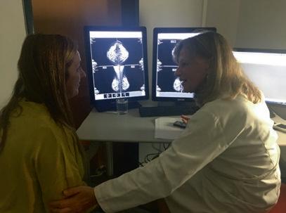
Fig. 3 Many different activities to promote emotional support for the patient can be performed, such as touching the patient on the hand or arm while giving bad news (Farber et al. 2002). Depicted in this figure: a face-to-face interaction between a patient and Dr. Teresa Guerra (with permission)
patient through the medical maze. Interventional radiologists are long known to have face-to-face interactions with patients due to the nature of their work, but radiologists specialized in breast imaging are in unique position to offer compassionate care and provide emotional support to patients when conveying bad news.
One study showed that in breast cancer survivors, anticipatory anxiety and pain catastrophizing were associated with a higher rate of not returning for mammograms (Shelby et al. 2012). Another study (Harvey et al. 2007) laid out a five-step approach in communicating bad news for radiologists specializing in breast imaging:
– Preparing for the encounter
– Disclosing the news
– Evaluating the patient’s response
– Discussing the next step
– Offering support (Fig. 3)
Even in the setting when a biopsy of a breast lesion yields benign results, there can still be high psychological burden in women. This does diminish with time but does not completely resolve (Schonberg et al. 2014). We recommend that the breast radiologist should convey the good news first to immediately relieve the anxiety, so that women will be better able to focus on further instructions.
4 Involvement of Family and Friends
Social support is a well-known, if not the most important, factor affecting one’s life satisfaction. Social support is particularly important when one faces adversities such as significant health morbidities which result in disability or significant change in lifestyle. Therefore, it is no surprise that patients will often share the diagnosis of their health calamities with family members and close friends. As the delivery of healthcare evolves, the inclusion of people important to the patients proves to be beneficial in affecting the outcomes of care. Besides tissue diagnosis of diseases, radiologists are often in a position to make the initial diagnosis through imaging, and at times a near-certain diagnosis of diseases including malignancy. Therefore, radiologists can improve the quality of care and patient satisfaction by including family members when providing information about imaging procedures or discussing abnormal findings (Itri 2015).
Harrison and Frampton (2016) argue that research design should also include engagement with patients and their families in an era of paradigm shift where patients are asked the question of “what matters most.”
5 The Quadruple Aim and the PFCC Model Coexistence
In the United States, a nonprofit, private organization called the National Committee for Quality Assurance (NCQA) provides accreditation and the “gold seal” for high-quality practices. NCQA started its model of high-quality care organizing around primary care in 2008, and in 2013 it broadened its scope to involve specialty practices called Patient-Centered Specialty Practice (PCSP) (NCQA 2018; Greene et al. 2017). The NCQA PCSP model has six pillars (NCQA 2018) that describe the core components of the PFCC framework:
– Provide access/communication
– Identify patient populations
– Track and coordinate referrals
– Plan and manage care
– Track and coordinate care
– Measure and improve performance
Key Points
• New PFCC (patient- and familycentered care) practices to pursue in radiology: Patient comfort, e.g., access and waiting times in imaging facilities, patient education, fear and anxiety alleviation, and involvement of all the stakeholders such as friends and caregivers.
• Increase visibility, increase value: Expanding the traditional field and tasks of radiology, like actively pursuing these PFCC practices, may be the most valuable weapon to fight the threat of commoditization of medical imaging.
Greene et al. illustrated the practical application of this model in the daily clinical practice of radiologists. Although the NCQA PCSP guidelines are well intended, the cost associated with implementation of new activities and the lack of increased payment from payers to offset the cost pose a real-life challenge (Greene et al. 2017). More rules and regulations can have unintended side effects to the medical practitioners. Increased bureaucracy without corresponding increase in clerical and other ancillary support can fuel job dissatisfaction and potentially lead to burnout.
Much debate has emerged in the last decade about the Triple Aim in Healthcare and using PFCC practices to achieve it. The Triple Aim was envisioned by Donald Berwick a decade ago (Berwick et al. 2008) to improve the patient experience of care, improve the health of populations, and reduce the cost of healthcare. Since then, with increasing reliance on metrics and methods to reduce cost, health practitioners are constantly under the pressure to increase productivity. The pursuit to maintain profitability
C. F. Silva et al.
by administrators and managers in the healthcare business also trickles down to the practitioners to treat more patients and perform more procedures. Bodenheimer et al. reminded us that there must be a fourth aim to balance the goals of the Triple Aim, i.e., to consider the wellbeing of health practitioners at and off work while keeping patient interest at the center of care (Bodenheimer and Sinsky 2014). This fourth aim was recently recognized and incorporated into the Declaration of Geneva (Hippocratic Oath) by the World Medical Association in 2017. The new sentence is “I will attend to my own health, well-being, and abilities in order to provide care of the highest standard” (BioEdge 2018; Parsa-Parsi 2017).
As the topic of physician burnout gains more attention in the lay media, a recent meta-analysis by Panagioti et al. in 2018 showed that the issue of physician burnout has trickle-down effect and may jeopardize patient care (Panagioti et al. 2018). In their analysis, patient safety incidents and suboptimal care owing to low professionalism were twice as likely to be related with burnout physicians, while receiving low satisfaction ratings from patients was three times more likely to occur with those affected physicians (Panagioti et al. 2018). Because of this untoward effect, Panagioti et al. suggested that healthcare organizations should invest in efforts to improve physician wellness, particularly for early-career physicians.
Radiologists are no exception to burnout. In fact, burnout in radiology was ranked above average when compared to other specialties. As physician burnout becomes more transparent in the medical community, some authors propose a seven-step solution which they call the Road to Wellness (Fishman et al. 2018). This includes acknowledging the problem, leadership commitment, finding solutions inside and outside of the workplace, and mindfulness of all involved. Engagement in leisure and outdoor activities, group relaxation practices, and social events with colleagues are some techniques to mitigate burnout (Fishman et al. 2018).
Key Points
• The (original) Triple Aim in Healthcare: improving the outcomes: (1) patient health, (2) patient satisfaction; and (3) reducing the costs.
• The (modern) Quadruple Aim in Healthcare: the mental and physical well-being of the physicians and other healthcare practitioners should be considered while improving the patient experience of care, improving the health of populations, and reducing the cost of healthcare.
• Hippocratic Oath (2017 version), NEW: “I will attend to my own health, well-being, and abilities in order to provide care of the highest standard.”
6 Summary
Medicine is in an era of transitioning from old practice of “paternalistic” medicine to modern practice where patients participate fully in their healthcare. PFCC practices are becomingly more mainstream and it is imperative for radiology practice to adapt as patients now have more choices than ever. This paradigm shift includes all facets of physician-patient encounter that can bring added value such as from the ease of scheduling an appointment, office visit and facility amenities, diagnostic testing, patient education, and active inclusion of all stakeholders important to the patient (family, friends, caregivers, etc.). The radiology profession should position itself to embark on this journey and actively participate in improving patient experience beyond generating imaging reports. In striving to achieve optimal experience, it should be noted that not all requests from patients are reasonable and some expectations can potentially be detrimental to health providers and their staff. Therefore, PFCC practices should be inclusive of everyone, and we should be mindful in
balancing the experience of patients and healthcare providers. Best health practices cannot be achieved by adopting a one-way street; best practices stem from mutual respect, mindfulness, and innate desire to help those in needs.
References
ACR (2018). https://www.acr.org/Media-Center/ ACR-News-Releases/2018/Nelson-Lung-CancerScreening-Study-Confirms-NLST-Results (Accessed 18 December 2018)
Berwick DM, Nolan TW, Whittington J (2008) The Triple Aim: care, health, and cost. Health Aff (Millwood) 27(3):759–769
BioEdge (2018). https://www.bioedge.org/bioethics/ new-hippocratic-oath-for-doctors-approved/12496 Accessed 9 December 2018
Bodenheimer T, Sinsky C (2014) From triple to quadruple aim: care of the patient requires care of the provider. Ann Fam Med 12(6):573–576
Boos J, Fang J, Snell A et al (2017) Electronic kiosks for patient satisfaction survey in radiology. AJR Am J Roentgenol 208(3):577–584
Farber NJ, Urban SY, Collier VU et al (2002) The good news about giving bad news to patients. J Gen Intern Med 17(12):914–922
Fishman MDC, Mehta TS, Siewert B et al (2018) The road to wellness: engagement strategies to help radiologists achieve joy at work. Radiographics 38(6):1651–1664
Greene AM, Bailey CR, Young M et al (2017) Applying the National Committee for quality assurance patient-
centered specialty practice framework to radiology. J Am Coll Radiol 14(9):1173–1176
Harrison J, Frampton S (2016 Dec) Patient and family engagement in research in era 3. J Am Coll Radiol 13(12 Pt B):1622–1624
Harvey JA, Cohen MA, Brenin DR et al (2007) Breaking bad news: a primer for radiologists in breast imaging. J Am Coll Radiol 4(11):800–808
Holbrook A, Glenn H Jr, Mahmood R et al (2016) Shorter perceived outpatient MRI wait times associated with higher patient satisfaction. J Am Coll Radiol 13(5):505–509
NCQA (2018). http://go.nationalpartnership.org/ site/DocServer/NCQA.SPR.FactSheet.2012. pdf?docID=11461. Accessed 9 December 2018
Panagioti M, Geraghty K, Johnson J et al (2018) Association between physician burnout and patient safety, professionalism, and patient satisfaction: a systematic review and meta-analysis. JAMA Intern Med 178:1317–1330
Parsa-Parsi RW (2017) The revised declaration of Geneva: a modern-day physician’s pledge. JAMA 318(20):1971–1972
RadiologyInfo (2018) (http://www.radiologyinfo.org). Accessed 9 December 2018
Schonberg MA, Silliman RA, Ngo LH et al (2014) Older women’s experience with a benign breast biopsy—a mixed methods study. J Gen Intern Med 29(12):1631–1640
Shelby RA, Scipio CD, Somers TJ et al (2012) Prospective study of factors predicting adherence to surveillance mammography in women treated for breast cancer. J Clin Oncol 30(8):813–819
C. F. Silva et al.
The Radiology Consult
Carlos Francisco Silva, Claus Peter Heussel, and Eduardo Mortani Barbosa Jr.
3 The University of Pennsylvania Embedded
C. F. Silva
Department of Diagnostic and Interventional Radiology, Translational Lung Research Center (TLRC), German Lung Research Center (DZL), University Hospital of Heidelberg, Heidelberg, Germany
C. P. Heussel
Department of Diagnostic and Interventional Radiology with Nuclear Medicine, Translational Lung Research Center (TLRC), German Lung Research Center (DZL), Thoraxklinik GmbH at University Hospital of Heidelberg, Heidelberg, Germany
E. Mortani Barbosa Jr. (*)
Director of Thoracic CT, Department of Radiology, University of Pennsylvania, Philadelphia, PA, USA e-mail: Eduardo.Barbosa@uphs.upenn.edu
A rise in radiology consult, in parallel with an ever-growing offer of value-based services, is currently increasing patient awareness of the radiologist’s role in clinical care. A German and a North American example of radiology consult are shown in this chapter. The German example, taken from the Radiology Department of the Thoraxklinik University Heidelberg, chaired by Prof. Dr. Claus Peter Heussel, will show different aspects like the workflow regarding severely immunocompromised patients being submitted to thoracic CT, the image-guided biopsy and re-biopsy of nodules or masses, regular tumor boards, and the Interstitial Lung Disease multidisciplinary conference (with pneumologist, radiologist, and pathologist). The University of Pennsylvania at Philadelphia embedded thoracic radiology reading room within an integrated Lung Center Clinic, is the North American example. A survey taken in this large tertiary academic medical center by Dr. Mortani Barbosa Jr. found an overwhelming positive response from the referring healthcare providers, and major positive impact on patient care and management. The most common reasons for consultation were to clarify interpretation of imaging studies and diagnoses, to assess for temporal changes, and in up to 25% of the cases to discuss management options. The radiology consult models we proposed can be implemented in most
19 Med Radiol Diagn Imaging (2019) https://doi.org/10.1007/174_2019_208,
Published Online: 02 July 2019
mid- to large-size hospitals. The radiologist as a consultant should be seen as the future but also a return to a past in which the interaction of radiologists and referring practitioners was the foundation of diagnosis and medical decision-making.
1 Introduction
Last years have witnessed the rise of radiology consult in parallel with the growing offer of value-based services, increasing patient awareness of the radiologist’s role in clinical care (Mangano et al. 2015; Gunn et al. 2015; Mortani Barbosa and Novak 2018). Wider availability and lower patient burden (short scan time resulting in seconds of breath-hold, lower radiation dose, and lower costs) caused higher acceptance of radiological services by patients. Joint image result interpretation together with the clinician taking the recent treatment into account to measure, i.e., oncological response, differential diagnosis including organ toxicities, and pseudo-progression, led to a higher value of imaging. Communication of examination findings directly to patients, explanation of interventional radiological procedures, and follow-up of these interventions are among the most frequent in breast, thoracic, and interventional radiology. These value added actions will probably survive the wide introduction of artificial intelligence applications in the radiological specialty. A European and a North American perspective of the pivotal aspects of the radiology consult are reviewed.
2 Radiology Consult:
The Thoraxklinik Heidelberg Experience
The practice of patient-centered care at the Radiology Department of Thoraxklinik University Heidelberg, chaired by Prof. Dr. Claus Peter Heussel, could be dated back to the 1990s when Heussel locally pioneered a workflow regarding severely immunocompromised
patients being submitted to thoracic CT (Heussel et al. 1997) instead of chest X-ray alone as done in immunocompetent ones. These immunocompromised patients deserved special attention, and as such a multidisciplinary discussion was set with the hemato-oncology team, regarding every single patient unique clinical features. Clearly a “one-size-fits-all” policy was not appropriate for the whole radiological care that was given to these frail patients, encompassing varied aspects such as CT protocol, reading, reporting, and communication. This workflow was well taken by clinicians and became standard of care nowadays.
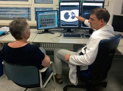
Since the beginning of this century, the imageguided biopsy of nodules or masses was becoming more and more frequent. Nowadays, re-biopsy of known tumors is adding further requests to interventional radiologists, as microbiological changes during treatment require additional attention. Therefore, a dedicated workflow was also set in motion since then. Every single patient that is submitted to interventional procedures in this department is beforehand subject to an interview with the attending thoracic radiologist that is going to ultimately perform the intervention on that respective patient. This establishes a personal relation between the interventional radiologist and patient, who later has to cooperate during the intervention as anesthesia is done locally only. Patients are presented with their own personal radiological findings on workstation screens (Fig. 1), which increases the awareness, motivation, and confidence for the intervention that will be performed.
A thorough explanation about the risks is given to every patient, including the major— pneumothorax, bleeding, death, stroke, and infection—as well as pertaining to patient anticoagulation, and of course about the benefits and safety of such procedures. In our experience, not a single patient that at the beginning was reluctant to being submitted to a biopsy or ablation remained reluctant or refused to do so after this consultation. After this informed consent, an informed consent is signed by both, and the patient is handed a copy including a self-explanatory CT image of the procedure, as well as a plan for which drug to continue or to stop
C. F. Silva
(anticoagulation), when to stop eating, when to appear in the hospital for the intervention, etc.
Besides regular tumor boards, which are nowadays integrated in all comprehensive cancer centers, the Interstitial Lung Disease (ILD) multidisciplinary conference (pneumologist, radiologist, pathologist) has been implemented. It takes place every week (Fig. 2), and that dates back to 2011 approximately, and we must say that once again the patient is the center of the care (Jo et al. 2016). We also have interdisciplinary conferences at the University of Pennsylvania for ILD and oncologic patients.
Fig. 1 A photo showing a radiology consult at Thoraxklinik Heidelberg. Prof. Dr. Claus Peter Heussel explains the patient where the nodule is located in her lung (with permission)
Every single case or thoracic CT is discussed with the referring pneumologist on site, and the patient (although not present in the room) knows precisely that his/her condition or disease is being submitted to a multispecialty analysis (pneumologist, radiologist, pathologist, thoracic surgeon, oncologist, radiation therapist) on that day, with the radiologist being a pivotal asset to assist the referring physician in the diagnosis and management of his/her illness. The protocol thereof becomes part of the patient’s record and is therefore transparent for patient and the entire treatment personnel.
3 The University of Pennsylvania Embedded Thoracic Radiology Reading Room Within an Integrated Lung Center Clinic
3.1 Background
In the United States, current decentralized healthcare reimbursement models compensate medical services through a resource-based relative value scale that assigns an arbitrary number of relative value units (RVUs) to every medical procedure or service, coded utilizing a system called current procedural terminology (CPT), in conjunction
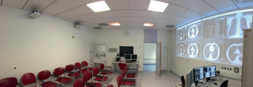
Fig. 2 A panoramic view of the multidisciplinary conference room at the Thoraxklinik. Two projectors held on the ceiling give the medical audience an excellent detail of what is depicted on the two radiologist’s high-resolution monitors. In the center of the image is also shown the
microscope and two small monitors for the pathologist. As the microscope can also be connected to one of the projectors, we can show microscopic and radiologic image side by side. Also clinical images (endoscopy, reports, lung function, etc.) can be shown side by side
with ICD-10 diagnostic codes. Each RVU has a monetary value that includes physician effort, practice costs, and geographic differences. This system is the so-called fee-for-service model, which strongly incentivizes volume and explicitly neither takes into consideration the quality of the services, nor patient outcomes.
Until recently, there were virtually no mechanisms to reward quality. In other words, the focus has historically been in producing more services, especially expensive ones, with little if any concern regarding the impact on patient outcomes or population health, therefore with little concern for value. We believe that the current US payment system is on an unsustainable course, given progressively rising costs to care for an aging population, and as physicians we ought to provide the highest possible value to society at large but at a reasonable cost, which necessarily implies making quality a centerpiece of future reimbursement models, at the same time we rein in costs. Measuring and promoting quality is not straightforward; however radiology can and should have a leading role in that endeavor.
Radiologists have traditionally practiced in relative isolation from other physicians, relying on their final product—the radiology report—as a means of communication with patients and referring physicians. While this is necessary for reimbursement and documentation, it is not sufficient to maximize our impact and the value we provide. Modern practice of medicine necessitates collaborative multidisciplinary discussions, and radiologists must become active consultants, providing useful guidance and assistance, to ensure our continued relevance in medicine. As we transition to valuebased care, radiologists must seek out ways to provide value beyond just generating a written report, by contributing to better quality patient care and outcomes, at a lower total cost. This can be done via in-person consultations, potentially conveying better information in a bidirectional fashion. How can this be accomplished in a busy, complex clinical environment? At the University of Pennsylvania, a large tertiary academic medical center in the Northeastern United States, we established a multidisciplinary Lung Center Clinic (LCC) encompassing clinic space and time for physicians seeing patients with thoracic diseases, with an integrated,
centrally located, radiology reading room staffed by thoracic radiologists and trainees throughout regular working hours (8 am to 5 pm), in which referring physicians can easily walk in at any time, without any appointment or bureaucracy, for in-person consultations. Sometimes, the patients themselves will come to review their images and discuss directly with their radiologist.
3.2 Our Clinical Setup and How We Measured Its Value and Impact on Workflow
The LCC-embedded radiology reading room was established nearly a decade ago, within a conference room in the clinic, and consists of two diagnostic radiology workstations, staffed by an attending thoracic radiologist and a resident or fellow through typical workday hours (8 am to 5 pm), on a daily basis as part of routine clinical schedule. Attending radiologists rotate through this location as well as the main reading room. The radiologist assigned to the LCC, whenever not engaged in consultations, reads examinations from the same work lists on PACS and shares the workload in a relatively balanced fashion with three or four additional thoracic radiologists, who are located in the main hospital reading room.
The University of Pennsylvania and the LCC in numbers
– The thoracic imaging section is staffed by ten subspecialty trained thoracic radiologists, four or five of them simultaneously on clinical service every day.
– Between 120 and 200 chest CTs and 300 and 500 chest radiographs daily.
– At the LCC, 1 attending radiologist and 1 fellow/resident provide between 5 and 30 consultations every day to physicians and advanced practitioners (nurses, physician assistants).
– The LCC practitioners see between 30 and 80 patients per day.
C. F. Silva et
While our qualitative experience with the LCC model is that it improves patient care and strengthens relationships between radiologists and referring physicians, we performed a study to quantitate how referring physicians assess the value of having continued, unhindered access to a thoracic radiologist while they are seeing patients, and documented their perceptions about this service. In parallel, we assessed the frequency, duration, and number of consultations and how these impact radiologist’s workflow.
In a recently published study (Mortani Barbosa and Novak 2018), we measured the utility and time commitment of our integrated thoracic radiologists in the multidisciplinary LCC with a consultancy log and a survey of referring physicians.
Over the course of 6 months, we measured the number, type, and duration of consultations in a consultancy log. We recorded 259 consultations over 44 clinical shifts (4 h each), in which 272 patients were discussed. Most consultations last between 2 and 5 min (75%), and in total consultations comprise approximately 10% of radiologist time, on average (though in busy days it can reach 25–30%).
The most common reasons for consultation:
– To clarify interpretation of imaging studies and diagnoses – To assess for temporal changes – In up to 25% of the times to discuss management options
In parallel, we ought to clarify the impact and value that we provide via a survey sent to the referring providers. The survey was comprised by the following questions:
Q1. How frequently do you review a case with a radiologist in the embedded LCC reading room?
Q2. How frequently do you review a case with a radiologist in the regular radiology reading room (main hospital)?
Q3. How useful is it to have a real-time inperson consultation with a thoracic radiologist during clinic?
Q4. What percent of these consultations benefit patient care or add clinical value, over and above receiving the dictated report alone?
Q5. What percent of these consultations change management, over and above receiving the dictated report alone?
Q6. What do you find most valuable about embedded thoracic radiologists in the LC?
We sent the survey to all qualified LCC healthcare practitioners, and obtained a response rate of 86.4% (51 out of 59 eligible providers), indicating that the referring providers feel strongly about and value the presence of a constantly available thoracic radiologist in the LCC.
The vast majority of providers (90.2%, n = 46) interact with thoracic radiologists in the embedded reading room at the LCC clinic at least once a week, and all respondents at least once a month. This demonstrates that this is a highly sought-after service. Not only do most providers seek the opinion of our thoracic radiologists frequently, but they also praise the quality of the service they get.
Key Results of the Survey
• Overwhelming positive response from the referring providers: unanimously rated the usefulness of this service as extremely high (90.2%) or high (9.8%).
• Major positive impact on patient care/management: 90.2% of providers believe that the consultations are beneficial in >60% of times; 51.0% think that these are beneficial in >80% of times; 86.2% of providers responded that their management changed because of the radiology consultation at least 50% of times.
• What providers found most valuable: rapid resolution of clinical questions or concerns (90.2%); deeper and higher level interaction with subspecialist chest radiologists (76.5%). The Radiology Consult
Multiple free-text comments were also entered, by 39.2% of respondents, including for example “This is an absolutely great program. Our radiology colleagues enable us to provide the best possible care to our patients. This service is EXTREMELY valuable to providing cutting edge, high quality care.” Qualitative analysis of free-text comments (which were provided by 20/51 respondents, 39.2%) revealed that all except one of the comments were highly positive and indicated a high level of satisfaction and praise for the service we provide.
In summary, our research demonstrates that having a radiologist in the LCC-embedded reading room always available for real-time consultations is deemed extremely valuable by the referring clinicians, who enthusiastically endorse the value we provide to patient care through optimal, expedited diagnosis and management, and hold our radiologist and the consultative services we provide in high esteem. At the same time, consultations comprise a significant proportion of radiologist time, which existing payment models in the United States do not account for.
3.3
Conclusions and Implications for the Future of Radiology
Our experience in our integrated LCC reading room over the past 9 years has been extremely rewarding. The overwhelming positive response and praise from providers in the LCC is a testament to the perceived substantial value generated by the on-site presence of a radiologist consultant integrated within the clinic. However, the value of having a radiologist embedded in the LCC is not compensated in any form; moreover, the numerous daily consultations take a substantial amount of time from the radiologist, diverting him/her from
RVU-producing activities, as the current feefor-service RVU-centric payment model in the United States does not account for radiology consultative services. In other words, the radiologists are penalized, from a compensation standpoint, for providing a service that is highly valued and praised by the referring physicians, who enthusiastically endorse their benefit to patient care and management. Clearly, there is misalignment of incentives.
Several alternative payment models, such as value-based purchasing, explicitly account for performance and quality metrics to either increase or decrease compensation, and mandates for implementation of such models will effect major changes in the healthcare landscape. Other models such as bundled payments or capitation are also being considered. Another possibility is a division of revenue from the clinic operation between the frontline physicians and the consultant radiologists. Payment models are currently in a state of flux and it is impossible to forecast what exactly will happen within the next decades; nonetheless, radiology will have to proactively demonstrate the value it can provide to frontline healthcare providers, patients, and payers, in order to defend and increase our relevance in the future. For that purpose, we strongly believe that becoming a consultant, who is actively, constantly, and directly involved in all facets of patient care—beyond merely generating a text report for an imaging study—will be the salvation of our specialty. We must continue to expand our scope of practice beyond an isolated reading room to integrate ourselves within clinical practices, wherever it may be from the hospital wards to the subspecialty clinics, to ensure we are relevant, helpful, and valuable to patients and referring providers and therefore ensuring the very best and most cost-effective care for all patients.
C. F. Silva et
Radiologists do much more than just generating text reports: – We are expert diagnosticians.
– We ensure optimal and safe utilization of imaging equipment by designing and overseeing imaging acquisition protocols.
– We foster appropriate imaging utilization utilizing best available evidence.
– We educate providers about most costeffective ways to diagnose and manage their patients utilizing imaging.
– We teach providers, from trainees to attending-level physicians, not only on imaging interpretation but also on complex diagnostic reasoning in challenging clinical settings.
– We provide consultations that impact patient diagnosis and management.
– We perform research that will change the future of medicine.
In the past, frontline physicians had to consult with radiologists primarily because they just did not have access to the images or reports on a timely fashion, but also because they thought that the consultations were positively impactful for their decision-making process. The difference is that nowadays, when we have nearly instantaneous access to images and very fast access to the reports (turnaround times were measured in weeks, rather than minutes, decades ago), we should strive to become consultants that will draw referring providers back to us, because the referring physicians want to directly discuss their patients with us, and not because they need to. And they would do it because they perceive that it is best for themselves and their patients, best for the quality of care they provide, best for expedite and accurate decision-making, and the
most cost-effective approach to diagnosis and management.
4 Summary
We have reviewed two different practices of radiology consult, both in the field of thoracic imaging subspecialty: the German, in the Thoraxklinik University Heidelberg, comprising not just diagnostic but also interventional consultancy services for both the patients and providers, and the North American, at the University of Pennsylvania in Philadelphia, where referring providers have an extremely positive opinion of the value provided by this consultative service as we have seen through the results of the survey our published research.
Nevertheless, current payment models do not incentivize this arrangement, and we must actively strive to change these. In the era of precision medicine and patient-centered care, we believe that our work suggests a pathway not only to better delivery of care, communication, and coordination among teams of healthcare practitioners, but also increases quality of care and may enable a brighter future for radiology and the continued relevance of our specialty, in spite of multiple threats. Our vision is that the radiologist should be an active, visible, available, crucial member of the healthcare team, practicing in the core of a patient-centered model that is organized and oriented towards enabling maximum quality of care with minimal delays, redundancy, and bureaucratic obstacles, by providing his/her expertise on a real-time basis. The integrated radiology practice setup we proposed can be implemented in most mid- to large-size hospitals. The radiologist as a consultant is at the same time the future of our specialty and also a return to a past in which the interaction of radiologists and referring practitioners was the foundation of diagnosis and medical decision-making.
References
Gunn AJ, Mangano MD, Choy G, Sahani DV (2015) Rethinking the role of the radiologist: enhancing visibility through both traditional and nontraditional reporting practices. Radiographics 35(2): 416–423
Heussel CP, Kauczor HU, Heussel G et al (1997) Early detection of pneumonia in febrile neutropenic patients: use of thin-section CT. AJR Am J Roentgenol 169(5):1347–1353
Jo HE, Glaspole IN, Levin KC et al (2016 Nov) Clinical impact of the interstitial lung disease multidisciplinary service. Respirology 21(8):1438–1444
Mangano MD, Bennett SE, Gunn AJ, Sahani DV, Choy G (2015) Creating a patient-centered radiology practice through the establishment of a diagnostic radiology consultation clinic. AJR Am J Roentgenol 205(1):95–99
Mortani Barbosa EJ Jr, Novak S (2018) The value of realtime thoracic radiology consulting in an Integrated Lung Center Clinic: bringing the radiologist to the Center of Multidisciplinary Health Care. J Thorac Imaging 33(4):260–265
C. F. Silva et al.
Value-Based Management of Incidental Findings
Sabine Weckbach and Oyunbileg von Stackelberg
S. Weckbach (*) · O. von Stackelberg
Diagnostic and Interventional Radiology, University Hospital Heidelberg, Heidelberg, Germany
Med Radiol Diagn Imaging (2019) https://doi.org/10.1007/174_2019_215,
Published Online: 02 July 2019
Radiological incidental findings (IFs) occur in research and diagnostic imaging. They are findings whose discovery is not intended. Due to a strong increase in high-tech imaging modalities the prevalence of IFs has risen. IFs regularly evoke discussions about accompanying ethical challenges. General principles to be considered in the management of IFs are responsibility for the well-being of the patient/ study participant and of the society. In order to avoid overdiagnosis and overtherapy and generate a high value for patients, radiologists as well as clinicians need to know how to manage IFs. Over the last years, important guidelines (“white papers”) for the handling of IFs have been published by different national and international imaging societies. Experiences from population-based research and measurement of value markers might trigger the further development and adaption of value-based guidelines.
1 Definition of Incidental Findings in Radiology
All findings could be called IFs when occurring in the context of medical diagnostics, potentially affecting the health of an individual and when not intended to be detected. However, it seems to be wrong to define an incidental finding only when
there is sufficient relevance for the health of a person. The concept of an incidental finding also includes marginal findings with no clinical relevance and false-positive findings (whose nature usually only becomes clear after further workup). IFs can occur in the context of research or while diagnostic methods are used to confirm or rule out the presence of a certain disease and also during follow-up examinations. Diagnostic findings that occur in the context of the doctor-patient relationship while searching for the cause of certain symptoms, but do not meet the doctor’s expectations, are not IFs. The occurrence of an incidental finding, in this way, is not unexpected. Nowadays it is basic knowledge that the use of high-resolution imaging yields a relatively high number of findings that were not aimed at detecting. IFs should therefore be characterized as unintended findings whose discovery was not intended by a treating physician or medical researcher (Wolf et al. 2008).
In the relevant literature, the concept of IFs is, however, often used in a narrower sense. Incidental findings are characterized by three features:
1. They occur in participants during a scientific study.
2. They potentially affect the health of the concerned participant.
3. They are findings the discovery of which was not intended in the context of the study’s aim.
Apart from research, an incidental finding can be described as a radiological finding not intentionally searched for, or an incidentally discovered mass or lesion, detected by an imaging modality, performed for an unrelated reason (Wolf et al. 2008 ; Schmücker 2012 ; Joerden et al. 2013 ).
Incidentally detected masses or tumorlike lesions are often called “incidentalomas,” especially if they affect the adrenal glands (adrenal incidentaloma). It is important to understand that the term incidentaloma is not a diagnosis, but only a description of how the lesion was detected. Not uncommonly, the term is incorrectly used to denote a benign finding. In fact, the
S. Weckbach and O. von Stackelberg
term incidentaloma says nothing about the character or etiology of the lesion found.
Key Points
–
Incidental findings occur in research and diagnostic imaging.
– They can be described as radiological findings not intentionally searched for, or an incidentally discovered mass or lesion, detected by an imaging modality, performed for an unrelated reason.
2 Ethical Aspects of Incidental Findings
Incidental findings regularly evoke a discussion about ethical challenges that accompany them. They don’t pose per se a critical ethical issue; they can be beneficial and lifesaving under certain circumstances. Under different circumstances though, such as when an incidental finding turns out to be a false positive, they can cause unnecessary followup or severe life-threatening complications (e.g., biopsy) and major distress (Schmidt et al. 2016). It is mainly the handling of IFs, then, that determines whether an incidental finding might be helpful or harmful, and therefore ethically critical or not (Erdmann 2015a, b; Fischer et al. 2015).
In December 2013, the Presidential Commission for the Study of Bioethical Issues (Bioethics Commission) released a report, Anticipate and Communicate: Ethical Management of Incidental and Secondary Findings in the Clinical, Research, and Directto-Consumer Contexts (Weiner 2014). The Bioethics Commission recommends that clinicians should have procedures in place including:
– Clear informed consent processes and communication strategies that convey the plan for managing IFs
– Use of tools to engage in shared decisionmaking with patients; strategies for effective communication about the risks associated with IFs and their follow-up
– Selective diagnostic testing to minimize the likelihood of IFs
– An awareness and understanding of existing clinical guidance for the management of incidental and secondary findings
3 General Principles to be Considered in the Management of Incidental Findings
• Responsibility for the well-being of the patient/study participant: A patient/study participant should be informed about health concerning IFs. This is in accordance with European and international ethical guidelines, e.g., Article 26 of the Additional Protocol to the Convention on Human Rights and Biomedicine, concerning Biomedical Research of the Council of Europe (Council of Europe 1999, 2007).
• Responsibility for the well-being of the society: The general population might be affected from undisclosed illnesses a participant might suffer from. This includes illnesses that might carry a risk for the patient/participant to cause a traffic accident. This is in accordance with Article 26 of the Convention for the Protection of Human Rights and Dignity of the Human Being with regard to the Application of Biology and Medicine: Convention on Human Rights and Biomedicine of the Council of Europe (Council of Europe 1999).
4 Frequency of Incidental
Findings
Generally, IFs can occur in clinical imaging and in clinical research. The frequency with which they occur naturally differs depending on the application of technique, the defined field of view, the purpose of the examination, as well as the expertise of the person who interprets the images. Since there has been a strong increase in the utilization and quality of high-tech imaging modalities as computed tomography (CT), magnetic
resonance (MR), and positron emission tomography (PET) imaging, IFs become increasingly prevalent. While whole-body MR scanning has become popular for large-scale population-based studies, the major cause for the detection of IFs in clinical imaging is CT scanning (Berlin 2011). In a recent retrospective study (based on radiology report review) of 1040 consecutive abdominal contrastenhanced CT examinations (mean age 66 years) relevant IFs, i.e., findings leading to further imaging, clinical evaluation, or follow-up, were found in 19% (Sconfienza et al. 2015). There was an increase with patient age. The distribution among the involved organs was kidneys (14%), gallbladder (14%), lung (12%), uterus (10%), adrenal (10%), and vessels (10%). In total, 39 different types of relevant IFs were made on the 1040 contrast-enhanced abdominal CT examinations.
Welch has summarized the appearance of IFs as follows: 50% in the lungs on chest CTs, 23% in the kidneys, 15% in the liver on abdominal CTs, and 67% in the thyroid gland on ultrasound of the neck (Gilbert Welch et al. 2011). His comprehensive review of the radiologic literature disclosed that less than 4% of lung nodules and overall less than 1% of IFS elsewhere evolve into a lethal carcinoma.
As examples of IFs in research imaging, the overall prevalence of IFs on brain MRI examinations in the Rotterdam Study (Bos et al. 2016) was 9.5% (549 findings in 5800 participants; mean age 64.9 years). Of these, most common are meningiomas and cerebral aneurysms. In the study of health in Pommerania (SHIP) there were 1330 IFs of potential clinical relevance in 904 subjects (36.2%). The abdominal organs (6.8%), the urinary tract (6.8%), and the skeletal system (6.0%) were affected most often. More than 50% of findings were of an unclear nature (Hegenscheid et al. 2013).
Key Points
– A strong increase in the utilization and quality of high-tech imaging modalities has led to an increasing prevalence of IFs.
– The major cause for IFs in clinical imaging is CT scanning.
5 Management of Incidental Findings in Research
Incidental findings in cohort studies is of course a rare subset of the whole IFS issue with only little effect on the healthcare system in contrast to the uncounted IFs in clinical imaging. Concerning value-based radiology, however, results from these cohort studies are very interesting and important in order to measure frequency and clinical relevance and set up appropriate guidelines and recommendations.
It is important to understand that there are different approaches concerning the management of IFs in different population-based imaging studies currently undertaken since so far no internationally obligatory guidelines exist. In this chapter, two quite different approaches to the management of IFs in the two largest population-based imaging studies currently undertaken in Europe are briefly depicted.
5.1 UK Biobank
The UK Biobank IF policy was developed following an extensive process which involved reviewing existing policies for the feedback of findings in UK Biobank participants, publishing evidence and guidance on IFs, receiving external legal advice on the scope of the duty of care, and consulting with the independent UK Biobank Ethics and Governance Council, UK Biobank’s major funders (Wellcome Trust and Medical Research Council), and with the Royal College of Radiologists and the Society and College of Radiographers. UK Biobank also consulted with relevant experts to explore the legal and ethical factors which were applicable to the development of the IF policy.
The UK Biobank IF protocol was developed from first principles as a pragmatic protocol that could be implemented on a large scale with the objective of striking the optimum balance of most net benefit and least net harm to 100,000 largely asymptomatic participants (Sudlow et al. 2015 ). Under this protocol, participants only receive feedback in specific, lim -
S. Weckbach and O. von Stackelberg
ited circumstances: when a radiographer identifies a potentially serious incidental finding during the acquisition or quality assessment of images during the imaging visit, and when a radiologist subsequently confirms the presence of a potentially serious IF. UK Biobank defines a potentially serious imaging incidental finding as “a finding which indicates the possibility of a condition which, if confirmed, would carry a real prospect of seriously threatening life span, or of having a substantial impact on major body functions or quality of life” (Sudlow et al. 2015 ).
5.2 German National Cohort (GNC)
The German National Cohort is a long-term, multicenter, population-based cohort study currently undertaken in Germany with the goal of investigating the development of common chronic diseases. As part of this investigation, 30,000 out of the total of 200,000 participants are being subjected to a whole-body 3-Tesla MR imaging without contrast agents. To help with the implementation of national and international ethical guidelines a system was developed to classify and report IFs that might be detected on imaging and possibly pose a risk to the participant’s health. This system focuses on guiding radiologists in the decision of reporting or not reporting a finding in an attempt to balance the risk of over- and underreporting, and thus to minimize false positives and false negatives. The cornerstone of that process is a list specifying findings and separating them into report-worthy and not-report-worthy. For defining IFs, studyspecific limitations and confounders had to be taken into account. Within this list, report-worthy IFs were classified into acutely relevant and nonacutely relevant findings. Acutely relevant findings were defined as suspected disease for which the participant should receive immediate clinical care. Examples include possible stroke, pneumothorax, and aortic dissection. These findings have to be reported in a timely manner not only for the benefit of the participant but also to avoid danger to the public, for example, from causing a traffic
accident. Non-acutely relevant findings are reported within 10 working days.
6 Management of Incidental Findings in Clinical Imaging
Before IFs even arise, radiologists have to decide about the appropriate imaging method: Is it possible to reduce IFs by choosing a different imaging method? When an incidental finding is then detected, we have to be aware that the description of an unexpected finding can trigger additional medical care including unnecessary tests, other diagnostic procedures, and treatments which in some cases may pose an additional risk to the patient and generate high costs or could be lifesaving in other cases.
We have to focus on value as well as outcomes and therefore ask the following questions:
1. Which IFs do we address?
2. How do we address IFs?
3. Do we need follow-up imaging or other diagnostic procedures?
4. Can undesirable consequences be avoided (Lumbreras et al. 2010)?
In order to especially avoid overdiagnosis and overtherapy and generate a high value for patients, radiologists as well as clinicians need to know how to deal with unexpected findings.
published several white papers on management of IFs on abdominal and pelvic CT and MRI, adrenal, vascular, splenic, nodal, gallbladder, biliary, thyroid, liver, renal and adrenal masses, pancreatic cyst, as well as pituitary findings and mediastinal and cardiovascular findings on thoracic CT (Berland et al. 2010; Gore et al. 2017; Heller et al. 2013; Herts et al. 2018; Khosa et al. 2013; Mayo-Smith et al. 2017; Megibow et al. 2017; Patel et al. 2013; Sebastian et al. 2013; Munden et al. 2018; Hoang et al. 2015, 2018; Berland 2013).
For further and detailed guidance, there have been several important guidelines published by different national and international imaging societies (“white papers”) for the handling of IFs in a daily clinical care over the last years. Since 2010 the ACR Incidental Findings Committee has
For example, the prevalence of noncalcified lung nodules on multi-detector CT (MDCT) has been reported as 33% (range 17–53%) and 13% (range 2–24%), in a screening and non-screening study population, respectively (Callister et al. 2015). However, the prevalence of malignancy in nodules measuring <5 mm is very low, ranging between 0% and 1% (Horeweg et al. 2014). Since the increase in the detection rate of small pulmonary nodules, the clinical significance of these findings represents a new challenge, and the optimal management of each case becomes pivotal and should be conducted according to the clinical setting (Larici et al. 2017). In 2017 the Fleischner Society has published a revised guideline for management of incidental pulmonary nodules. In this guideline the minimum threshold size for routine followup has been increased, and recommended follow-up intervals are now given as a range rather than as a precise time period to give radiologists, clinicians, and patients greater discretion to accommodate individual risk factors and preferences (MacMahon et al. 2017). Most of the guidelines however target radiologist and the recommendations assume a level of radiological expertise (O’Sullivan et al. 2018). Clinicians, particularly primary care physicians, are unsure which incidentalomas require urgent further investigation, nor are they sure which incidentalomas are likely to be benign (Sexton 2014). Systematic reviews on prevalence and malignancy of incidentalomas, such as the review by O’Sallivan et al., aid clinicians and patients to weigh up the pros and cons of requesting imaging scans and will help with
management decisions after an incidentaloma diagnosis (O’Sullivan et al. 2018).
Over the last years, the awareness for valuebased healthcare has risen. The Image Wisely® and Image Gently® initiatives (https://qpp.cms. gov/mips/explore-measures/quality-measures? py=2018&specialtyMeasureSet=Diagnostic%20 Radiology) have the goal to address radiation safety concerns and to decrease the exposure to the patient as well as to eliminate unnecessary procedures. It is important that radiologists are available for referring physicians to collaborate and advise how to handle IFs. The other aspect, which is extremely important, is to put always the patient at the center of care. A team-based approach with the referring doctor, the radiologist, the patient, and the administration will improve healthcare and continue to provide value.
Recently, the American Medical Association has started to publish criteria for value markers and to specify how value is measured referring also to IFs (https://qpp.cms.gov/mips/exploremeasures/quality-measures?py=2018&specialty MeasureSet=Diagnostic%20Radiology). This approach is new and the results have to be closely watched for the development and adaption of value-based guidelines in the future.
References
S. Weckbach and O. von Stackelberg
Key Points
– Overdiagnosis and overtherapy of IFs have to be avoided.
– IFs should be categorized into actionable (urgently/non-urgently) and non-actionable.
– Radiologists and clinicians need to know how to manage IFs.
– Detailed guidance is provided by several important guidelines published by different national and international imaging societies (“white papers).
– A team-based approach with the referring physician, the radiologist, and the patient with the patient in the center of care will lead to a successful, valuebased management of IFs.
Berland LL (2013) Overview of white papers of the ACR incidental findings committee ii on adnexal, vascular, splenic, nodal, gallbladder, and biliary findings. J Am Coll Radiol 10(9):672–674
Berland LL, Silverman SG, Gore RM, Mayo-Smith WW, Megibow AJ, Yee J, Brink JA, Baker ME, Federle MP, Foley WD et al (2010) Managing incidental findings on abdominal CT: white paper of the ACR incidental findings committee. J Am Coll Radiol 7(10):754–773
Berlin L (2011) The incidentaloma: a medicolegal dilemma. Radiol Clin N Am 49(2):245–255
Bos D, Poels MM, Adams HH, Akoudad S, Cremers LG, Zonneveld HI, Hoogendam YY, Verhaaren BF, Verlinden VJ, Verbruggen JG et al (2016) Prevalence, clinical management, and natural course of incidental findings on brain MR images: the population-based Rotterdam scan study. Radiology 281(2):507–515
Callister ME, Baldwin DR, Akram AR, Barnard S, Cane P, Draffan J, Franks K, Gleeson F, Graham R, Malhotra P et al (2015) British Thoracic Society guidelines for the investigation and management of pulmonary nodules. Thorax 70(Suppl 2):ii1–ii54
Council of Europe (1999) Convention for the protection of human rights and dignity of the human being with regard to the application of biology and medicine: convention on human rights and biomedicine. Council of Europe, Strasbourg, France
Council of Europe (2007) Additional Protocol to the Convention on Human Rights and Biomedicine, concerning Biomedical Research. Council of Europe Steering Committee on Bioethics, Strasbourg, France
Erdmann P (2015a) Zufallsbefunde aus bildgebenden Verfahren in populationsbasierter Forschung. Mentis, Münster
Erdmann P (2015b) Handling incidental findings from imaging within IM related research—results from an empirical-ethical study. In: Fischer T, Langanke M, Marschall P, Michl S (eds) Individualized medicine: ethical, economical and historical perspectives. Springer International Publishing, Cham, pp 231–250
Fischer T, Langanke M, Marschall P, Michl S (2015) Individualized medicine. Ethical, economical and historical perspectives. Springer, Heidelberg, pp 231–250
Gilbert Welch H, Schwartz LM, Woloshin S (2011) Overdiagnosed: making people sick in the pursuit of health. Beacon Press, Boston, MA
Gore RM, Pickhardt PJ, Mortele KJ, Fishman EK, Horowitz JM, Fimmel CJ, Talamonti MS, Berland LL, Pandharipande PV (2017) Management of incidental liver lesions on CT: a white paper of the ACR incidental findings committee. J Am Coll Radiol 14(11):1429–1437
Hegenscheid K, Seipel R, Schmidt CO, Volzke H, Kuhn JP, Biffar R, Kroemer HK, Hosten N, Puls R (2013) Potentially relevant incidental findings on research whole-body MRI in the general adult population: frequencies and management. Eur Radiol 23(3):816–826
Heller MT, Harisinghani M, Neitlich JD, Yeghiayan P, Berland LL (2013) Managing incidental findings on abdominal and pelvic CT and MRI, part 3: white paper of the ACR incidental findings committee II on splenic and nodal findings. J Am Coll Radiol 10(11):833–839
Herts BR, Silverman SG, Hindman NM, Uzzo RG, Hartman RP, Israel GM, Baumgarten DA, Berland LL, Pandharipande PV (2018) Management of the incidental renal mass on CT: a white paper of the ACR incidental findings committee. J Am Coll Radiol 15(2):264–273
Hoang JK, Langer JE, Middleton WD, Wu CC, Hammers LW, Cronan JJ, Tessler FN, Grant EG, Berland LL (2015) Managing incidental thyroid nodules detected on imaging: white paper of the ACR incidental thyroid findings committee. J Am Coll Radiol 12(2):143–150
Hoang JK, Hoffman AR, Gonzalez RG, Wintermark M, Glenn BJ, Pandharipande PV, Berland LL, Seidenwurm DJ (2018) Management of incidental pituitary findings on CT, MRI, and (18)F-Fluorodeoxyglucose PET: a white paper of the ACR incidental findings committee. J Am Coll Radiol 15(7):966–972
Horeweg N, van Rosmalen J, Heuvelmans MA, van der Aalst CM, Vliegenthart R, Scholten ET, ten Haaf K, Nackaerts K, Lammers JW, Weenink C et al (2014) Lung cancer probability in patients with CT-detected pulmonary nodules: a prespecified analysis of data from the NELSON trial of low-dose CT screening. Lancet Oncol 15(12):1332–1341
Joerden JC, Hilgendorf E, Thiele F (2013) Menschenwürde und Medizin. Duncker & Humblot, Berlin
Khosa F, Krinsky G, Macari M, Yucel EK, Berland LL (2013) Managing incidental findings on abdominal and pelvic CT and MRI, part 2: white paper of the ACR incidental findings committee II on vascular findings. J Am Coll Radiol 10(10):789–794
Larici AR, Farchione A, Franchi P, Ciliberto M, Cicchetti G, Calandriello L, Del Ciello A, Bonomo L (2017) Lung nodules: size still matters. Eur Respir Rev 26(146)
Lumbreras B, Donat L, Hernandez-Aguado I (2010) Incidental findings in imaging diagnostic tests: a systematic review. Br J Radiol 83(988):276–289
MacMahon H, Naidich DP, Goo JM, Lee KS, Leung ANC, Mayo JR, Mehta AC, Ohno Y, Powell CA, Prokop M et al (2017) Guidelines for management of incidental pulmonary nodules detected on CT images: from the Fleischner Society 2017. Radiology 284(1):228–243
Mayo-Smith WW, Song JH, Boland GL, Francis IR, Israel GM, Mazzaglia PJ, Berland LL, Pandharipande PV (2017) Management of incidental adrenal masses: a white paper of the ACR incidental findings committee. J Am Coll Radiol 14(8):1038–1044
Megibow AJ, Baker ME, Morgan DE, Kamel IR, Sahani DV, Newman E, Brugge WR, Berland LL, Pandharipande PV (2017) Management of incidental pancreatic cysts: a white paper of the ACR incidental findings committee. J Am Coll Radiol 14(7):911–923
Munden RF, Carter BW, Chiles C, MacMahon H, Black WC, Ko JP, McAdams HP, Rossi SE, Leung AN, Boiselle PM et al (2018) Managing incidental findings on thoracic CT: mediastinal and cardiovascular findings. A white paper of the ACR incidental findings committee. J Am Coll Radiol 15(8):1087–1096
O’Sullivan JW, Muntinga T, Grigg S, Ioannidis JPA (2018) Prevalence and outcomes of incidental imaging findings: umbrella review. BMJ 361:k2387
Patel MD, Ascher SM, Paspulati RM, Shanbhogue AK, Siegelman ES, Stein MW, Berland LL (2013) Managing incidental findings on abdominal and pelvic CT and MRI, part 1: white paper of the ACR incidental findings committee II on adnexal findings. J Am Coll Radiol 10(9):675–681
Schmidt CO, Sierocinski E, Hegenscheid K, Baumeister SE, Grabe HJ, Volzke H (2016) Impact of whole-body MRI in a general population study. Eur J Epidemiol 31(1):31–39
Schmücker R (2012) Zufallsbefunde – was gebietet die Menschenwürde? In: Preprints and working papers of the centre for advanced study in bioethics. WWU, Munster
Sconfienza LM, Mauri G, Muzzupappa C, Poloni A, Bandirali M, Esseridou A, Tritella S, Secchi F, Di Leo G, Sardanelli F (2015) Relevant incidental findings at abdominal multi-detector contrast-enhanced computed tomography: a collateral screening? World J Radiol 7(10):350–356
Sebastian S, Araujo C, Neitlich JD, Berland LL (2013) Managing incidental findings on abdominal and pelvic CT and MRI, part 4: white paper of the ACR incidental findings committee II on gallbladder and biliary findings. J Am Coll Radiol 10(12):953–956
Sexton SM (2014) How should we manage incidentalomas? Am Fam Physician 90(11):758–759
Sudlow C, Gallacher J, Allen N, Beral V, Burton P, Danesh J, Downey P, Elliott P, Green J, Landray M et al (2015) UK Biobank: an open access resource for identifying the causes of a wide range of complex diseases of middle and old age. PLoS Med 12(3):e1001779
Weiner C (2014) Anticipate and communicate: Ethical management of incidental and secondary findings in the clinical, research, and direct-to-consumer contexts (December 2013 report of the presidential commission for the study of bioethical issues). Am J Epidemiol 180(6):562–564
Wolf SM, Lawrenz FP, Nelson CA, Kahn JP, Cho MK, Clayton EW, Fletcher JG, Georgieff MK, Hammerschmidt D, Hudson K et al (2008) Managing incidental findings in human subjects research: analysis and recommendations. J Law Med Ethics 36(2):219–248, 211
The Value in Artificial Intelligence
Ramandeep Singh, Fatemeh Homayounieh, Rachel Vining, Subba R. Digumarthy, and Mannudeep K. Kalra
R. Singh · F. Homayounieh · S. R. Digumarthy
M. K. Kalra (*)
Division of Thoracic Imaging, Massachusetts General Hospital, Boston, MA, USA
Harvard Medical School, Boston, MA, USA
e-mail: mkalra@mgh.harvard.edu
R. Vining
Division of Thoracic Imaging, Massachusetts General Hospital, Boston, MA, USA
Past 5 years have seen burgeoning applications of machine learning (ML) in diverse radiological domains including thoracic radiology, neuroimaging, abdominal imaging, musculoskeletal imaging, and breast imaging. Deep learning technologies have been applied to improve image resolution at ultralow radiation dose. Publications abound on ML in chest CT have focused on detection and characterization of pulmonary nodules, as well as for rib and spine straightening and labeling, vessel segmentation, and estimation of CT fractional flow reserve. ML has also been applied for detecting lines, tubes, pneumothorax, pleural effusions, cardiomegaly, and pneumonia, on chest radiographs. Applications of ML in cerebral hemorrhage detection and prediction of stroke outcomes, appendicitis and renal colic prediction, hand bone age calculation or rib unfolding for fracture detection, and characterization of breast macro-calcifications and masses are also shown. We review fundamentals, applications, and limitations of machine learning in thoracic radiology, neuroimaging, abdominal imaging, musculoskeletal imaging, and breast imaging.
35 Med Radiol Diagn Imaging (2019) https://doi.org/10.1007/174_2018_193,
Published Online: 19 February 2019
Introduction
Artificial intelligence (AI), a term used for computers with cognitive functions akin to human intelligence, incorporates learning by perception decision-making for solving issues. Machine learning (ML) refers to the ability of computers to learn with human-devised algorithms as well as to improve from experience without being explicitly programmed (Erickson et al. 2017).
Alan Turing introduced the concept of machine learning in the seminal paper on “Computing machinery and intelligence” in 1950. He described the “Turing test” to determine if a computer has intelligence. In the test, a judge types questions for the two entities—a person and a computer—and then deciphers responses for human and computer identities. If the judge is wrong, the computer passes the Turing test and is deemed as intelligent. Almost 70 years later, a reversed form of Turing test is still used on the Internet, known as the CAPTCHA test, to determine whether the user is a human or a computer.
Key Points
–
Artificial intelligence: is a subspecialty of computer science devoted to creating systems to perform tasks that commonly require human intelligence.
– Machine learning: is a subfield of AI, referring to the ability of computers to learn with human-devised algorithms, as well as to improve from experience, without being explicitly programmed.
The credit for coining the term “computeraided diagnosis (CAD)” in the year 1975 goes to a remarkable radiologist, Gwilym S. Lodwick, who introduced application for image modeling, and computer-aided diagnosis for interpreting bone tumors on radiographs. Traditionally, CAD systems are classified as computer-aided detection (CADe) and computer-aided diagnosis
R. Singh et al.
(CADx). CADe localizes lesions, and CADx characterizes them (Firmino et al. 2016). Computer-aided detection/diagnosis (CAD) is based on image processing and interpretation using a large amount of information. A complex pattern recognition uses AI and computer vision. CAD server analyzes image data in a sequential manner for preprocessing noise and artifacts, and segmentation for differentiating anatomical structures and regions of interest for compactness and grey value. Finally, it uses different algorithms for classification/character analysis. The first report on CAD in imaging was published in 1963, and described the role of chest radiographs in predicting prognosis of primary lung cancer (Lodwick et al. 1963). The radiographic findings were converted into numerical codes for lesion location, size, shape, contour, density, and calcification, and then interpreted for malignancy by a computer. Limitations of CAD without ML included high false-positive rates which prolonged interpretation times, and at times led to additional biopsies (Firmino et al. 2016).
Key Points
– CAD: introduced by Gwilym S. Lodwick in 1975, a radiologist who was nominated for Nobel Prize, has serious limitations without machine learning.
In radiology, ML algorithms use radiological images, reports, and annotations as training datasets to generate a model with learning methods that can be either unsupervised or supervised. The input and output statistical probabilities are termed as nodes with included numeric weights known as hyperparameters. If the output data ensemble continuous values they denote a regression. The output data with categorized value are termed as classification (Chartrand et al. 2017). Training data are forward fed into a computation system, and then backpropagated over several cycles to form a