Research Paper
Botany
E-ISSN No : 2454-9916 | Volume : 3 | Issue : 5 | May 2017
ISOLATATION OF FUNGI FROM ENICOSTEMMA AXILLARE (LAM.) A.RAYNAL 1
Dr. Anil N. Korpenwar | Mrs. Deepmala A.Gaikwad 1 2
2
Principal, Rashtrapita Mahatma Gandhi Science and Arts College, Nagbhid, Dist. Chandrapur- 441205. (MS). India. Research Scholar, Shri Shivaji Science and Arts College Chikhli, Dist. Buldana- 443201. (MS). India.
ABSTRACT Enicostemma axillare (Lam.) A. Raynal. is commonly known as Katvinaye. The leaves used as a raw material for the preparation of some important drugs for curing various human diseases. Unscientific methods of storage causing fungal contamination .The fungal contamination affects on the chemical composition of raw materials and thereby decreases potency of drugs. Regarding the above fact the present study was conducted that the maximum 21 fungal species viz. Alternaria alternate, A. solani, Aspergillus flavus, A. niger, A. fumigat , A. nidulance, A. terreus, A. ustus,Curvularia lunata, Colletotrichum sp., Cladosporium sp., Drechslera sp.,Fusarium oxysporum, F. equiseti, Helminthosporium sp., Mucor globsus, Phoma .sp, Penicillium notatum, P. citrinum, Rhizopus stolonifer, Trichoderma viride were isolated from six months old authentic stored leaf sample and 12 from fresh fruit sample on Potato Dextrose Agar (PDA) method as compared with Czapek Dox Agar (CZA) medium and Moist Blotter method. KEY WORDS: Enicostemma axillare, Isolation, Fungi. INTRODUCTION: Enicostemma axillare (Lam.) A. Raynal ,belongs to family Gentianaceae , It is a glabrous perennial herb found throughout the greater part of India. The bittery natured plant acts as a laxative, helps in curing fever, rheumatism, skin diseases, abdominal disorders, and helps to regulate blood sugar levels (Snehlata , 2008),( Kirtikar and Basu,1999) The plant is traditionally used in the treatment of hepatic diseases and as a blood purifier. It also acts as ethnomedicine for snakebite . The leaves are fed to cattle to increase appetite. (Garg ,2000). Medicinal plants may be associated with a microbial contaminants, represented by bacteria, fungi and viruses. Inevitably, this microbiological background depends on several environmental factors and exerts an important impact on the overall quality of herbal products and preparations. The traditional methods of collection , storage and marketing coupled with humid climatic condition make them victim to the fungal contamination. (Masoumeh and Deokule, 2013) (Muntanola,1987) and (Durakovic et al., 1989) studied that the fungal contaminates has been reported to affect the chemical composition of the raw materials and thereby, decreases the medicinal potency of the plant material whereas mycotoxins produced by these fungal contaminants causes several effects on liver, kidney, genital organs, digestive tract, nervous system, skin, respiratory organs etc. The unscientific methods of harvesting, collection, storage of raw materials, post harvest processing, transport and storage of herbal drugs in unhygienic conditions, are the main causes considered to make both, raw materials as well as herbal drugs prone to microbial infections leading to deterioration in safety and quality and can also cause health hazard to consumer in spite to cure the disease (Pinkey, 2014), Many researchers have reported that the presence of potential contaminants in herbal preparations viz. (Czech et al., 2001),(Idu et al., 2011) (Martins et al., 2001), (Kulshrestha et al., 2008),(Alwakeel 2008),( Kosalec et al., 2009). The manufacturers should ensure the lowest possible level of microorganisms in the raw material, finished dosage forms and the packaging components to maintain appropriate quality, safety and efficacy of the natural products. ( Okunlola et al., 2007). According to the WHO, about 80% of the population of the world depends on traditional medicine, mostly herbal remedies, for their primary health care needs (Moerman ,1996) . Various pathogens adversely affect the medicinal plant parts and decrease the medicinal value of the part. It may be harmful to the human body while using these infected parts as a medicine. (Hamayun et al.,2004) So present investigation is an attempt to identify the mycoflora associated with the leaf sample of Enicostemma axillare (Lam.) A. Raynal.
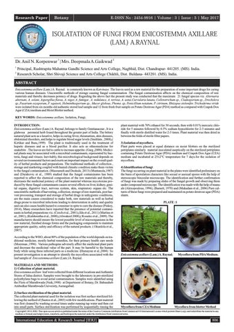
plant material with 70% ethanol for 30 seconds, then with 0.01% mercuric chloride for 5 minutes followed by 0.5% sodium hypochlorite for 2-3 minutes and finally with sterile distilled water for 2-3 times. Plant material was then dried in between the folds of sterile filter papers. 3) Isolation of mycoflora. Plant parts were placed at equal distance on moist blotters on the sterilized petriplates similarly material inoculated aseptically on the sterilized petriplates containing Potato Dextrose Agar (PDA) medium and Czapek Dox Agar (CZA) medium and incubated at 25±2°C temperature for 7 days.for the isolation of mycoflora. 4) Identification of fungi The fungi occurring on plant material in the plates were identified preliminary on the basis of sporulation characters like sexual or asexual spores with the help of stereoscopic binocular microscope. The identification and further confirmation of fungi was made by preparing slides of the fungal growth and observing them under compound microscope. The identification was made with the help of manuals (Alexopoulous, 1996), (Barnett, 1970) and (Mukadam et al., 2006) Pure cultures of these fungi were prepared and maintained on potato dextrose agar (PDA) slants. PLATE :1
Enicostemma axillare (Lam.) A. Raynal.
Mycoflora from PDA Medium.
MATERIALS AND METHODS: 1) Collection of plant material. Enicostemma axillare leaf were collected from different locations and Authentic Stores of Jalna district. Samples were brought to the laboratory in pre-sterilized polyethylene bags to avoid aerial contamination. Samples were identified using the Flora of Marathwada (Naik,1998) at Department of Botany, Dr. Babasaheb Ambedkar Marathwada University, Aurangabad. 2) Surface sterilization of the plant material. The collected plant material used for the isolation was first surface sterilized following the method of (Santos et.al., 2003) with few modifications. Plant material was first cleaned by washing several times under running tap water and then cut into small parts. Surface sterilization was performed by sequentially rinsing the
Mycoflora from CZA Medium
Mycoflora from blotter Method
Copyright© 2016, IERJ. This open-access article is published under the terms of the Creative Commons Attribution-NonCommercial 4.0 International License which permits Share (copy and redistribute the material in any medium or format) and Adapt (remix, transform, and build upon the material) under the Attribution-NonCommercial terms.
International Education & Research Journal [IERJ]
806
Research Paper
E-ISSN No : 2454-9916 | Volume : 3 | Issue : 5 | May 2017 Table. 1 Isolation of fungi from Enicostemma axillare (Leaf) . Media
Fungi
PDA Medium
CZA Medium
SMB Method
Ea1
Ea 2
Ea 1
Ea 2
Ea 1
Ea 2
Alternaria alternata
+
+
+
+
+
+
Alternaria solani
-
+
-
+
-
-
Aspergillus flavus
+
+
+
+
+
+
Aspergillus niger
+
+
+
+
+
+
Aspergillus fumigates
+
+
-
+
-
-
Aspergillus nidulance
-
+
-
+
-
+
Aspergillus terreus
+
+
+
+
+
+
Aspergillus ustus
-
+
+
+
-
+
Curvularia lunata
-
+
-
+
-
-
Colletotrichum sp.
+
+
-
-
-
+
Cladosporium sp.
-
+
-
+
-
+
Drechslera sp.
-
+
-
-
-
-
Fusarium oxysporum
+
+
+
+
+
+
Fusarium equiseti
-
+
-
+
-
Helminthosporium sp.
-
+
-
-
-
-
Mucor globsus
+
+
+
+
+
+ -
Phoma .sp
+
+
-
-
-
Penicillium notatum
-
+
+
+
-
-
Penicillium citrinum
+
+
-
+
+
+
Rhizopus stolonifer
+
+
+
-
-
-
Trichoderma viride
+
+
-
+
+
+
Total no. of isolates
12
09
16
08
12
+ = Fungi Present.
21
- = Fungi Absent.
Ea 1= Fresh Leaf Sample. Ea 2 = Stored Leaf Sample (six months old plant). RESULTS & DISCUSSION: In the present study the maximum fungal species were associated with six months old authentic Stores leaf sample of Enicostemma axillare as compaired with fresh fruit sample .The data presented indicate that maximum 21 fungal species viz. Alternaria alternate, A. solani , Aspergillus. flavus, A. niger, A. fumigates, A. nidulance, ,A. terreus, A. ustus, Curvularia lunata ,Colletotrichum sp., Cladosporium sp., Drechslera sp.,Fusarium oxysporum, F.equiseti, Helminthosporium sp., Mucor globsus, Phoma .sp, Penicillium notatum, P. citrinum, Rhizopus stolonifer,Trichoderma viride were isolated from six months old authentic stored leaf sample and 12 from fresh leaf sample on Potato Dextrose Agar (PDA) Method . Similarly in case of Czapek Dox Agar (CZA) method maximum 16 fungi were isolated viz. Alternaria alternate, A. solani, A. flavus, A. niger, A. fumigates, Aspergillus nidulance, A. terreus, A. ustus, Curvularia lunata, Cladosporium sp., Fusarium oxysporum, F. equiseti, Mucor globsus, Penicillium notatum ,p. citrinum, Trichoderma viride and 09 fungi on fresh sample as shown in table no.1.It is clear from table that 12 fungal species were observed from authentic Stores leaf sample and 08 from fresh sample on Standard Moist Blotter (SMB) Method. All fungi were identified on the basis of their cultural and morphological characteristics. (Roy, 2003) studied that the frequent occurrence of Aspergillus, Fusarium and Penicillium species on different crude herbal drugs. Total of one hundred and thirty two endophytes were isolated from different healthy parts such as leaves, stem, fruits and roots of the four ethno medicinal plants (Maroof,2012). (Dhale, 2013) observed that 45 fungi were recorded on the Blotter and Agar plate methods . All samples of plant material showed maximum infestation of A. niger and Aspergillus spp. (Sumanth et al., 2010) who isolated fungal genera from tested spices, found that the most common fungi isolated were Aspergillus spp. followed by Alternaria alternata, Cladosporium, Curvularia, Fusarium spp., Helminthosporium and Trichoderma show maximum incidence on Agar Plate Method.The some herbs are good substrate for Aspergillus flavus infestation and production of aflatoxins with potential hazard to the health of consumers ( Sharma et al., 2013) . (Jalgaonwala et.al. 2010) isolated 78 bacterial and 142 fungal endophytes from aerial and underground parts of various medicinal plants. (Santhosh et al., 2011) observed 41 endophytic fungi from 195 samples of healthy leaves and stem of a red listed endangered medicinal plant Coscinium fenestratum. The herbal preparations had the presence of fungal contaminants with predominance of Aspergillus spp. and Penicillium spp. (Kumar et al., 2009).similarly from the fruits of Trichosanthes tricuspidata 22 fungal species viz. Alternaria alternate, A. solani, A. flavus, A.
807
niger, A. fumigates, A. paraciticus, A. terreus, A. ustus, Curvularia lunata, Colletotrichum sp., Cladosporium sp., Drechslera sp., Fusarium oxysporum, F.equiseti, F. moniliforme , Helminthosporium sp., Mucor globsus, Phoma .sp, Penicillium citrinum, Rhizopus stolonifer, Trichoderma viride,Verticellium sp.were isolated from one year old authentic stored fruit sample and16 from fresh fruit sample on Potato Dextrose Agar (PDA) Method (Gaikwad and Korpenwar, 2017) .The fungal deterioration adversely affects the chemical composition of the raw materials and thereby decreases the medicinal potency of herbal drugs.respectively, supporting the findings of present investigations. In general, fresh fruit material showed decrease in the growth and incidence of fungi as compared with one year old authentic Stores fruit material of Enicostemma axillare . It was found both the Potato Dextrose Agar (PDA) Method and Czapek Dox Agar (CZA) Method are effective, routinely, consistently applicable and provide reliable results. CONCLUSION: The present study suggests that the methods of harvesting, collection, preparing and storage of medicinal plants must be improved for reducing percentage incidence of mycoflora and mycotoxins contaminations. ACKNOWLEDGEMENT: The authors express their sincere thanks to Ex. Prof & Head. Dr. S.R Manik, Department Of Botany, Sant Gadge Baba Amravati University, Amravati .We also thankful to Principal, Shri Shivaji Science And Arts College, Chikhli. for providing the necessary laboratory facilities. REFERENCES: 1. 2. 3. 4. 5. 6. 7.
Alexopolous, C. J. (1996): Introductory To Mycology, John Wiley and Sons, Inc.Publication, New York Winchester, Brisbane ,Tornoto And Singapore. Alwakeel. S.S. (2008). Microbial and heavy metals contamination of herbal medicines. Research Journal of Microbiology., 3(12), 683-691. Barnett. H.C. (1970): Illustrated genera of Fungi . imPerfecti, Burges Publication, Minn ( USA). Czech, E., Kneifel, W., and Kopp, B. (2001). Microbiological status of commercially available medicinal herbal drugs- A screening study, Planta Medica., 67, 263-269. Dhale, D . (2013). surface mycoflora of stored part of herbal medicine Int J pharm Bio Sci ., 4(3): (B) 568 – 574. Durakovic ,S., Galic, J. and Pajnovic, P.( 1989). Toxic and cancer metabolites of moulds in food and fodder. Hrana Iishrana., 30, 71-100. Gaikwad, Deepmala A. and Korpenwar, Anil. N.(2017). Mycoflora associated with the
International Education & Research Journal [IERJ]
Research Paper 8. 9. 10. 11. 12. 13.
14. 15. 16. 17. 18. 19. 20. 21. 23. 24. 25. 26. 27. 28. 29. 30.
E-ISSN No : 2454-9916 | Volume : 3 | Issue : 5 | May 2017
fruit of Trichosanthes tricuspidata (Lour.) during storage Bioscience Discovery, 8(2): 280-284. Garg, S .C. (2000). Ethnomedicine for Snake Bite, J Med Arom Plant Sci, 22(4a) & 23(1a), 546-553. Hamayun, M., Khan, M.A.and Begum, S. (2004).Marketing of medicinal plants of Utror-Gabral Valleys, Swat, Pakistan. Ethnobotanical Leaflets. Idu, M., Erhabor, J.O. and Idele, S.O. (2011). Microbial load of some medicinal plants sold in local markets of Benin City, Nigeria. International Journal of Medicinal and Aromatic Plants., 1(3): 272-277. Jalgaonwala, R.E., Mohite, B.V.and Mahajan, R.T.( 2010). Int. J. Pharmaceut. Biomed. Res., 1, 136-41. Kulshrestha, R., Gupta, C.P., Shukla, G., Kundu, M .G., Bhatnagar, S.P and Katiyar, C.K. (2008). The effect of water activity and storage temperature on the growth of Aspergillus flavus in medicinal herbs. Planta Medica.,74, 1308-1315. Kumar, A., Shukla, R., Singh, P and Dubey, N,K. (2009). Biodeterioration of some herbal raw materials by storage fungi and aflatoxin and assessment of ymbopogon flexuosus essential oil and its components as antifungal. International Biodeterioration &Biodegradation., 63: 712-716. Kosalec, I., Cvek, J and Tomic, S. (2009). Contaminants of medicinal herbs and herbal products. Archives of Industrial Hygiene and Toxicology., 60, 485-501. Kirtikar, K. R and Basu, B.D. (1999). Indian Medicinal Plants, Bishen Sing, 2 nd edition. Mahendra Pal Sing publication, Dehradun; 1655-1656 Martins, H.M, Martins, M.L, Dias, M.I and Bernardo, F. (2001). Evaluation of microbiological quality of medicinal plants used in natural infusions. International Journal of Food Microbiology., 68, 149-153. Maroof, Ahmed., Muzaffer, Hussain., Manoj, K. Dhar and Sanjana, Kaul.( 2012) . Isolation of microbial endophytes from some ethnomedicinal plants of Jammu and Kashmir. J. Nat. Prod. Plant Resour. , 2 (2):215-220. Masoumeh, Rashidi and Deokule, S. S. (2013). Associated fungal and aflatoxins contamination in some fresh and market herbal drugs. J. Microbiol. Biotech. Res., 3 (1):23-31. Moerman, D.E. (1996) An Analysis of the Food Plants and Drug Plants of Native North America. Journal of Ethnopharmacology; 52:1-22. Mukadam, D.S., Patil, M.S., Chavan, A.M, Patil, A.R.(2006):The Illustrations Of Fungi.Saraswati Printing Press.Aurangabad.(M.S) India.1-254. Muntanola, M. (1987). General mycology, Beograd: NIRO. Knjez evne novine Pp 25726922. Naik, V.N. (1998): Flora of Marathwada, Amrut Prakashan, Aurangabad (M. S.) India. Okunlola, A., Adewoyin, B.A and Odeku, A.O.(2007). Evaluation of pharmaceutical and microbial qualities of some herbal medicinal products in South Western Nigeria.Tropical Journal of Pharmaceutical Research., 6, 661-670. Pinkey, Khati. (2014).Mycoflora And Aflatoxin Assessment Of Crude Herbal Drugs During Storage in Haridwar, Uttarakhand, India Indian Phytopathology., 67 (4) : 407411 . Roy, A.K. (2003). Mycological problems of crude herbal drugs: Overviewand challenges. Indian Phytopath .,56: 1-13. Santos, R.M.G, Rodrigues-Fo, E., Rocha, W.C. (2003). MFS Teixeira; World J. Microbiol. Biotech., 19, 767-770. Santhosh .(2011) . Eurasia. J. Biosci., 5, 48-53. Sharma, Sumedha., Dimple, Gupta and Sharma, Y.P. (2013). Aflatoxin Contamination In Chilgoza Pine Nuts (Pinus gerardiana Wall.) Commercially Available In Retail Markets of Jammu, India. Int J Pharm Bio Sci Apr; 4(2): (B) 751 – 759. Snehlata, Bhandari., Upma, Dobhal., Mamta, Sajwan and Bisht, N.S. (2008). Trichosanthes Tricuspidata: A Medicinally Important Plant. Trees for Life Journal, www.TFLJournal.org,1-4. Sumanth, G.T., Wagmare, B.M and Shinde, S.R., (2010). Incidence of mycoflora from the seeds of Indian main spices. Afr. J. Agric. Res., 5(22): 3122-3125.
International Education & Research Journal [IERJ]
808