Research Paper Biological Science
E-ISSN No : 2454-9916 | Volume : 4 | Issue : 2 | Feb 2018
TRICHINELLA SPIRALIS BREAKOUTS IN THE BUENOS AIRES, ARGENTINA PROVINCE. A PENDING SOLUTION MATTER 1
1
1
2
José Lino Zumaquero Rios | Jorge Sarracent Pérez | Ygnacio Martínez Laguna |Pablo Aguirre | Ezequiel Scialfa 2 | Jorge Bolpe 2 1 2
Benemérita Universidad Autónoma de Puebla. Biological Science Faculty. Rural Zoonosis Department Ministry of Health of Buenos Aires, Argentina´s province.
ABSTRACT The following work conducts a research about three trichinosis outbreaks that took place in the province of Buenos Aires, Argentina in the year 2016. An inquiry was made to determine the risk factors associated with the epidemic outbreaks. The products elaborated with pig meat coming from the presumably infested swine, an enzymatic digestion was performed in order to find the cause of the outbreaks in humans. All the patients that stated they had consumed meat products elaborated from presumably infected animals and presented any symptoms, were included in the study. Two techniques for antibody detection in patients serum were used; IFI and ELISA. Furthermore, an antigen detection technique that allowed to determine the infection before the antibody determination studies, was evaluated. It is propound to include major sensibility and specificity techniques that allow an efficient and quick diagnose in epidemic outbreaks. At the same time, improve the sanitary education among population with a view to avoid the ingestion of uncertified meat or sub products. KEY WORDS: Epidemic outbreaks, Trichinosis, ELISA, IIF, Patients. INTRODUCTION: Trichinosis is a mammal parasite disease caused by the nematode Trichinella spp. It is a zoonosis of wide world distribution prevalent in China, India, Spain and other European countries; In America: Argentina, Canada and Chile frequent outbreaks have been reported ((Pozio & Darwin Murrel, 2006; Darwin Murrel & Pozio, 2011). It is produced when raw or inadequately cooked meat from infected animals is consumed by humans. Infection is more common in omnivores (horses, humans, pigs and rats) and carnivores (cats, dogs, and seals). Pigs and rodents play an important part in disease´s epidemiology (Gottstein et al, 2009). Trichinosis expresses symptoms and manifestations associated with other parasite entities (Ribicich et al, 2005 ; Calcagno et al 2014).
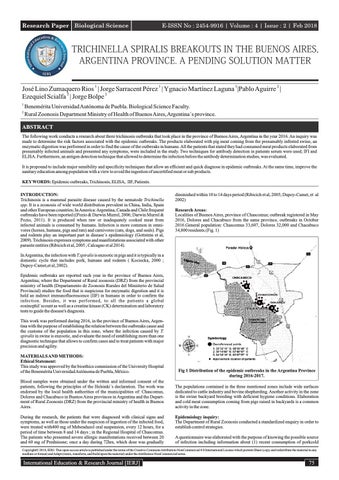
diminished within 10 to 14 days period (Ribicich et al, 2005; Dupoy-Camet, et al 2002) Research Areas: Localities of Buenos Aires, province of Chascomus; outbreak registered in May 2016, Dolores and Chacabuco from the same province, outbreaks in October 2016.General population: Chascomus 33,607, Dolores 32,000 and Chacabuco 34,800 residents.(Fig. 1)
In Argentina, the infection with T.spiralis is enzootic in pigs and it is typically in a domestic cycle that includes pork, humans and rodents ( Kociecka, 2000 ; Dupoy-Camet,et al, 2002). Epidemic outbreaks are reported each year in the province of Buenos Aires, Argentina; where the Department of Rural zoonosis (DRZ) from the provincial ministry of health (Departamento de Zoonosis Rurales del Ministerio de Salud Provincial) studies the food that is suspicious for enzymatic digestion and it is held an indirect immunofluorescence (IIF) in humans in order to confirm the infection. Besides, it was performed, to all the patients a global eosinophil´scount as well as a creatine kinase (CK) determination and laboratory tests to guide the disease's diagnosis. This work was performed during 2016, in the province of Buenos Aires, Argentina with the purpose of establishing the relation between the outbreaks cause and the customs of the population in this zone, where the infection caused by T. spiralis in swine is enzootic, and evaluate the need of establishing more than one diagnostic technique that allows to confirm cases and to treat patients with major precision and agility. MATERIALS AND METHODS: Ethical Statement: This study was approved by the bioethics commission of the University Hospital of the Benemérita Universidad Autónoma de Puebla, México. Blood samples were obtained under the written and informed consent of the patients, following the principles of the Helsinki´s declaration. The work was endorsed by the local health authorities of the municipalities of: Chascomus, Dolores and Chacabuco in Buenos Aires provinces in Argentina and the Department of Rural Zoonosis (DRZ) from the provincial ministry of health in Buenos Aires. During the research, the patients that were diagnosed with clinical signs and symptoms, as well as those under the suspicion of ingestion of the infected food, were treated with400 mg of Mebendazol oral suspension, every 12 hours, for a period of time between 8 and 14 days ; in the Regional Hospital of Chascomus. The patients who presented severe allergic manifestations received between 20 and 60 mg of Prednisone; once a day during 72hrs, which dose was gradually
Fig 1 Distribution of the epidemic outbreaks in the Argentina Province during 2016-2017. The populations contained in the three mentioned zones include wide surfaces dedicated to cattle industry and bovine shepherding. Another activity in the zone is the swine backyard breeding with deficient hygiene conditions. Elaboration and cold meat consumption coming from pigs raised in backyards is a common activity in the zone. Epidemiology inquiry: The Department of Rural Zoonosis conducted a standardized enquiry in order to establish control strategies. A questionnaire was elaborated with the purpose of knowing the possible source of infection including information about (1) recent consumption of porkcold
Copyright© 2018, IERJ. This open-access article is published under the terms of the Creative Commons Attribution-NonCommercial 4.0 International License which permits Share (copy and redistribute the material in any medium or format) and Adapt (remix, transform, and build upon the material) under the Attribution-NonCommercial terms.
International Education & Research Journal [IERJ]
75
Research Paper
E-ISSN No : 2454-9916 | Volume : 4 | Issue : 2 | Feb 2018
meat (2) if the meat came from a safe source or if it came from backyard raised and slaughter pigs. Several answers were obtained by most of the inquired persons. After the first evidences coming from patients with disease manifestations, meat sub product samples were collected in different corner stores to which enzymatic digestion tests were applied to detect larvae. In Chascomus case, the zone where the cases showed up, an inspection was conducted; the slaughtered animal´s owner was localized and samples from meat elaborated products were taken, from which a determination was made. In the same manner, the inquiries made to the persons in these localities ( Chacabuco and Dolores) pointed out that there was the presence of commercial activity without an official certification coming from meat pork sub products, activity that was referred by patients and sanitary authorities. Enzymatic digestion of meat sub products: Were taken 5gr of alimentary samples, cold and dry pork meat (chacinados) that were artificially digested in pepsin 1% (1:10,000 USA national standard formula) and hydrochloric acid 1%, employing a magnetic agitator(Gamble et al, 2000).Larvae were found and conserved in ethanol for further studies. Selection and study sample taking: All the patients that presented the symptoms were informed in a written way about the study´s nature. Personal data (Name, sex and age) was registered in each blood sample tube; 5ml of blood were taken from each patient or person that was considered suspicious of being infected. A total of 128 persons turned to the Hospital Municipal de Chascomus and presented first symptoms related to the outbreak of the disease, blood samples were taken to each one of them. From this total, some of these persons were asymptomatic at the moment when the first sample was taken, but some serologic tests were also made, because they had consumed cold meat coming from the infected animals. The blood samples obtained were incubated at 37°C for an hour and it was centrifuged at 1000g per 10 minutes to obtain the serum that was conserved at -80°C. To gather a second sample collection, persons where called back four weeks after to repeat the studies. In this occasion a total of 92 clinical samples were collected.
for an hour. After the recovering and after each incubation at 37°C, plates were washed 4 times with washing solution (PBS, Tween 20, 0.05%), the blocking of unspecific sites was made with 300 µL of PBS- albumin of bovine serum (BSA Sigma, A 4503, USA) at 2.5% per well for an hour at 37°C. Concluding the blocking time, 25 µL of PBS, Tween 20, 0.05% plus 75 µL of each person´s serum were added to evaluate per well, and it was incubated for an hour. After the washing, 2 µg/mL of monoclonal antibody against excretionsecretion antigen was added and incubated for an hour. At the end of the established incubation period and after the washing, 100 µL of peroxidase conjugated produced in sheep, anti mouse IgG (whole molecule)(Sigma, A 6782, USA) diluted 1/ 1000 in PBS, Tween 20, 0.05% was added. Following, to each well 100 µL substrateof Orto - Phenylenediamine dihydrochloride (OPD, Sigma, USA)in citrate-phosphate buffer were added for revealing purposes and plaques were maintained in darkness at environment temperature for 15 minutes. The reaction was detained by adding50 µL of H2SO4 2.5 M solution and the optic density was read at 492 nm though a micro plates reader (BioTek instruments, USA). Antibody in patients´ serum determination by indirect immunofluorescence: Muscular larvae of infected rats T. spiralis was obtained as described in Gamble et al (2000). Parasites were included in paraffin and incisions of 5µm were performed. The cuts were placed over the slides and then a dewaxing procedure was performed first with xylol at 60°C and xylol at room temperature, absolute ethanol at 97%, 70% and finally a distilled water rinse. Slides were left to dry at room temperature. Serial dilutions in patients' serum with PBS pH 7.2 were made and placed over the slides at 37°C for 20 minutes. Expiring the incubation period, 3 washes were made with PBS during 7 minutes each and a final rinse with distilled water, slides were left to dry at room temperature. Following the ´procedure, a conjugation of immunoglobulin total human anti marked with fluorescence was added. Samples were observed in the fluorescence (Leitz Dialux 20) microscope; with 25X y 40X magnification. Serum with fluorescence in dilution 1/32 were considered as probable positives and as positives 1/64 there on.
Determination of the eosinophilia and creatine-kinase: A determination of creatine-kinase (CK) was made to the patient´s serum through the commercial laboratory kit M Wiener UV unitest, Rosario, Argentina. The determinations were made at a temperature of 25°C and longitudinal wave of 340 nm. Besides, the presence of parasite´s antigens and antibodies against excretion-secretion antigens of T. spiralis by ELISA and by IFI were determined. The eosinophilia was determined by the eosinophil's global counting technique, expressed by normal values between 1 and 6%, that represent up to 350 leucocytes per microliters.
Patient´s treatment: As a general, two weeks after the infection´s (infected meat ingestion) persons who manifested the symptoms attended the hospital. Blood clinical samples for serologic studies were taken and it was decided to examine all the persons who had consumed meat and other products elaborated from the suspicious animals. The positive diagnosed patients were treated with Mebendazol 400 mg oral suspension twice a day for a period of 8 to 14 days.
Obtaining of secretion-excretion antigens: The secretion-excretion antigens from muscular T.spiralis larvae used in the ELISA of antibodies, were obtained by using the protocol described by De-laRosa et al. ( De-la-Rosa et al 2012).
The ones who presented allergic reactions and sever inflammation Prednisona between 20 and 60 mg was administrated once a day in a period of 3 or 4 days, which dose was gradually dimished between 10 to 14 days period (Ribicich et al, 2005; Dupoy-Camet et al, 2002)
Antibodies against excretion-secretion antigens in patients serum determined by ELISA: For the antibody IgG determination in patients´ serum against muscular larvae antigens of T. spiralis. MaxiSorp plates (Nunc, USA) were covered with 1 microgram/ mL of excretion secretion antigen diluted in carbonate-bicarbonate buffer ((NaHCO3 35 mM, Na2 CO3, 11 mM, pH 9.4-9.6),these were incubated at 37°C for 2 hours in a humid camera. Subsequently, plateswere washed 4 times with a washing solution (PBS, Tween 20, 0.05%), unspecificsites were blocked with 300 µL per well of PBS- albumin of bovine serum (BSA Sigma, A 4503, USA) at 2% for an hour at 37°C. Once passing the established blocking period, they were washed 4 times with washing solution and subsequently serum samples were added, 100µL per well. To find the best serum´s dilution, previous clinical trials were made and it was determined that the dilution 1:50 was the most adequate.
ELISA normal values: To establish the normal values of the non-infected population,100 students from the Faculty of Biological Science from the Benemérita Universidad Autonoma de Puebla, Mexico healthy without any illness symptoms, to which values of antibodies and T. spiralis antigen were determined by Elisa techniques The cut-off point was established as media ± 2 standard deviations. Obtaining the following values: Antigens, n= 100 Media ± 2DS
O.118 ±0.014
All the serums with optic density ≥ 0.150 were considered as positive. After an hour incubation period at 37°C in humid camera, plates were washed and 100 µL of anti-human IgG was added per well conjugated with peroxidase (Sigma, A6029, USA), diluted in 1:1000 in PBS, Tween 20, 0.05% PBS, bovine´s serum albumin 0.5%. Plates were incubated for an hour at 37°C in humid camera, after they were washed 6 times. Antibody binding was detected by addition of ortho-phenilendiamine in citrate-phosphate buffer pH 5, plates were maintained in darkness at room temperature for 15 minutes. The reaction was stopped when adding 50 µL of H2SO4 2.5 M solution, and the optic density was read at 492 nm through a micro plaque reader (BioTek instruments, USA). Results were expressed in Optic Density units (DO).
Antibodies, n= 100. Media± 2DS
0.210±0.019
All the serums with optic density ≥0.250 were considered as positive.
Antigen of excretion-secretion in patient´s serum presence determination: The antigen determination was made as it is described in Zumaquero-Rios et al (Zumaquero-Rios et al, 2012) with slight modifications.
RESULTS: In the present paper three illness outbreaks were notified, in localities from the Buenos Aires province in the municipalities of Chascomus in May 2016, and in Chacabuco and Dolores in October the same year. The interrogation to the patients demonstrated that the ingestion of artisanal products coming from pig´s meat like: (chorizo seco, ondiola and pancheta)were involved in the manifestations of trichinosis shown by the first patients who presented the symptoms.
Briefly, the Maxisorp plates were covered with 100 µL/mL per well of a solution that contained 10 µg/mL of rabbit polyclonal antibody against total muscular larvae of the parasite antigens, diluted in carbonate-bicarbonate buffer (pH 9.4-9.6)
Within the unspecific signs of the disease, retro-orbital pain, myalgia and high fever of 39°C (38.8%) were common. The palpebral edema and articular pains were mentioned by more than 90% of patients. Fig 2. Only 8 of them manifested
76
International Education & Research Journal [IERJ]
Research Paper
E-ISSN No : 2454-9916 | Volume : 4 | Issue : 2 | Feb 2018
long term diarrhea which appeared after the cold meat consumption. Table 1
DISCUSSION: European migration to Argentina in the beginning of the XX century, introduced the consumption of cold pork raw meat, as a result an increase in the number of trichinosis cases in humans has been presented (Ribicich et al, 2005). The 21% of the survey respondents, pointed out to know more than one person who in previous periods had been treated for trichinosis, and even though the 87% knew or had heard about the disease, they did not take the proposed measures to prevent the infection, because of the difficulty of not knowing the origin of the pig´s meat that they consumed. The rural activities are associated with the epidemic outbreaks that occur every year in the province of Buenos Aires, although the inspections and opportune diagnosis from the sanitary authorities in general, allows in the majority of the cases, the reference of manifestations and symptoms in a short term and it is well known, that in that zone there haven't been any cases of death related to trichinosis. Although, according to the information gathered in the inquiry, every social sector of the population, without making a difference between gender and age, there is a risk involving the possibility of contracting the infection because of the consumption of these high demand products. Until now, there is a lack of an effective vaccine in the market, against T. spiralis (Ortega Pires et al, 2015)
Fig 2.Male patient with positive antigen and elevated antibody titers showing bipalpebral edema Table 1: Clinical manifestations and symptoms among the patients of the 3 epidemic outbreaks of the Province of Buenos Aires. May- October 2016 Localidad Chacabuco n=44 Dolores n=14 Chascomus n=70
Cefalea Vómito Mialgia 6 13.6%
0
4 1 28.57% 7.14% 18 25.71%
0
Fiebre
Diarrea
Edema bipalpebral
14 31.8%
8 18.18%
5 11.36%
14 31.8%
3 21.42
0
1 7.14
2 14.28
20 28.57%
27 38.57%
17 24.28%
14 20%
The 100 percent of the presented cases with or without symptoms, showed an increment in the eosinophilia values, between 20 – 90%.The CPK was highly elevated between 60-85% detected in the positive cases within more than 100 and 1000u/l. Immunodiagnostic results. The values coming from the determination by the ELISA of antibodies and antigens outbreak are reflected individually (Table 2). Emphasizing that out of 24 patients from the first sample from the IIF, 10 turned out to be positive with a dilution of 1 / 32. However, there were other cases in superior dilutions. In the second sample the majority of positive cases showed high titles that reached the 1:256. The Western blot described by Zumaquero-Rios et al ( Zumaquero-Rios et al, 2012), was used as a gold test for a small amount of positive samples by ELISA of antibody (D.O. ≥ 1.5) against excretion-secretion antigens. The antigen detection showed evident diagnose superiority, since with the first sample 24 cases were detected with antibodies by IIF, while detecting the antigens by the ELISA technique, more than the double of the cases were found (Table 2). Table 2: Results of Trichinella spiralis antigen and antibody determinations among patients of epidemic outbreaks SEROLOGICS TEST LOCALITIES
IIF
ELISA (antigens)
Sample 2da 1ra 1ra 2da Chascomus number (n=70) (n=53) (n=70) (n=53) 17 48 46 Positives 6 6(1/32) 4(1/32) Dolores
Sample 1ra 2da number (n=14) (n=13) Positives
0
5 3(1/32)
2da 1ra (n=14) (n=13) 5
2
Sample 2da 2da 1ra 1ra Chacabuco number (n=44) (n=26) (n=44) (n=26) 7 15 Positives 2 15 4(1/32) 4(1/32)
ELISA (antibodies) IgG 1ra (n=70)
2da (n=53)
20
45
1ra (n=14)
2da (n=13)
4
4
1ra (n=44)
2da (n=26)
13
16
International Education & Research Journal [IERJ]
Previous studies performed in other epidemic breakouts worldwide, showed as guiding diagnosis this illness, the global eosinophil count (O'Connell & Nutman, 2015). There are numerous parasite infections where the eosinophil values are raised in an abrupt manner during the course of the infection, however in this work, patients did not belong to areas where parasite highly elevated illnesses (Paragonimiasis, Sarcocystosis, Fascioliasis, Schistosomiasis, and Gnathostomiasis) are reported. In a useful diagnose, it must be known if the eosinophilia has developed in a chronic or intense form. Parameter evaluation values should have previously been taken because it tends to diminish to normal values after being treated for 6 months. In the infections caused by helminthes, and in the case of trichinosis, eosinophil becomes pronounced at the beginning of the infection, allowing the larvae migration through the tissue which diminishes slowly with time. The determination of aminotransferases (TGP) constitutes an important element that could not be evolved in patients because it was not previously indicated and the values did not appear registered in the patient files, restricting aspect for the possible interpretation of tissue damage that could be presented. Despite in the file there was not a reference towards hepatomegalies or other disorders, there was not jaundice in any of the presented cases that could justify the use of all the recommended techniques. The increment of the CPK could be an indicative of intense myocardial infarction episodes, but it could also be indicated in infections caused by Trichinella sp, could be used depending on the epidemic conditions (Sharma et al, 2014). It was demonstrated in the serologic studies that the excretion-secretion antigens determination using the monoclonal antibody class IgM is effective at the beginning of the parasite´s infection (when patients arrive to the clinic, some days after the cold meat ingestion) and the serum antigen level values between 5 to 6 weeks drop in the majority of patients. In rodents, when the antigen´s kinetic was studied using the same technique, these were kept high for a longer period of time ( Zumaquero-Rios et al, 2012) This makes this technique convenient for the infections confirmation, when the first symptoms appear and allow to confirm the outbreak and initiate the investigation and control actions. The antibody class IgG determination by ELISA against secretion-excretion parasite antigens, is the only proved trial for surveillance and epidemiologic research in infections and outbreaks in domestic animals and wild life, (Gottstein et al, 2009), (Xue et al 2017) although the IIF is accepted to detect antibodies against the parasite as a second option (Gottstein et al, 2009). In this work, the ELISA of antibody determination was also superior to the IFI, which is a technique that rely on the observer's subjectivity, overall, when the antibody title is low (1 / 32). Furthermore, The IIF used by the Centro de Zoonosis de la provincial de Buenos Aires employs total antigens of T. spiralis, fact that may rise the number of false positives because a crossed reaction with other parasites; and requires from the study additionally second sets of samples during advanced illness stages in order to confirm the seroconversion cases. On the other hand, the antibody determinations from both methods (IIFand ELISA) possess as an inconvenient their low sensibility at the beginning of the infection, where it is only possible to detect a few cases when they were compared with the antigen detection. Although the antibody detection against excretion-secretion antigens from muscular larvae of the parasite by ELISA is the most recognized method for the trichinosis (Gottstein et al 2009)diagnostic, its disadvantages are the false negatives In early stages of infection and the crossed reaction with other helminthiasis. (Wang et al, 2013), (Yang et al, 2016) Several proteins resulting from infectious larvae and excretion-secretion anti-
77
Research Paper
E-ISSN No : 2454-9916 | Volume : 4 | Issue : 2 | Feb 2018
gens of adult worms have been identified as possible antigens for early diagnose of trichinosis (Wang et al, 2017), (Sun et al, 2015) (Liu et al, 2016) with the intention of solving the early detection problem in the parasite infection. There has also been explored the ADN detection in feces through PCR (Liu et al, 2017). CONCLUSION: This research shows the need to include another method to strengthen the diagnose capacity and disease's confirmation, for the research, control and specific treatment for humans, in the beginning of the outbreak, at the same time it allows to suggest the passive surveillance based in hospital records, it is probable to underestimate the real load of trichinosis in the zone because of the lack of notification from some patients that do not attend to the health care system. These outbreaks emphasize the need of implementing an educational program in the population about the risks of acquiring this disease, and the importance of cooking meat adequately. Furthermore, authorities and swine owners should be recommended to conduct parasite surveillance, an effective rodent control and improve swine breeding. REFERENCES: 1.
Calcagno MA, Bourlot I, Taus R, Saracino MP & Venturiello SM (2014). Description of an outbreak of human trichinellosis in an area of Argentina historically regarded as Trichinella free: the importance of surveillance studies. Veterinary Parasitology. 200: 251-256.
2.
Darwin Murrell K & Pozio E (2011). Worldwide occurrence and impact of human trichinellosis, 1986–2009.Emerging Infectious Diseases 17: 2194–2202.
3.
De-la-Rosa-Arana JL, Campos-Rodríguez RV, Rivera-Aguilar V, Escobar-Gutierrez A, Millar-Garcia A, Herrera-Gonzalez NE & Jarillo-Luna A (2012). Comparative effects of levamisole, Staphylococcus and Freund´s adjuvant on rat immunization with excretory and secretory antigens of Trichinella spiralis muscle larvae. Parasitology Research. 111: 1599-1605.
4.
Dupoy-Camet, J, Kociecka W, Bruschi F, Bolas-Fernandez F & Pozio, E (2002). Opinion on the diagnosis and treatment of human trichinellosis. Expert Opinion on Pharmacotherapy3: 1117-1130.
5.
Gamble HR, Bessonov AS, Cuperlovic K, Gajadhar AA, Van Knapen F, Noeckler K, Schenone H & Zhu X (2000). International Commission on Trichinellosis: recommendations on methods for the control of Trichinella in domestic animals intended for human consumption. Veterinary Parasitology 93: 393-408.
6.
Gottstein B, Pozio E & Nockler, K (2009). Epidemiology, diagnosis, treatment and control of trichinellosis. Clinical Microbiology Review. 22: 127-145.
7.
Kociecka W (2000). Trichinellosis: human disease, diagnosis and treatment. Veterinary Parasitology 93: 365-383.
8.
Liu RD, Jiang P, Wen H, Duan JY, Wang LA, Li JF, Liu CY, Sun GG, Wang ZQ & Cui J. (2016). Screening and characterization of early diagnostic antigens in excretory secretory proteins from Trichinella spiralis intestinal infective larvae by inmunoproteomics. Parasitology Research. 115: 615-622.
9.
Liu XL, Ren HN, Shi YL, Hu CX, Song YY, Duan JY, Zhang HP, Du XR, Liu RD, Jiang P, Wang ZQ & Cui J (2017). Early detection of Trichinella spiralis DNA in the feces of experimentally infected mice by using PCR. Acta Tropica 166: 351-355.
10. O'Connell EM & Nutman TB (2015). Eosinophilia in Infection Disease. Immunology and Allergy Clinics of North America. 35:493-522. 11. Ortega-Pires G, Vaquero-Vera A, Fonseca-Liñan R, Bermudez- Cruz RM, & ArguelloGarcia, R (2015). Induction of protection in murine experimental models against Trichinella spiralis: an up-to-date-review. Journal of Helminthology .89: 526-539. 12. Pozio E, & Darwin Murrel, K (2006). Systematics and epidemiology of Trichinella. Advances in Parasitology. 63: 367-439. 13. Ribicich M, Gamble HR, Rosa A, Bolpe J, & Franco A (2005). Trichinellosis in Argentina: an historical review. Veterinary Parasitology 132: 137-142. 14. Sharma RK, Raghavendra N, Mohanty S, Tripathi, BK, Gupta B, & Goel A (2014). Clinical and biochemical profile of trichinellosis outbreak in north India. Indian Journal of Medical Research 140: 414–419. 15. Sun GG, Liu RD, Wang ZQ, Jiang P, Wang L, Liu XL, Liu CY, Zhang X, & Cui J( 2015). New diagnostic antigens for early trichinellosis: the excretory secretory antigens of Trichinella spiralis intestinal infective larvae. Parasitology Research. 114: 4637-4644. 16. Wang L, Wang ZQ, Hu DD & Cui J (2013) Proteomic analysis of Trichinella spiralis muscle larval excretory-secretory proteins recognized by early infection sera. BioMed Research International 2013, Article ID 139745, 7 pages, http://dx.doi.org/10.1155/2013/139745 17. Wang ZQ, Liu RD, Sun GG, Song YY, Jiang P, Zhang X, & Cui J (2017). Proteomic analysis of Trichinella spiralis adult worm excretory- secretory proteins recognized by sera of patients with early trichinellosis. Frontiers in Microbiology 8, 986, doi 10.3389/fmicb.2017.00986. 18. Xue B, Xiaoxiang H, Xiaolei L, Bin, T, & Mingyuan L (2017).Current research of trichinellosis in China. Frontiers in Microbiology 8, 1472, doi 10.3389/fmicb 19. Yang Y, Cai YN, Tong MW, Sun N, Xuan YH, Kang YJ, Vallee I, Boireau P, Cheng SP, & Liu MY (2016) Serological tools for detection of Trichinella infection in animals and humans. One Health. 4: 25-30. 20. Zumaquero-Rios JL, Garcia-Juarez J, De-la-Rosa-Arana JL, Marcet R, & SarracentPerez J (2012) Trichinella spiralis: monoclonal antibody against the muscular larvae for the detection of circulating and fecal antigens in experimentally infected rats. Experimental Parasitology. 132: 444-449
78
International Education & Research Journal [IERJ]