Research Paper
Health Science
E-ISSN No : 2454-9916 | Volume : 3 | Issue : 6 | June 2017
PRIMARY MANDIBULAR 2ND MOLAR WITH FOUR ROOTS AND FIVE ROOT CANALS: A RARE ENTITY 1
2
3
4
Dr. Noopur Kaushik | Dr. Nikhil Srivastava | Dr. Vivek Rana | Dr. Neha Sharma | Dr. Priyadarshini Bhatia
5
1
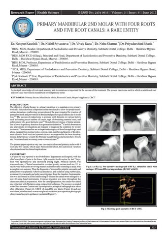
BDS , MDS, Reader, Department of Paedodontics and Preventive Dentistry, Subharti Dental College, Delhi – Haridwar Bypass Road, Meerut – 250005. 2 BDS, MDS FICD (Glasg), Principal and Head, Department of Paedodontics and Preventive Dentistry, Subharti Dental College, Delhi – Haridwar Bypass Road, Meerut – 250005. 3 BDS, MDS, Professor, Department of Paedodontics and Preventive Dentistry, Subharti Dental College, Delhi – Haridwar Bypass Road, Meerut – 250005. 4 BDS, MDS, Department of Paedodontics and Preventive Dentistry, Subharti Dental College, Delhi – Haridwar Bypass Road, Meerut – 250005. 5 Post Graduate 3rd Year, Department of Paedodontics and Preventive Dentistry, Subharti Dental College, Delhi – Haridwar Bypass Road, Meerut – 250005. ABSTRACT An in depth knowledge of root canal anatomy and its variations is important for the success of the treatment. The present case is one such in which an additional root and root canal was found in mandibular second primary molar. KEYWORDS: Primary Second Mandibular Molar, Five root Canals, Mayne's appliance, CBCT. INTRODUCTION: The objective of pulp therapy in primary dentition is to maintain every primary tooth as a fully functional component in the dental arch to allow for proper mastication, phonation, swallowing, preservation of the space required for eruption of permanent teeth and prevention of detrimental psychological effects due to tooth loss.[1,2] The success of pulpectomy in primary teeth depends on various factors such as locating exact number of canals, type of obturating material used, and achievement of a good hermetic seal.[3] Though the prevalence of dental anomalies is lower in primary dentition than permanent dentition.[4] Careful observation and appropriate investigations are required to diagnose the condition for proper treatment. These anomalies are an important category of dental morphologic variations ranging from normal color, contour, size, number and degree of development of teeth. Both local and systemic factors are responsible for these developmental disturbances. Usually the Primary mandibular second molars have 2 roots and 3 root canals and accessory roots existence is rare.[5] The present paper reports a very rare case report of second primary molar with 4 roots and five canals, which urges Paedodontist about, the anatomical variation of root canals and its clinical implications. CASE REPORT A 5-year old boy reported to the Pedodontics department and hospital, with the chief complaint of pain in the lower right posterior tooth region for last 7 days. Pain was spontaneous and increased during night. Medical history was noncontributory. Clinical examination revealed grossly carious tooth no. 85, xray showed deep caries involving enamel and dentine and extending to pulp, with a complex root anatomy. Thus the diagnosis of irreversible pulpitis was made and pulpectomy was planned. After local anesthesia and isolation using rubber dam, access cavity was made and pulp was extirpated from the chamber. Instrumentation was performed in all the canals using H-file and the canals were enlarged to a size 40 using hand instruments. Copious irrigation was done throughout the chemomechanical preparation. After the canal preparation, the canals were dried and obturated with Metapex using compaction technique. The access was sealed with Glass ionomer Cement and a postoperative periapical radiograph was taken after obturation (Figure 2). CBCT of mandible was taken (Figure 2) and one week later, stainless steel crown was placed and a final post operative photograph of crown and mayne's appliance was taken (Figure 3).
Fig 1: (A-B) (A). Pre operative radiograph of 85 b,c. obturated canal with metapex 85 from different angulations. (B) SSC with 85.
Fig 2: Showing post operative CBCT of 85.
Copyright© 2016, IERJ. This open-access article is published under the terms of the Creative Commons Attribution-NonCommercial 4.0 International License which permits Share (copy and redistribute the material in any medium or format) and Adapt (remix, transform, and build upon the material) under the Attribution-NonCommercial terms.
International Education & Research Journal [IERJ]
303
Research Paper
E-ISSN No : 2454-9916 | Volume : 3 | Issue : 6 | June 2017 mandibular molars usually have three root canals, viz. Mesiobuccal, mesiolingual and distal canal but Naser et al conducted a study and found that all mandibular primary second molar have four canal: Mesiobuccal, mesiolingual, distobuccal and distolingual.[11-14] Based on the above classification, case presented could be considered as variant 6 had four roots (Mesiolingual, mesiobuccal, distobuccal and distolingual) and five root canals (two mesial canals and three root canals on two distal roots). As the incidence of variation is low, paediatric dentist should be cautious and observant while performing clinical procedures.
(A)
(B)
Fig 3 : (A-B) (A) Pre operative photograph. (B) crown and mayne's appliance i.r.t. 85. DISCUSSION A huge number of variations may occur in size, shape, number of roots and accessory canals. Therefore thorough knowledge of root canal anatomy and its variation is essential for the success of pulpectomy. The prevalence of dental anomalies and variations are more common in primary than in permanent dentition as reported by W.C Baker.[6] The frequency of three rooted mandibular molars has been found to be less than one percent in primary dentition (Tratman 1938) but occurrence of a four rooted primary molar with five canals is very infrequent.[7] When there anatomical variations occur, clinicians often encounter difficulty in locating, preparing and obturating these root canals. The use of Routine intraoral radiographs with different angulations aid in detecting the presence of additional roots.[4] Certain Indian and Mongoloid populations have occurrence of an extra distal root in primary second molar and are considered as a racial characteristic. 8 Rana et al. (2011) stated a case of three roots with five root canals in grossly decayed primary second mandibular molar.[9] Yang et al. (2013) assessed 487 primary second mandibular molars using CBCT and found seven categories of variants in the root canal anatomy of primary mandibular second molars (Figure 4).[10]
CONCLUSIONS The success of endodontic treatment is dependent upon correct diagnosis for which through knowledge of root canal anatomy and possible awareness absolute awareness of the radiographic limitations and instrumentation procedure etc is required. Knowledge of complexity of primary mandibular molar like in present case is important as failure of obturation of undiagnosed canals can lead to failure of root canal procedure. REFERENCES 1.
Dummett CO Jr, Kopel HM. Pediatric endodontics. In: Ingle JI, Bakland LK, editors. Endodontics. 5thed. Hamilton: BC Decker Inc.; 2002. p. 861‐902.
2.
Ounsi HF, Debaybo D, Salameh Z, Chebaro A, Bassam H. Endodontic considerations in pediatric dentistry: A clinical perspective. Int Dent South Afr 2009;11:40‐50.
3.
Gupta D, Grewal N. Root canal configuration of deciduous mandibular first molars--an in vitro study. J Indian SocPedodPrev Dent. 2005;23(3):134-7.
4.
Selvakumar H, Kavitha S, Bharathan R, Varghese JS. Five canalled and three-rooted primary second mandibular molar. Case Rep Dent. 2014;2014:216491.
5.
Winkler MP, Ahmad R. Multirooted anomalies in the primary dentition of Native Americans. J Am Dent Assoc. 1997 Jul;128(7):1009-11.
6.
Barker BC. Dental anthropology: some variations and anomalies in human tooth form. Aust Dent J. 1973 Jun;18(3):132-40.
7.
Tratman EK. Three rooted lower molars in man and their racial distribution. Br Dent J. 1938;64:264–74.
8.
Mayhall JT. Three-rooted deciduous mandibular second molars. J Can Dent Assoc.1981 May;47(5):319-21.
9.
Rana V, Shafi S, Gambhir N, Rehani U. Deciduous Mandibular Second Molar with Supernumerary Roots and Root Canals Associated with Missing Mandibular Permanent Premolar. Int J Clin Pediatr Dent. 2011 May-Aug;4(2):167-9.
10. Yang R, Yang C, Liu Y, Hu Y, Zou J. Evaluate root and canal morphology of primary mandibular second molars in Chinese individuals by using cone-beam computed tomography. J Formos Med Assoc. 2013 Jul;112(7):390-5. 11.
Simpson WJ. An examination of root canal anatomy of primary teeth. J Can Dent Assoc (Tor). 1973 Sep;39(9):637-40.
12. Skillan WG. Morphology of root canal. JADA 1932;19(5):719-35 13. WINTER GB. Abscess formation in connexion with deciduous molar teeth. Arch Oral Biol. 1962 May-Jun;7:373-9. 14. Naser A, Ramin MZ, Esrafil B. Study of root canal accessibility in human primary molars. Journal of Oral Science 2008;50(1):69-74.
(A)
(B)
Figure 3 : The categorization of the seven variants in primary mandibular second molars. (A) Illustrations showing the seven variants. (B) Cone-beam computed tomography images showing the seven variants in PMSMs found in this study. The white circles indicate the tooth examined. Variant 1: Two separate roots, a mesial and a distal root, with one canal in each root Variant 2: Two separate roots, with two canals in the mesial root and one canal in the distal root. Variant 3: Two separate roots, with two canals in the mesial root and two canals in the distal root. Variant 4: Three separate roota mesial, a distobuccal and a distolingual root with one canal in each root. Variant 5: Three separate roots, with two canals in the mesial root and one canal in each of the distobuccal and distolingual roots. Variant 6: Three separate roots, with two canals in the mesial root, two canals in the distobuccal root, and one canal in the distolingual root. Variant 7: Four separate roots, with one canal in each root. Variations in the root canals pose difficulty in endodontic procedures. Primary
304
International Education & Research Journal [IERJ]