Research Paper
Medical Science
E-ISSN No : 2454-9916 | Volume : 3 | Issue : 9 | Sep 2017
PARTIAL ANOMALOUS PULMONARY VENOUS CONNECTION Dr. Shilpa Deshmukh Kadam MD. DM (Cardiology), Assistant Professor, Department of Cardiology, MGM Medical College and Hospital, Kamothe, Navi Mumbai, MGM New Hospital Vashi, Navi Mumbai. ABSTRACT Anomalous Pulmonary Venous Connection is a rare, life threatening congenital heart disease presenting in early life. It may be Total [ TAPVC] or Partial[PAPVC]. KEYWORDS: PAPVC, Partial Anomalous Pulmonary Venous Return. INTRODUCTION: PAPVC is found in 0.4 – 0.7 % of the autopsies. In most cases, the anomalous venous connection involves the right pulmonary veins that drain in to the SVC or RA. Left sided pulmonary veins connect anomalously to the derivatives of the left Cardinal system i.e. Coronary sinus and the Left Innominate vein. [1] In a normal person, each pulmonary vein contributes to an average of 25% of total pulmonary blood flow. [1,2] PAPVC adversely affects the cardio-pulmonary physiology by presenting as a left to right shunt, aggravated by frequent coexistent ASDs. Anomalous pulmonary venous connections occur in 10 – 15% of Ostium Secundum ASDs and 85% of patients with sinus venosus ASDs.[1,3] Patients with PAPVC eventually develop right sided volume overload that leads to PAH and right heart failure. PA pressure increases with increasing age. [1] CASE REPORT: A 30 year old male presented with dyspnoea on exertion [NYHA Class II] since 6 months. Patient was tall [170 cms] weighing 44kgs, with pulse rate of 75/min, BP of 110/ 70 in the right upper limb in supine position. On general examination, patient was found to have Grade II clubbing and cyanosis. On Cardiovascular examination, patient had diffuse apical impulse in left 5th ICS in anterior axillary line, parasternal heave was present. On auscultation, patient had normal S1, fixed split second heart sound in pulmonary area. P2 was normal in intensity. Short systolic murmur was present in left 5th parasternal space, increasing with inspiration. ECG showed Heart rate of 75/min, Right axis 90 degrees, RVH and RBBB, ST depression in II, III and aVF and T inversion in V1 to V3. Chest X – ray showed prominent Main pulmonary artery and Right pulmonary artery and pulmonary plethora. 2 D Echocardiogram shows Supracardiac PAPVC, moderate sized Secundum ASD and mild PAH. Cardiac Catheterisation was done to accurately measure PA pressure, which revealed unobstructed supracardiac PAPVC. Right pulmonary veins draining in to LA. Left pulmonary veins draining via confluence to left vertical vein to innominate bridge to Right SVC to RA. Mean pulmonary artery pressure was 36mmHg. Coronaries were normal. Ultrasound of the abdomen was done to rule out aplenia/ polysplenia, which was found to be normal.
ance. [2] Identification of a common chamber with echocardiographic evidence of Right ventricular Diastolic volume overload is strongly suggestive of TAPVC irrespective of the site of drainage.[7, 8] TEE is highly diagnostic for PAPVC and can obviate angiography. [3] The main strength of Right Heart Catheterisation is for accurate evaluation of Pulmonary Hypertension. [1] TAPVC or PAPVC leads to volume overloading of the pulmonary vascular bed, predisposing to mild to moderate PAH.[9] Lung infection can precipitate severe PAH crisis leading to early post-operative RV dysfunction. A study quotes moderate to severe PAH in 74.4% of pts in their study. Accurate diagnosis and early correction improves surgical outcome. [10] PHOTOGRAPHS:
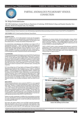
Grade II clubbing and cyanosis in Upper limbs
DISCUSSION: Partial Anomalous pulmonary venous connection [PAPVC] is a rare congenital anomaly in which one or more, but not all pulmonary veins are connected to systemic veins.[2] Winslow is credited with the first description of this anomaly in 1939.[4] PAPVC was first demonstrated during Cardiac Catheterisation by Dotter et al in 1949.[5] Patients with visceral heterotaxy and polysplenia have high incidence of partial anomalous pulmonary venous drainage.[2] Recent reports have described a significant incidence of PAPVC in association with Turner and Noonan syndrome. Anomalous pulmonary veins are frequently associated with congenital heart anomalies, predominantly atrial septal defects.[1 ]A rare but clinically important association is that of PAPVC with TOF [0.6%].[6] Patients are usually asymptomatic in childhood, but some dyspnoea on exertion may occur. The frequency of Cyanosis increases during third and fourth decades as a result of changes in pulmonary vascular bed, pulmonary hypertension and increasing right to left shunt.2 ECG shows rsR' pattern, peaked p waves and Right ventricular hypertrophy. When left innominate vein is the site of connection of left pulmonary veins, Chest X Ray shows a prominent supracardiac shadow with vertical vein on the left, innominate vein above and SVC on the right giving a 'Snowman' appear-
Patient with clubbing in all Four limbs
Copyright© 2016, IERJ. This open-access article is published under the terms of the Creative Commons Attribution-NonCommercial 4.0 International License which permits Share (copy and redistribute the material in any medium or format) and Adapt (remix, transform, and build upon the material) under the Attribution-NonCommercial terms.
International Education & Research Journal [IERJ]
20