Research Paper
Medical Science
E-ISSN No : 2454-9916 | Volume : 3 | Issue : 9 | Sep 2017
DIAGNOSIS OF UNICYSTIC AMELOBLASTOMA USING CALRETININ - CASE REPORT 1
2
2
Hina Mehrotra | Monica Mehendiratta | Mohit Sharma | Puneet Ahuja
3
1
PG Student, Department of Oral Pathology and Microbiology, ITS Dental College and Hospital, Greater Noida. Reader, Department of Oral Pathology and Microbiology, ITS Dental College and Hospital, Greater Noida. 3 Zonal Head, Clove Dental, Preet Vihar, New Delhi. 2
ABSTRACT Ameloblastomas are the commonest occurring tumors. More than 80% of all ameloblastomas are solid or multicystic variants (SMA), in which unicystic ameloblastoma (UA) is an important clinicopathologic variety. It occupies the remaining 20% of the cases. UA clinicopathologically resembles an odontogenic cyst but with few exceptions. KCOT, characterized histologically, by a palisaded basal cell layer of basophilic columnar cells and a surface of corrugated parakeratin, sometimes with spongiosis. This resembles very closely with the stellate reticulum like cells. If the tissue sample is small and if the neoplastic epithelium displays reactive changes induced by inflammation, it can closely resemble unicystic ameloblastoma histologically. Thus, at times, both lesions become histologically indistinguishable. As Calretinin is used as a specific immunohistochemical marker for neoplastic ameloblastic epithelium. We report a case of UA exhibiting histologically disconnected islands in the capsule without accompanied bony invasion seen in left premolar and molar region of mandible in a 38 year old female patient. The final diagnosis in this case was confirmed by using this marker. The final diagnosis of Unicystic Ameloblastoma with disconnected active intra-mural ameloblastomatous follicle was given. KEYWORDS: Ameloblastoma, Unicystic Ameloblastoma, KCOT, Calretinin. INTRODUCTION: Ameloblastomas are the commonest occurring tumors. More than 80% of all ameloblastomas are solid or multicystic variants (SMA), in which unicystic ameloblastoma(UA) is an important clinicopathologic variety. It occupys the remaining 20% of the cases (Nagalaxmi et al. 2013). The concept of unicystic ameloblastoma(UA) was first introduced by Robinson and Martinez in 1977 and there were various names given such as mural, monocystic, intracystic, cystogenic, or cystic ameloblastoma (Singh et al., 2010; Eversole et al. 1984). UA clinicopathologically resembles an odontogenic cyst but with few exceptions. The radiographic representation is commonly unilocular (especially impaction associated ones) but may be multilocular. It clinically often appears at a yonger age than SMA. Histologically, it is cystic in architecture.
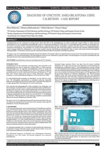
Incisional biopsy specimen (Fig.2) was taken from the buccal vestibular edentulous 34-35 region and was sent to the department of Oral Pathology for histopathologic examination (Fig3-7). The histopathological sections revealed cyst with ameloblastomatous epithelial lining and stellate reticulum like superficial cells exhibiting large keratinizing squames resembling ghost cells. Although there were no distinct calcifications in it but the ghost cell –like appearance led to the provisional diagnosis of Calcifying Odontogenic Cyst. The basal lining exhibited Vicker and Gorlin positive ameloblast like cells with superficial stellate reticulum like cells with surface squamous differentiation and areas of parakeratin formation. Focal areas of juxta-epithelial hyalinization could also be observed. The capsule was highly fibro- cellular with an area exhibiting disconnected active ameloblastomatous island.
Thus clinically and radiographically it often resembles of an odontogenic cyst but in histologic examination show a typical ameloblastomatous epithelium lining part of the cyst cavity, with or without luminal and/or mural tumor proliferation. Males are slightly more often affected than females (1.5:1) when UAs associated with an impacted tooth are considered. On the other hand when the tumour is impaction-associated, the ratio seems to be reversed (M: F=1:1.8) (Philipsen and Reichart, 1998).4 Irrespective to the impaction, mandible is predominantly affected over maxilla (Philipsen and Reichart, 1998). We report a case of UA exhibiting histologically disconnected islands in the capsule without accompanied bony invasion seen in left premolar- molar region of mandible in a 38 year old female patient. CASE REPORT : A 38-year-old female patient came to the Department of Oral Medicine and Radiology of ITS Dental College, Greater Noida complaining of pain and swelling in the left lower back tooth region since 3 years. Patient was apparently well 3 years back when she started feeling mild pain and swelling intermittently. Patient had undergone the extraction of lower left first and second premolar and first molar (34, 35, 36) 6 months ago. During intraoral inspection mild smooth surfaced swelling was present in buccal and lingual vestibule irt 34 & 35 and was measuring approximately 1.5x1.5cms causing mild buccal vestibular obliterartion. The swelling was soft and tender with no rise in local temperature and few bleeding points were present. The patient did not report any systemic health problems.The extraoral examination did not reveal any abnormality.
Fig.1: orthopantemogram exhibiting multilocular radiolucency in 33 region at the lateral border of 32
A
B
C
An orthopantomographic (Fig.1) evaluation revealed multilocular radiolucency in edentulous 34 region with radio-opaque smooth sclerotic border. 33 was root canal treated and exhibited root resorption apically. The differential diagnosis of Residual cyst, Odontogenic keratocyst, Dentigerous cyst were made. Provisional diagnosis was unicystic ameloblastoma.
Fig. 2: Biopsy specimen received: A, B: soft tissue specimens received in 10% normal buffered formalin. It is greyish brown in color and soft in consistency. It has smooth surface and irregular borders.; C: hard tissue is a bit of bone which was taken for decalcification.
CopyrightŠ 2016, IERJ. This open-access article is published under the terms of the Creative Commons Attribution-NonCommercial 4.0 International License which permits Share (copy and redistribute the material in any medium or format) and Adapt (remix, transform, and build upon the material) under the Attribution-NonCommercial terms.
International Education & Research Journal [IERJ]
8