Research Paper
Medicine
E-ISSN No : 2454-9916 | Volume : 3 | Issue : 11 | Nov 2017
A STUDY OF DIABETIC FOOT ULCER AND ITS CORRELATION WITH NEUROPATHY, PERIPHERAL VASCULAR DISEASE AND GLYCEMIC CONTROL 1
2
Dr. Kunal Bhuta | Dr. Sandeep Rai | Dr. Satish Chavan
3
1
Junior Resident, Department of Medicine, MGM Medical College, Navi-Mumbai. Professor of Medicine, Department of Medicine, MGM Medical College, Navi-Mumbai. 3 Senior Resident, Department of Medicine, MGM Medical College, Navi-Mumbai.
2
ABSTRACT Diabetes is one of the endocrine disorders that reached epidemic proportions worldwide. The metabolic deregulation associated with diabetes mellitus (DM) causes secondary path physiologic changes in multiple organ systems that impose a tremendous burden on the individual with diabetes. Diabetic foot ulcer is becoming major concern of diabetic patients. The vascular insufficiency and neuropathy accompanying the diabetic foot most often necessitate amputation of the limb. With the above considerations we undertook the study of 50 patients of diabetic foot admitted in our institute over a period of 2 years and 6 months. The mean age of study subjects was 53.18 ± 12.89 years. Male predominance was observed in study subjects with male to female ratio of approximately 5:1. 52% had BMI above 30. Clinical examination of ulcers for grading revealed that the percentage of grade IV lesion was highest (44%) followed by grades III (24%). Maximum diabetic foot ulcers patients had duration between 8 -12 years. 36% of diabetic foot ulcer patients had HBA1c between 8.6-9.5%. There is strong correlation between various grades of diabetic foot ulcers and peripheral neuropathy. Males (76%) had higher signs and symptoms of peripheral neuropathy as compared to females (24%). The most common clinical presentation of peripheral neuropathy was intermittent claudication (28%), paresthesia (26%) and numbness. Our study also identified some important risk factors for diabetic foot ulcers including older age, long duration of diabetes, lack of awareness of this disease, poor glycemic control and rural residence of patients. Sensory neuropathy was also found to be important risk factors for diabetic foot complications. KEYWORDS: diabetic foot, peripheral neuropathy, ulcer, diabetes mellitus, peripheral vascular disease. INTRODUCTION: Diabetes, once considered as a disease of developed countries, is one of the endocrine disorders that reached epidemic proportions worldwide now [1]. The metabolic deregulation associated with diabetes mellitus (DM) causes secondary path physiologic changes in multiple organ systems that impose a tremendous burden on the individual with diabetes and on the health care system [2]. Overall all 15% of individuals with diabetes mellitus will have foot ulcer during their lifetime and the annual incidence is 2-3% [3]. Diabetic foot ulcer is becoming major concern of diabetic patients and those who treat them from quality of life, social and economical stand point. Diabetic foot is a frequent complication involving the foot of diabetic patient. The vascular insufficiency and neuropathy accompanying the diabetic foot most often necessitate amputation of the limb. With the above considerations we undertook the study of 50 patients of diabetic foot admitted in our institute over a period of 2 years and 6 months. An emphasis was laid on determination of neuropathy and vascular status and to study in detail the risk factors responsible for development of diabetic foot ulcers and subsequent lower extremity amputation in patients of diabetic foot. AIMS AND OBJECTIVES: Ÿ To study risk factors in development of diabetic foot ulcers. Ÿ
To study the correlation of diabetic foot ulcer with HbA1c and glycemic control
Ÿ
To study the incidence of neuropathy and peripheral vascular disease in patients of diabetic foot ulcers.
Ÿ
To study the outcome of diabetic foot ulcers with available treatment in a tertiary care hospital.
MATERIALS AND METHODOLOGY: A cross-sectional descriptive hospital based study consisting of 50 consecutive adults diagnosed with diabetes having diabetic foot ulcer at Diabetes Specialty Clinic in MGM Hospital, Kamothe, Navi Mumbai for a period of 2 Years (Oct 2013 to Nov 2015).
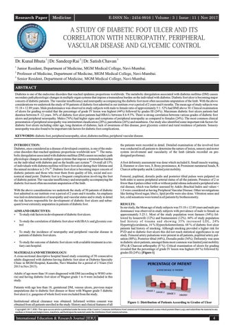
the patients were recorded in detail. Detailed examination of the involved foot was conducted in all patients to determine the nature of lesion, sensory and motor system involvement and vascularity of the limb (details recorded on predesigned proforma). A foot deformity assessment was done which included 1. Small muscle wasting, 2. Hammer or claw toes, 3. Bony prominence, 4. Prominent metatarsal heads, 5. Charcot arthropathy and 6. Limited joint mobility. Femoral, popliteal, dorsalis pedis and posterior tibial pulses were palpated on both sides to assess peripheral arterial status of all the patients. Presence of 2 or fewer than 4 pulses either with or without pedal edema indicated a peripheral arterial disease, which was further assessed by Ankle–Brachial Index and values < 1.0 were considered as having Peripheral Vascular Disease. Other investigations like fasting blood sugar, hba1c, lipid profile were also done. Vibration sense and hot, cold sensations were tested in all patients by biothesiometry. RESULTS: In our study, the Mean age of study subjects was 53.18 ± 12.89 years and male predominance was observed in study subjects with prevalence of male to female as approximately 5.25:1. Most of the study population were farmers (54%) followed by housewife (12%) and businessmen (12%). 66% of study population had history of trauma and showing 35% increased LDL, 24% Hypertriglyceridemia, 16 % Hypercholesterolemia. 68 % of diabetic foot ulcer patients had history of smoking. Although smoking provided a higher risk for PVD and to diabetic foot ulcers this did not reach statistical significance in our study. Femoral artery pulsations were present in all patients, popliteal artery pulsation (88%), Posterior tibial (44%), Dorsalis pedis (36%). Deformity was seen in diabetic ulcer patients, amongst them most common was limited joint mobility (8%) & Charcoat arthropathy (8 %). Critical examination of ulcers for grading revealed that the percentage of grade IV lesion was highest (44 %) followed by grades III (24%). [Figure 1]
Adults of age more than 18 years diagnosed with DM (according to WHO criteria) and having diabetic foot ulcer of Wagner grade 1 to 4 were included in this study. Patients with age less than 18, gestational DM, venous ulcers, previous major amputations due to diabetic foot disease or those with Wagner grade 5 diabetic foot ulcer (i.e. gangrene of whole foot) were excluded from the study. Institutional ethical clearance was obtained. Informed written consent was obtained from all patients enrolled in the study. History and clinical features of all
Figure 1: Distribution of Patients According to Grades of Ulcer
Copyright© 2017, IERJ. This open-access article is published under the terms of the Creative Commons Attribution-NonCommercial 4.0 International License which permits Share (copy and redistribute the material in any medium or format) and Adapt (remix, transform, and build upon the material) under the Attribution-NonCommercial terms.
International Education & Research Journal [IERJ]
4