
Patients As Art Philip A. Mackowiak
Visit to download the full and correct content document: https://ebookmass.com/product/patients-as-art-philip-a-mackowiak/

Patients as Art
Patients as Art
Forty Thousand Years of Medical History in Drawings, Paintings, and Sculpture
PHILIP A. MACKOWIAK, MD, MBA, MACP
EMERITUS PROFESSOR OF MEDICINE
CAROLYN FRENKIL AND SELVIN PASSEN HISTORY OF MEDICINE SCHOLAR- IN- RESIDENCE
UNIVERSITY OF MARYLAND SCHOOL OF MEDICINE
BALTIMORE, MARYLAND
Oxford University Press is a department of the University of Oxford. It furthers the University’s objective of excellence in research, scholarship, and education by publishing worldwide. Oxford is a registered trade mark of Oxford University Press in the UK and certain other countries.
Published in the United States of America by Oxford University Press 198 Madison Avenue, New York, NY 10016, United States of America.
© Oxford University Press 2019
All rights reserved. No part of this publication may be reproduced, stored in a retrieval system, or transmitted, in any form or by any means, without the prior permission in writing of Oxford University Press, or as expressly permitted by law, by license, or under terms agreed with the appropriate reproduction rights organization. Inquiries concerning reproduction outside the scope of the above should be sent to the Rights Department, Oxford University Press, at the address above.
You must not circulate this work in any other form and you must impose this same condition on any acquirer.
CIP data is on file at the Library of Congress
ISBN 978–0–19–085821–6
9 8 7 6 5 4 3 2 1
Printed by Sheridan Books, Inc., United States of America
For Connie, wife, mother, and grandmother nonpareil
There are aspects of sickness and health and life and death that can never be explained by science alone—humanistic aspects of the patient experience that can’t be measured or weighed or dissected. Art can help fill in the gaps in what medical science does for our understanding of such things. Art can provide glimpses below the surface of patients’ physical ills into the psychology, sociology, and sometimes even the biology of the human condition. This is because artists view the world through a unique lens, one that gives them a more nuanced and richer view of patients and their disorders than scientists. Artists find complexity in simplicity that enables them to endow their depictions of sickness and health and life and death with an authenticity that medical science alone is never quite able to capture.
Art offers an equally compelling perspective on the history of medicine. Long before humans could write, before they had a medical science or possibly even a religion, they had art. Some of the earliest art took the form of stunningly beautiful cave paintings and mysterious stone figures. What the artists’ intent was in creating these works is uncertain. Even so, the works have much to say about the artists who created them and the times in which they lived. The works mirror not only the eras in which they were created, but in concert with the works of later artists, mirror all times, as reflections of the dynamic nature of the human condition.
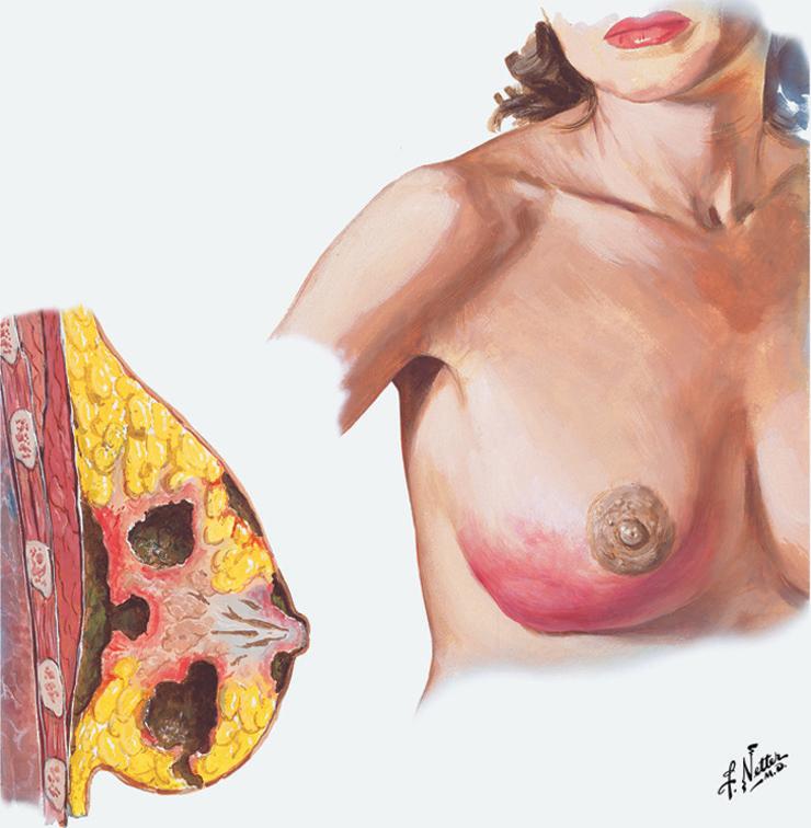
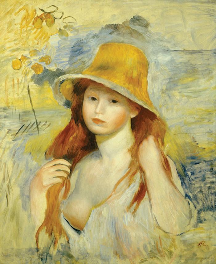
A “work of art” generally implies a certain degree of complexity. In contrast to illustrations, (visual) works of art express something more than the outward appearance of the objects depicted. They reveal unique and private things about the objects’ character. Compare, for example, Netter’s illustration of acute mastitis with Renoir’s portrait of a Young Girl with the Straw Hat (Figure 0.1). Both works are beautifully executed. Both depict a young woman with a breast abnormality (note the reddish lump at the lower border of the exposed breast in Renoir’s portrait). Netter’s illustration is scientifically informative, although impersonal. Renoir’s is intimate. It is more pleasing esthetically than Netter’s illustration, and although less informative scientifically, raises numerous questions of potential value in expanding the viewer’s appreciation of the subject’s apparent mastitis beyond those stimulated by Netter’s illustration. What, for example, did Renoir make of his model’s breast abnormality? Was the abnormality real or simply an artistic distortion added to the portrait for esthetic reasons? And if real, what was its cause? How was the abnormality affecting Renoir’s model? How long had it been there, and what became of it? And what was known of the
pathophysiology of cystic mastitis in 1884 when Renoir painted the portrait and the seminal work of pathologist luminaries of the likes of Remak, Rokatanski, and Virchow was just beginning to influence medical thought?
Art, in fact, can complement medical science in myriad ways. Art is a universal language capable of communicating ideas beyond the limits imposed by English as the dominant scientific language. Art can inspire those working in the medical field. It can reveal aspects of the mysteries of health and disease yet to be considered by science, and provide a humanizing perspective on the medical profession’s struggle to delay death’s inevitability and lighten the burden of life’s pain and suffering. Art can be a medium for celebrating and preserving medicine’s triumphs and follies, as well as for communicating recent advances by the profession to the public. Art can be used to sharpen the observational skills of physicians-in-training; and perhaps most important, art can complement the science of medicine by revealing the ebb and flow of the profession’s long history in ways that resonate with the lives, physical struggles, and mortality of ordinary people.
Given tomorrow’s uncertainty, the best we can do in contemplating the future is study the past in search of context and perspective. Medicine’s history, especially as preserved in art, is a case in point. Hidden among images created by artists, even before the dawn of civilization, are lessons learned and then forgotten that remain applicable today in the ongoing battle with shadowy enemies of health that are forever slain only to rise again. History teaches that time present and times past are inseparable one from the other and also from time future. It shows how closely tied the onward tendency of human accomplishments is to prior generations and, in the words of Sir William Osler, how like the ever-changing waters of a storm-tossed ocean “the philosophies of one age become the absurdities of the next, and the foolishness of yesterday becomes the wisdom of tomorrow.” Art reminds us of these things. It impresses upon us the ways in which history is alive in the present. Where medical history is concerned, art humanizes the stark scientific realities of clinical practice by depicting the care of patients through the ages in all its trials and tragedies
and wonder and beauty. And where clinicians themselves are concerned, medical history as revealed in art is an effective antidote to the profession’s pride in dogma that refuses to be embarrassed.
In that art is fundamentally a special mode of communication, when a work of art is contemplated, questions naturally arise as to the work’s meaning. What is the work’s message? What is the artist trying to communicate through the work? These are difficult questions at best, and at worst, ones for which answers can never be known.
The meaning of any work of art is complicated by the fact that great works of art, such as those featured in this book, are intended to stretch the viewer’s mind some distance beyond understanding. Sometimes the artist succeeds in doing so consciously and at others intuitively. Moreover, since such works invariably involve outward expressions of artists’ innermost feelings and personal prejudices, their interpretation is anything but straightforward. Consequently, all too often one is left with the impression that a work of art simply means whatever the artist intended it to mean. However, the viewer has a role in determining the meaning of a drawing, painting, or piece of sculpture no less important than the artist or, for that matter, a professional critic trained in art history. Each brings a different and equally valuable perspective to the interpretation.
The visual works of art included in this book offer a pictorial review of medical history stretching from Paleolithic times to the present. Each features a patient, some several patients. Most are prominent works that have been analyzed repeatedly by experts as to their outward appearance and inner significance, the extent to which they reflect the ideals and sensibilities of the time in which they were created, and the formal, spiritual, and/or scientific values they appear to communicate. Rarely have experts considered the potential clinical implications of the works or their collective value as an archive of medical history.
A great many prominent works of art have depicted aspects of medicine’s long struggle against ignorance, superstition, and religious and political dogma to emerge as one of mankind’s greatest achievements. The works included in this book were chosen both for their esthetic appeal and for the skill with which they depict important developments in medicine over
time. In analyzing the works and interpreting their meaning vis-à-vis medical history, I have brought the perspective of an internist with over four decades of experience caring for patients, teaching doctors-in-training, and conducting clinical research. I have also brought over twenty years of experience as a medical historian. Given this background, my particular focus in analyzing these works concerns what they have to say about the status of the “art of medicine” at the time they were created and its relationship to the medicine of today. I also speculate on current diagnoses that might be applied to the conditions featured in the works. In some cases, diagnoses are given by artists in the titles of their works. In others, they are so obvious as to be indisputable, and in others still, necessarily tentative owing to the absence of critical information, such as the subjects’ medical histories, physical examinations, and diagnostic test results. And of course, there is the problem of “artistic license.” For as Plato observed in the Republic over two thousand years ago: “Artworks present only an appearance of an appearance of what is really real.
ACKNOWLEDGMENTS
I owe an immense debt of gratitude to a host of friends and associates for giving generously their time and expertise in providing insight and perspective that enhanced my understanding of the artwork presented in this book and helped enrich my brief review of medical history. I am particularly indebted to Dr. Frank Calia (former Vice-Dean and Chairman of Medicine at the University of Maryland School of Medicine), Mrs. Eleanor Herman (New York Times best-selling author of Sex with Kings, Mistress of the Vatican, and The Royal Art of Poison), and Mr. Wayne Millan (classics scholar) for reading every word, analyzing every work of art, and checking every citation in making copious insightful recommendations. Dr. David Nalin (Professor Emeritus, Albany Medical College) offered valuable recommendations that enhanced the Public Health chapter. Dr. Will Carpenter (former Director of the Maryland Psychiatric Research Center) provided a masterful critique of the Mental Health chapter, as did Drs. Miriam Blitzer and Carole Greene (Department of Pediatrics, University of Maryland School of Medicine) the Genetics chapter, and Dr. Dale Smith (Professor of Military Medicine & History at the Uniform Services University of the Health Sciences) and Dr. Edward R. McDevitt CAPT MC USN-R the chapter on Military Medicine. Mrs. Frederic Billings kindly assisted me by translating Spanish phrases into English. Mr. Larry Pitrof (Executive Director of the Medical Alumni Association of the University of Maryland) provided general advice and continued encouragement and
support during the many long months I labored over this project. I am grateful to Mrs. Carolyn Frenkil and Dr. Selvin Passen for the privilege of occupying the history of medicine scholar-in-residence position they endowed at the University of Maryland School of Medicine in 2013 as one of many generous contributions to my medical school. Most of all, I am grateful to my wife Connie for the love, support, and forbearance that has allowed me to pursue my passionate interest in the past, while insulated from the day-to-day necessities of living in the present.
Nutrition
Throughout much of history, humans have struggled to find enough food to grow and to reproduce. We assume that in the distant past, food was scarce but that a meager diet had at least one advantage. That is, if not killed prematurely by infection, a wild beast, accident, or armed opponent, our early ancestors would have avoided the present-day scourges of overnutrition (diabetes, heart disease, cancer, and such). Some of the earliest works of art, however, suggest that the nutritional status of our ancient ancestors might have been more varied and complicated than is generally believed.
OVERNUTRITION
The Venus of Willendorf, also known as the Woman of Willendorf (Figure 1.1), is an 11.1 cm (4.4 in) high Paleolithic statuette of a massively obese woman discovered in 1908 at an archeological site near the Austrian
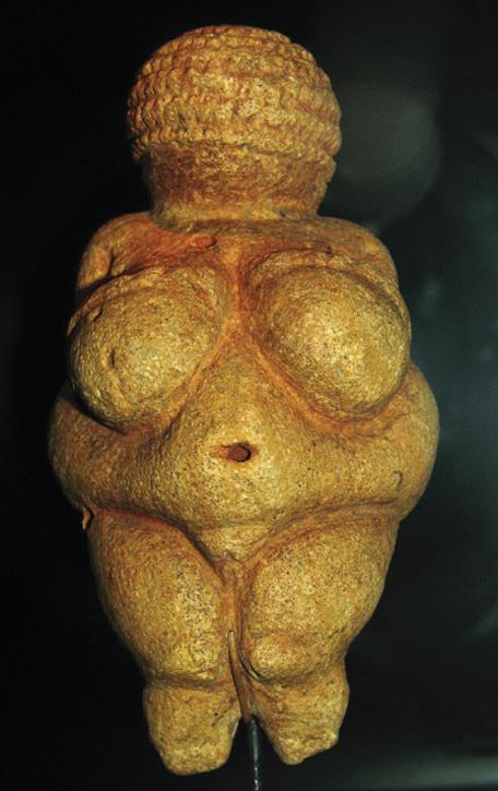
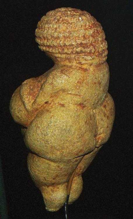
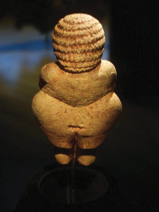
Figure 1.1. Venus of Willendorf statuette, oolitic limestone, 28,000 B.C.E. by an unknown artist, Naturhistorisches Museum, Vienna, Austria. As is typical of primitive works of art, the Willendorf figure lacks important anatomical details (in this case a face, feet, and fully developed arms and hands) possibly to emphasize the figure’s symbolic purpose (1). The sculptor’s identity and purpose in creating the figure are forever lost in the blur and mists of that long-vanished time. It has been speculated that the statuette served a ritual or symbolic function, was a religious figure, a self-depiction by a female artist, or simply erotic art (2). Whatever its purpose, the statuette’s portrayal of massive obesity is so anatomically correct, it seems certain that the artist who created it had seen obesity of such magnitude, and that it existed among Paleolithic peoples, no matter how harsh their lives might have been.
village of Willendorf. The statuette is estimated to have been created between 27,000 and 30,000 years ago during the Paleolithic or “Old Stone” Age (2). If the statuette is realistic, which seems likely, rather than simply symbolic, it raises several interesting questions: Was the Paleolithic diet conducive to a level of obesity that today would be designated “morbid” based on its numerous associated medical complications? Did Paleolithic persons who became as obese as the Venus of Willendorf, and whose lives were not cut short by infection, a complication of childbirth, or trauma, develop type 2 diabetes, heart disease, or the various cancers associated with morbid obesity today? And exactly how prevalent was obesity among Paleolithic humans?
Early humans, like today’s peoples, consumed an assortment of diets, which varied both over time and among different groups. They were opportunistic omnivores, with some appearing to have consumed large amounts of meat, while others apparently subsisted primarily on a plantbased diet, consisting primarily of carbohydrate-rich tubers. Because the human population of the Paleolithic period was small relative to the biomass of edible fauna, the Willendorf woman’s people likely enjoyed an abundance of animal protein. However, the nutritional quality of the animal protein consumed (wild game, especially gregarious ungulate herbivores, such as deer, bison, horses, and mammoths) differed from that found in the modern American supermarket. The latter meat has much more saturated fat, less eicosapentoenoic acid (a fatty acid shown to protect against coronary heart disease), and more calories per unit weight than the meat of wild game. Although the Paleolithic environment almost certainly had a greater abundance both of game and plant foods than current hunter-gatherer environments, Paleolithic humans would have experienced occasional shortages of food sufficient to threaten survival in the absence of adequate adipose reserves. Therefore, consuming excess calories to generate fat stores during times of abundance would have been adaptive (3, 4). Whether the resultant obesity would have been maladaptive then, as it is now, if a person as fat as the Willendorf woman survived beyond her 5th decade, or she would have been spared the adverse effects of morbid obesity because her diet was more wholesome than that being consumed today, is a question whose answer will lie forever lost in the moldering gap between her age and ours.
How prevalent morbid obesity was among pre- historic humans is equally difficult to know. All that can be said is that images with bodies similar to that of the Willendorf woman number in the hundreds, cover a period from 40,000 to 11,000 years ago, and have been uncovered at sites ranging from the Pyrenees to the plains of Siberia (Figure 1.2) (2).
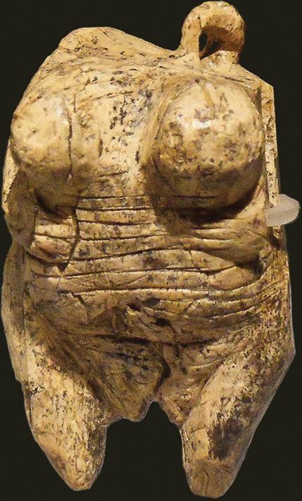
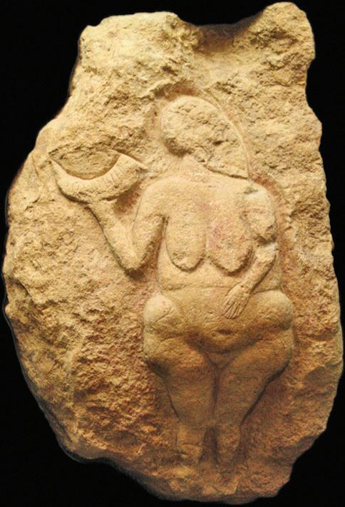
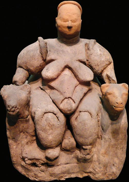
Figure 1.2. Venus of Hohle Fels statuette, mammoth ivory, 38,000–33,000 B.C.E. by an unknown artist, University of Tubingen, Germany (a). Venus of Laussel, bas-relief, limestone, c. 23,000 B.C.E. by an unknown artist, Musée d’ Aquitaine, Bordeaux, France (b). Mother Goddess of Çatal Hüyük statuette (with restored head), baked clay, c. 23,000 B.C.E. by an unknown artist, Museum of Anatolian Civilizations, Ankara, Turkey (c).
Images from more recent periods testify to the fact that morbid obesity was also present among residents of some of the earliest civilizations. A bas-relief in Hatshepsut’s mortuary temple in Luxor, for example, features the grossly overweight queen of the Chief of Punt (Figure 1.3), who lived during the time of Egypt’s 18th dynasty (c. 1543–1292 B.C.E.).
The queen of Punt and the Willendorf woman were both massively obese with what appears to have been similarly elevated body-mass indices (BMI, weight in kg/height in m2), the screening measurement commonly used today to estimate degrees of obesity. Based on their size, these two ancient women would have had BMIs of at least 40, with normal being 18.5–24.9. The BMI has the advantage of simplicity but, unfortunately, does not distinguish between fat and lean body mass (e.g., muscle), nor does it distinguish between abdominal obesity (so-called apple-shaped obesity) and obesity typified by that of the queen of Punt (so-called pearshaped obesity). The former, also known as “central obesity,” has been more closely associated in some studies, though not all, with cardiovascular
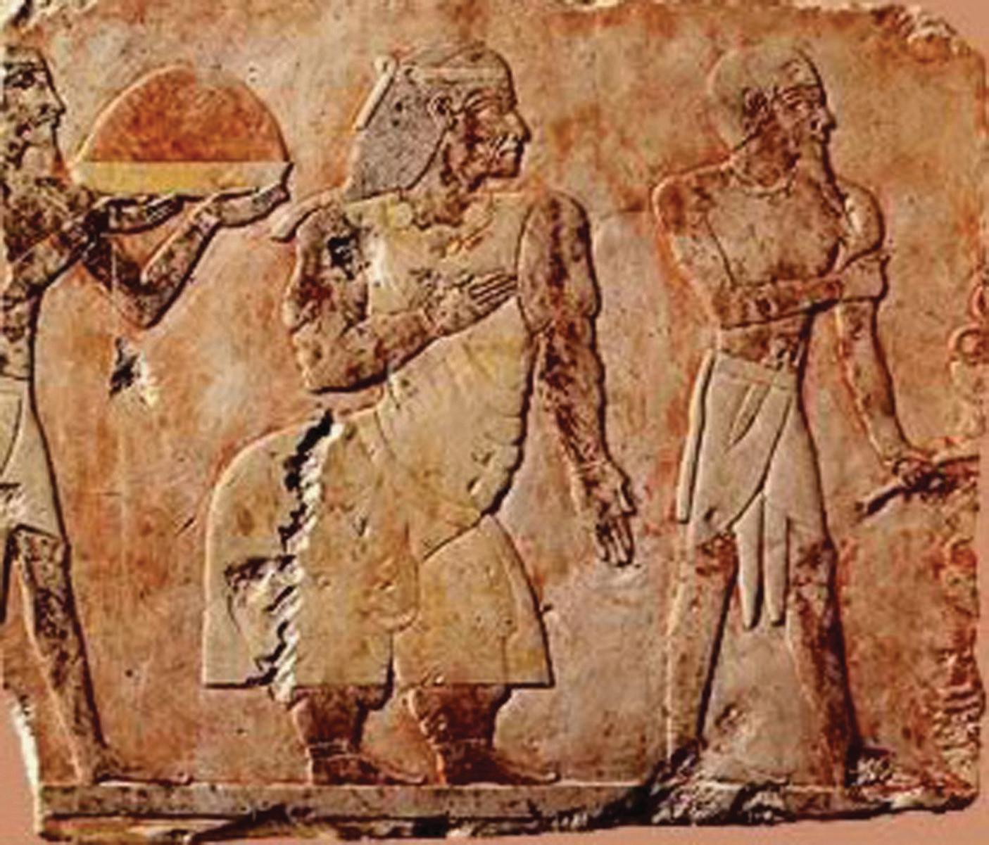
Figure 1.3. Chief of Punt accompanied by his excessively obese queen, sandstone basrelief, c. 1450 B.C.E., Hatsehepsut Mortuary Temple, Valley of the Kings, Egypt. This image is of interest not only because of its antiquity, but also because it depicts a pattern of obesity different from that exhibited by the Willendorf woman—one in which adipose tissue predominates in the hips, buttocks and thighs, rather than the waist. This particular pattern of obesity, called steatopygia (from the Greek stéar, “tallow” and pugē, “rump”), is a genetic characteristic prevalent among women of African origin, most notably those from the Khoisan region (5).
disease and the other complications of morbid obesity than the latter (6). Given these observations, the queen of Punt might have suffered fewer adverse health effects due to her overnutrition than the Willendorf woman if both had survived beyond middle age, even though they had similarly elevated BMIs.
Numerous other stone reliefs, as well as studies of the skin folds of royal mummies, document the presence of obesity among the ancient Egyptian elite, although not its prevalence. Moreover, the Ebers papyrus,
written c. 1500 B.C.E., suggests that physicians of that era recognized the symptoms of diabetes (7). Although there is no evidence that they understood the importance of obesity as a risk factor for the disorder, they did appreciate the influence of diet on health in general. Like the Chinese before them, who devised dietary principles for longevity based on Taoist teachings, the physicians of ancient Egypt advocated a frugal diet of fish, bread, fruits, and vegetables (8).
Two portraits painted 3,000 years later reveal other pernicious aspects of obesity with particular relevance for some modern societies. They are portraits of 5-year-old Eugenia Martínez Vallejo (Figure 1.4) painted by Juan Carreño de Miranda in 1680 C.E. (9). They speak to the physical, social, and psychological burden of childhood obesity, a problem that has reached epidemic proportions in many of today’s societies.
Aside from enduring the potential long-term adverse effects of childhood obesity on health, overweight children also face the stigma of being viewed as abnormal by many societies (11, 12). How Eugenia, a poor peasant girl who died when only 25 years old, happened to be so obese at such a young age apparently was of no concern to Charles II of Spain (no less odd in appearance himself, see Chapter 8), who commissioned the portraits. Nor did the medical profession have any knowledge then of the various genetic disorders associated with such early obesity (e.g., Prader-Willi syndrome, Bardt-Giedl syndrome, Cohen syndrome, etc.), or alterations in fetal gene expression caused by maternal malnutrition that are thought to cause a child to become obese, or the myriad ways in which poverty itself predisposes to obesity (13).
One-and-a-half centuries later, William Wadd produced an engraving of a grossly overweight woman, seated in a chair and seemingly about to drift into the arms of Morpheus (Figure 1.5). Although physicians since at least the time of Hippocrates have been concerned with the adverse effects of obesity on well-being, not until the 18th century C.E. did the condition become common enough, especially among the privileged classes, for the medical profession to begin to take notice of it as a serious threat to public health. Wadd, a surgeon, was one
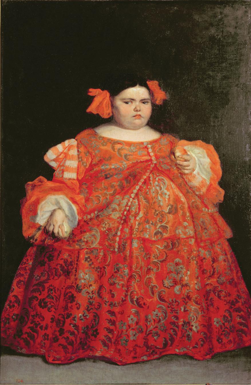
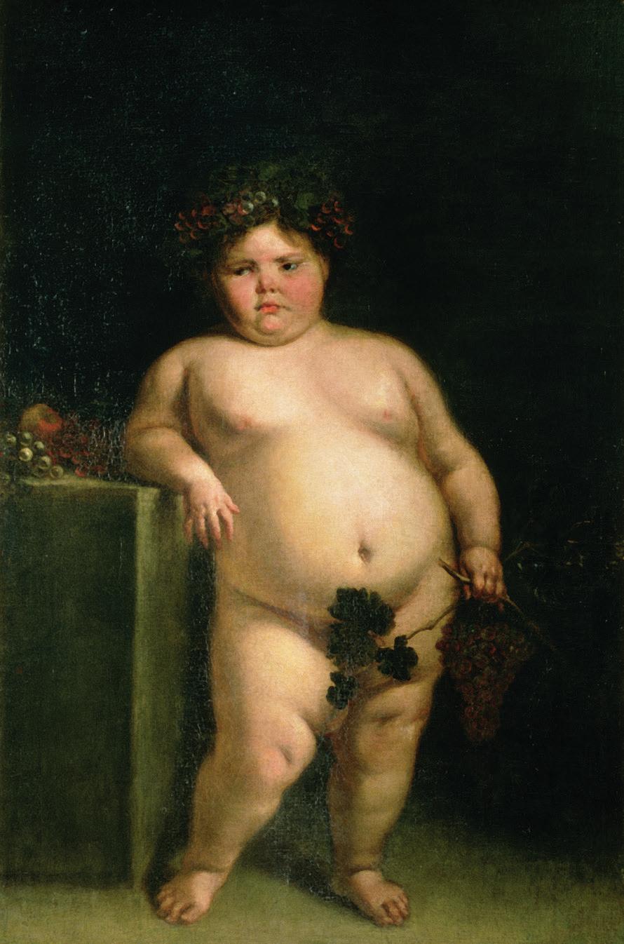
Figure 1.4. La Monstrua Vestida (a) and La Monstrua Desnuda (b), oil on canvas, 1680 C.E. by Juan Caruño de Miranda, Museo Nacional del Prado, Madrid, Spain. Caruño de Miranda (1614–1685 C.E.) succeeded Sebastian de Herrera as court painter to the Spanish queen in 1671 C.E. He is known mainly for his portraits of the royal family and court (10).
The two portraits of Eugenia Martínez give subtle expression to the psychological effects of negative stereotyping on the overweight child. On close inspection of the Vestida painting, the hint of a tear is visible in the child’s right eye. The tear is especially touching, given how young and defenseless she was when Carreño de Miranda saw fit to violate the angelic confidence of a small child without refuge or appeal by portraying her more as an object of derision—a Monstrua (Monster) —than a human being.
of the earliest and most influential physicians to do so. He maintained that the cause was obvious—“an over-indulgence at the table”—and the solution simple: “a sensible approach to food” (14). The results of numerous modern investigations argue otherwise. They indicate that the cause of the current obesity epidemic is far from obvious and its solution much more complicated than Wadd or physicians of his era could have imagined.
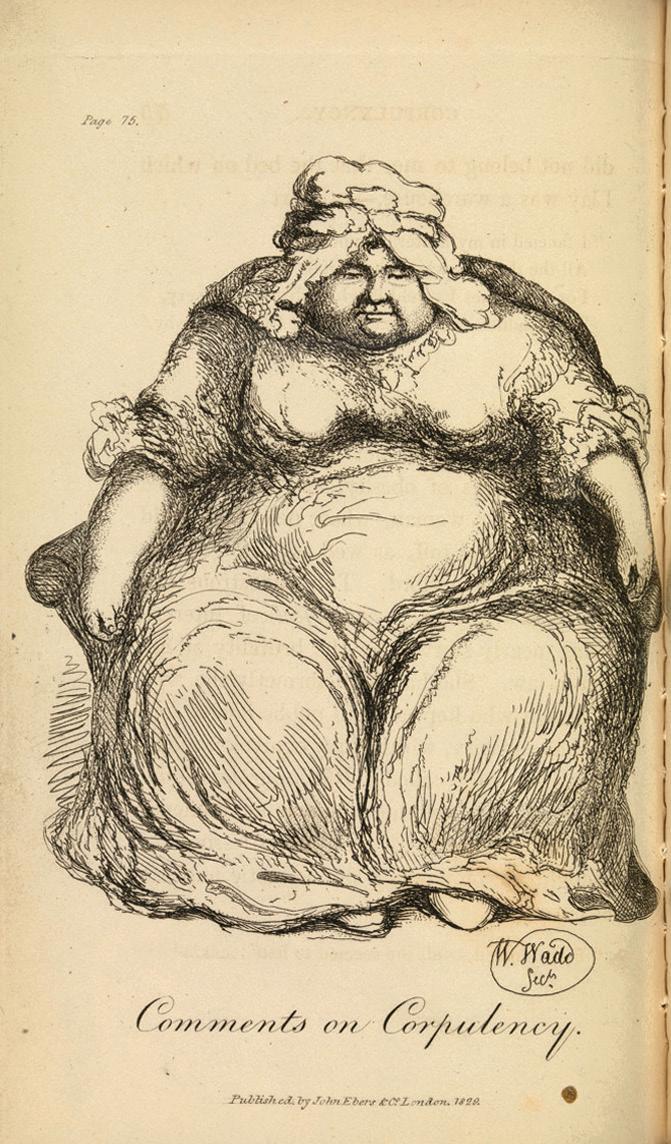
Figure 1.5. Obese Woman Seated, engraving, 1829 C.E. by William Wadd, Wellcome Library, London, England.
William Wadd (1776–1829 C.E.), was a surgeon as well as a skilled draughtsman, who personally drew and engraved this image and the others featured in his Comments on Corpulency (1829) (14). Wadd’s illustration brings to mind Joe, “the fat and red faced boy in a state of somnolency” in Charles Dickens’ Pickwick Papers. It further recalls the association of extreme obesity with uncontrollable sleepiness, hypoventilation, and right-sided heart failure, which in 1956 C.E. was given the name “Pickwickian syndrome” based on Dickens’ prescient description of the disorder in his novel. In its advanced stages, the syndrome is one of the most pernicious complications of extreme obesity (15).
If a picture is worth a thousand words, Lucian Freud’s Benefits Supervisor
Sleeping (Figure 1.6) tells a great deal about just how complicated the causes and control of the current obesity epidemic are. Historically, obese persons were thought to be low energy expenders, that is to say that they conserve energy (and thereby calories) to a greater extent than non-obese persons by moving less (16). This was believed to be the reason, at least in
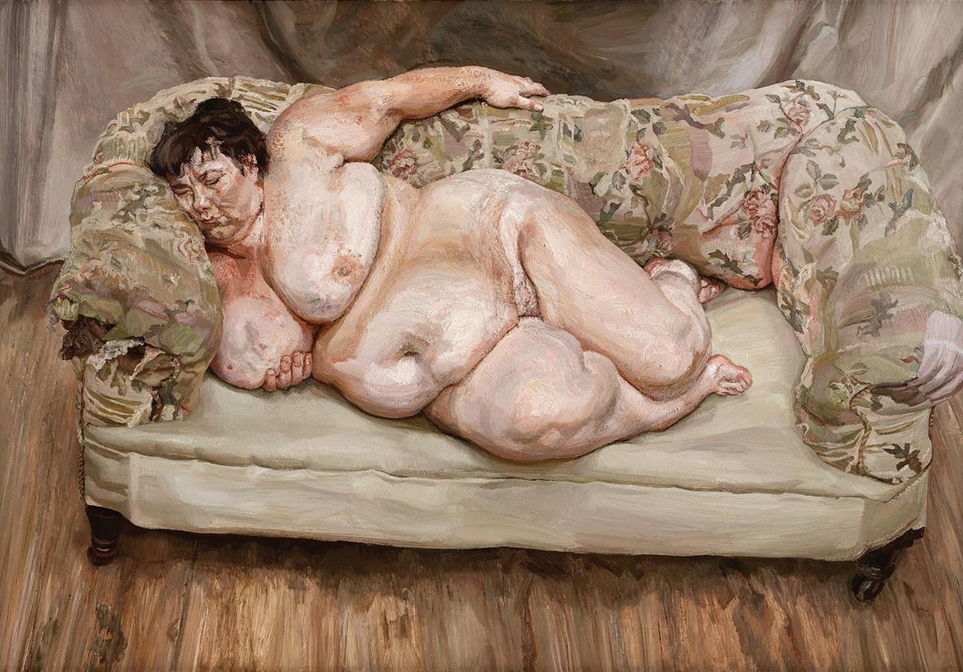
Figure 1.6. Benefits Supervisor Sleeping, oil on canvas, 1995 C.E., by Lucian Freud, private collection, Europe.
Lucien Freud (1922–2011, C.E.), a grandson of Sigmund Freud, fled Nazi Germany with his family in 1933 and settled in London. At the time of his death, he was considered Britain’s pre-eminent artist, known chiefly for impasto portraits , such as this one with its characteristic thick layers of paint. Like the ideal physician, he spent an inordinate number of hours studying his subjects to detect inner secrets, which he revealed with brutal objectivity in his works (17). Benefits Supervisor Sleeping sold for $33.6 million at Christie’s in 2008 C.E., setting a world record auction price for a work by a living artist (18).
part, why so often obese persons failed to lose weight when dieting. Freud’s portrait of the woman he called “big Sue,” and whose body he described as “flesh without muscle” (18), shines a lurid and seemingly confirmatory light on the concept. Although recent evidence fails to support this hypothesis, exercise is, nevertheless, currently considered an essential component of the comprehensive lifestyle modification included in virtually all obesity management programs.
If Freud’s “big Sue” were to adhere to the various interventions promoted in current weight loss programs, she might lose weight,
possibly even a substantial amount of weight, but only if she committed completely for an indefinite period to the program. Unfortunately, losing weight is easier for morbidly obese persons than maintaining the weight loss. Few patients of “big Sue’s” size succeed over the longhaul. For many, safeguarding one’s health at the cost of too strict a diet becomes an illness more tedious than the obesity, and they give up. In others, the battle is lost, in part because their weight loss activates some mysterious internal mechanism that produces a countervailing drop in metabolic rate seemingly designed to pull them back to their original level of obesity (19).
UNDERNUTRITION
There is perhaps no better clinical description of the devastating effects of starvation on the human body than that of the Buddha’s condition at the end of his 6-year fast recorded in the Pali Canon. According to the account:
The Buddha, having consumed “as little as one spoonful of bean soup a day, a single sesame seed, and a single grain of rice [became so emaciated his] rib cage was like an old stable with its sides caved in; [his] head withered until it looked old and wrinkled and dry; his eyeballs were sunken, and he had difficulty seeing. When he placed his hand on his abdomen he could feel his spine; when he tried to stand he fell backwards. His beautiful skin turned as black as the color of the madgura fish, and all the hairs came away from his body . . . . when the sun fell on him, he did not move into the shade, and from the shade he did not move into the sun. He did not seek refuge from the wind, sun, or rain; he did not chase away horseflies, mosquitoes, or snakes. He did not excrete urine or excrement or spittle or nasal secretions . . . . [he became] so weak, so feeble and thin that when they put grass and cotton in the openings of his ears, it came out through his nostrils. (20)
The second-century B.C.E. bronze casting of the Starving Buddha by an unknown artist (Figure 1.7) offers an equally arresting image of the ravages of prolonged starvation. However, it tells us nothing about starvation’s prevalence in India during the Buddha’s lifetime. We can only guess what it might have been based on how common it is today in areas of the Indian subcontinent lacking adequate nutrition. Today undernutrition no less severe than that resulting from the Buddha’s self-imposed fast is a terrifying torture of necessity for some 19 million children under the age of 5 years in certain areas of the world

The Buddha was a young man when he resorted to self-mortification through extreme fasting in his quest for perfect enlightenment (20). The effects of prolonged starvation, so graphically illustrated in this bronze casting, gave him the appearance of a man much further along in life’s journey than his age would suggest.
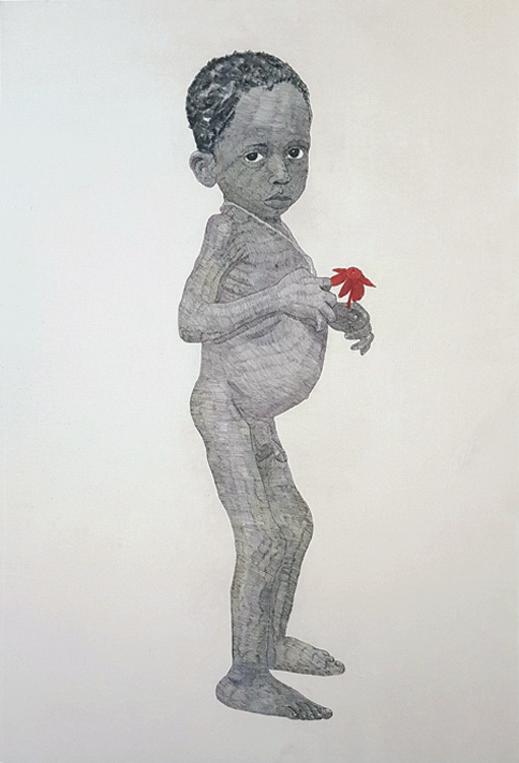
Figure 1.8. Impact of Malnutrition, brush pen on cotton, 2016 C.E. by Pierre Mukeba, artist’s personal collection. Mukeba, a self-taught artist, was born in the shadow of civil war in the eastern Democratic Republic of Congo. He had personal experience with food shortages severe enough to cause the signs of starvation exhibited by this child—spindly arms, bloated belly and edematous lower legs. In his works, Mukeba restricts his palette to just three colors—red (for war), blue (piety), and yellow (happiness). The small red flower in this child’s hand speaks not to piety or to happiness, but to war’s role in creating food shortages responsible for such severe malnutrition (22).
(21). Pierre Mukeba’s Impact of Malnutrition (Figure 1.8) captures the loneliness, hopelessness, and terror of starvation that has fastened its grip on vast areas of sub-Saharan Africa, as well as places in Asia where the Buddha preached moral enlightenment through selfless endeavor so long ago.
Kwashiorkor
Although the causes of starvation (e.g., war, draught, governmental mismanagement) are rarely medical per se, starvation’s clinical consequences are profound and highly variable, depending on the age of the victim and the particular nutrient that is most lacking. If the deficient nutrient is
primarily protein, the deficiency is severe, and the victim is a young child, a syndrome known as “Kwashiorkor” can develop. The name, derived from the Ga dialect of West Africa, is shorthand for “the sickness the older child gets when the next baby is born” (23). In certain areas of subSaharan Africa, the older child sickens (i.e., becomes profoundly protein deficient) with the arrival of the second child, because he/she is weaned from protein-rich mother’s milk to a diet consisting nearly exclusively of carbohydrates. As the deficiency progresses, the older child becomes weak, apathetic, and peevish (Figure 1.8). The child’s muscles waste away, growth ceases, and edema and anemia set in. The skin turns dry and scaly. The hair changes from black to red, and the eyes lose their luster. The belly becomes bloated.
Treatment is simple in principle, a nutritious diet containing adequate carbohydrates, fats, and vitamins, along with protein introduced in gradually increasing amounts. Unfortunately, treatment is considerably more complicated in practice than in theory, because Kwashiorkor is a disorder of poor countries whose ability to provide proper nutritional support to their people is compromised further by war, natural disasters, and/or political unrest.
Iodine Deficiency
When iodine is the deficient nutrient, the resulting physiological damage may be relatively minor or catastrophic depending on the extent of the deficiency and the age of the victim. In the adult, the result may be nothing more severe than the prominent goiter (enlargement of the thyroid gland) visible in Albrecht Dürer’s portrait of Elsbeth Tucher (Figure 1.9). If, however, the victim is a fetus developing in the womb of a markedly iodine-deficient woman, the effect can be catastrophic. Iodine is essential for the production of thyroid hormone. In its absence, synthesis of the hormone is impaired, resulting in a multisystem disorder known as “hypothyroidism” (25). When severe, the disorder is particularly hard on
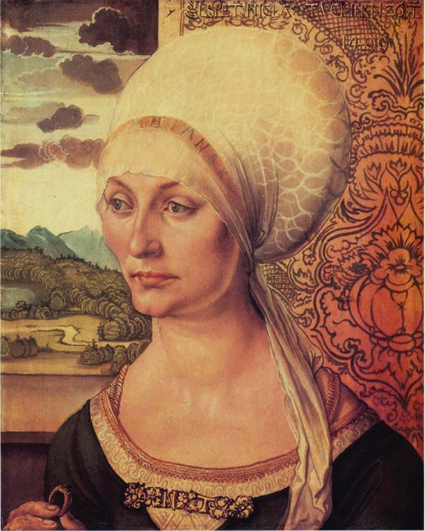
Figure 1.9.
Albrecht Dürer (1471–1528 C.E.) is widely regarded as one of the most important figures of the Northern Renaissance for his work as a painter, printmaker, and theorist (24). His theories of “ideal beauty” based on variety are reflected in this portrait of a beautiful woman with a large goiter (note the prominent rounded mass in her neck) and a strange tubular deformity extending from the left ear to the lower chin for which there is no anatomical basis.
the developing fetus, producing a concatenation of mental and physical deformities, known as “cretinism” (Figure 1.10).
The consequences of iodine deficiency (goiter and fetal abnormalities) have been recognized by physicians since the dawn of the profession and featured in works of art for nearly as long. Chinese medical writings dating to approximately 3600 B.C.E. record decreases in the size of goiters with the ingestion of seaweed and burnt sea sponge. Iodide, the key ingredient of these foods, was not discovered until 1811, when French chemist, Bernard Courtois, identified the element while extracting sodium salts necessary for the manufacture of gun powder. By the 1920s, hypothyroidism due to iodine deficiency was eliminated in most countries following adoption of the Swiss practice of adding sodium iodide or potassium iodide to table salt (27).
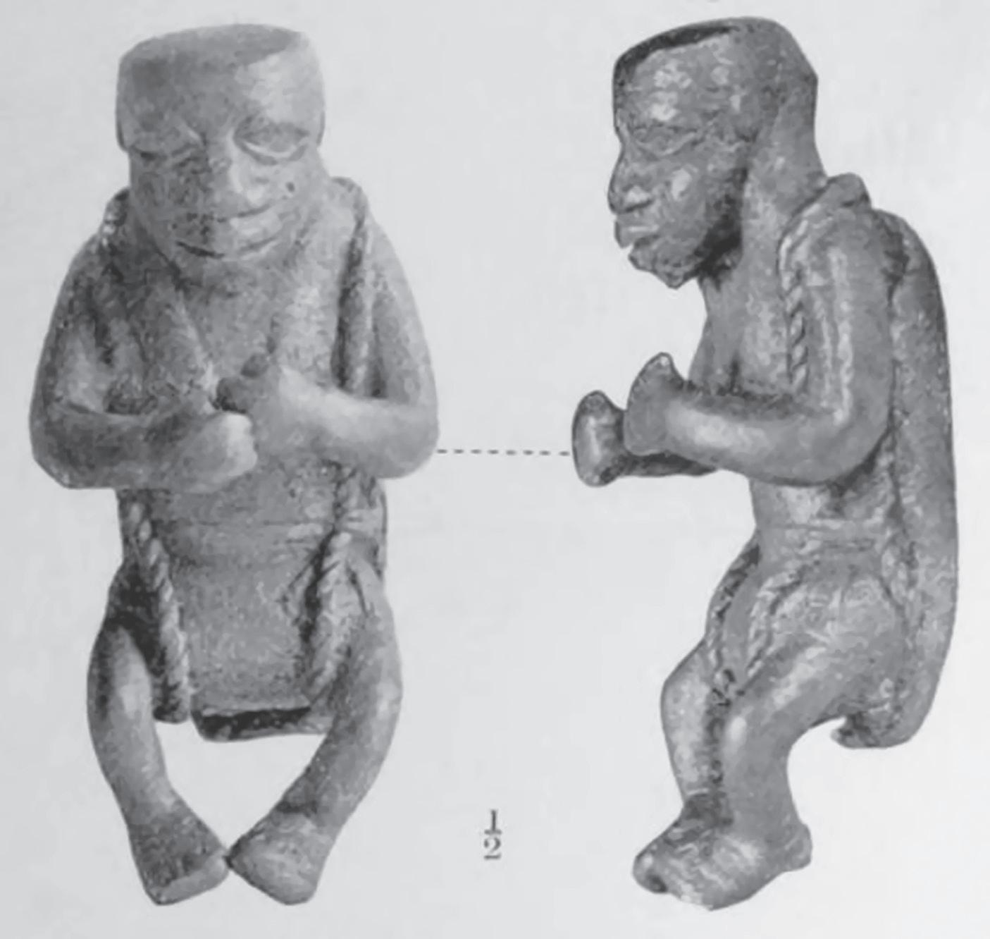
Figure 1.10. Naked Cretin, bronze figure (a thick plaster covering the back is believed to have represented a device designed as a cure for cretinism), c. 1700 C.E. by an unknown Benin artist, British Museum, London, England. This dwarf-like figure, with its misshapen head, vacuous expression, coarse facial features, and flaccid-appearing limbs is a stunning representation of a child with classic features of cretinism. The figure was one of many removed from the King of Benin’s compound by the British in 1897 C.E. during a punitive expedition. Several of the figures were covered with blood, indicating their use during religious sacrifices. British scholars of the early 20th century speculated that this and other such figures represent an advanced stage of Benin art developed under Portuguese influence during the 16th century C.E. (26).
Scurvy
During the Age of Exploration, the medical profession encountered a previously unknown disorder among sailors forced to subsist on diets devoid of fruits and vegetables during prolonged voyages. The early phase of the disorder was marked by weakness and fatigue. Then victims lost their appetite and began losing weight. Next their muscles and joints began to ache. Small hemorrhages erupted around their hair follicles from which cork-screw shaped hairs emerged. The gums of affected sailors began to swell and bleed; then their teeth loosened and fell out. Their legs swelled; wounds would not heal. The worst affected developed cardiorespiratory failure and died (28, 29). Nikolai Getman captured several of the essential features of the disorder in a painting depicting the suffering of Prisoners in Forced Labor Camps in Siberia and Kolyma (Figure 1.11).
The cause of scurvy remained a mystery until 1912 C.E., when Frederic Gowland Hopkins published a landmark paper in the Journal of Physiology describing feeding experiments that demonstrated the importance of “accessory” factors other than fat, protein, and carbohydrate in maintaining health (31). That same year Casimir Funk, working at the Lister Institute in London, identified the first such factor—thiamine—and coined the term “vital amine” or vitamin (32). In time, vitamin C, another “accessory factor,” was identified as the key ingredient of fruits and vegetables that cure scurvy.
Beriberi
Severe thiamine (vitamin B1) deficiency causes a different, though equally disabling syndrome known as “beriberi.” Western physicians first encountered the disorder in the Far East during the 17th century C.E. among patients having an appearance similar to those featured in Leslie Cole’s portrait of starving prisoners of war of the Japanese during World War II (Figure 1.12). The disorder produced swollen feet and
Figure 1.11. Prisoners in Forced Labor Camps in Siberia and Kolyma, oil on canvas by Nikolai Getman, the Jamestown Foundation, Washington, D.C., United States. Getman (1917–2004 C.E.), a prisoner of both forced labor camps from 1946 to 1953 C.E., was one of only a few artists who recorded the life of prisoners in the Soviet Gulag. He risked further torture and death to produce works such as this one, “convinced that it was [his] duty to leave behind a testimony to the fate of millions of prisoners who died” (30). Signs of scurvy in the man facing the viewer include his emaciation, absent teeth, gasping respiration (possibly due to a failing heart), and the purple discoloration below his right knee, possibly representing subcutaneous hemorrhage.
caused legs to become so numb and weak that victims walked with an unsteady, tottering gait. Some developed palsies (paralysis), particularly of the legs. In others, their hearts began to fail, occasionally resulting in sudden death. General malnutrition contributed to this host of agonies. However, near total lack of thiamine in a diet consisting almost exclusively of polished white rice was the principal cause, in that the process of power milling involved in the production of white rice robs the grain of most of its content of thiamine and other vitamins (34).