
Synthesis, DFT Calculations, DNA Interaction, and Antimicrobial Studies of Some Mixed Ligand Complexes of Oxalic Acid and Schiff Base Trimethoprim with Various Metal Ions
Eid Abdalrazaq1* , Abdel Aziz Qasem Jbarah1 , Taghreed Hashim Al-Noor2 , GassanThabitShinain2,and MohammedMahdiJawad3
1Department of Chemistry, College of Science, Al Hussein Bin Talal University, Ma’an 71111, Jordan
2Department of Chemistry, Education for Pure Science College Ibn Al Haitham, University of Baghdad, Baghdad 10071, Iraq
3Department of Biology, Education College Ibn Al Haitham, University of Baghdad, Baghdad 10071, Iraq
* Corresponding author:
tel: +962 796862267 email: eidalzooby@yahoo.com
Received: April 9, 2022
Accepted: July 19, 2022
DOI: 10.22146/ijc.74020
Abstract: Mixed ligand metal complexes are synthesized from oxalic acid with Schiff base, and the Schiff base was obtained from trimethoprim and acetylacetone. The synthesized complexes were of the type [M(L1)(L2)], where the metal, M, is Ni(II), Cu(II), Cr(III), and Zn(II), L1 corresponds to the trimethoprim ((Z) 4 ((4 amino 5 (3,4,5 trimethoxybenzyl)pyrimidine 2 yl)imino)pentane 2 one) as the first ligand and L2 represent the oxalate anion (��2��2 ) as a second ligand. Characterization of the prepared compounds was performed by elemental analysis, molar conductivity, magnetic measurements, 1H NMR, 13C NMR, FT IR, and Ultraviolet visible (UV Vis) spectral studies. The recorded infrared data is reinforced with density functional theory (DFT) calculations. Also, the recorded and calculated IR spectra of the complexes suggested that the coordination of Schiff base is a bidentate ligand with Cu and Ni complexes and a tridentate ligand with Co, Cr, and Zn complexes. The electronic structures of the complexes were investigated by DFT calculations, showing several degrees of HOMO LUMO energy gaps between complexes. The complexes were studied for their DNA interaction activities. The synthesized ligand and its metal complexes were evaluated for antimicrobial properties against bacterial strains of Bacillus subtilis (G+), Enterobacter cloacae (G ), and Staphylococcus aureus (G+). These complexes considered in this study showed good antimicrobial activity.
Keywords: trimethoprim; oxalic acid complexes; acetyl acetone; Schiff base; antimicrobial activity
■ INTRODUCTION
Schiff base compounds showed a unique role as ligands with transition metals and main groups in bioinorganic and coordination chemistry [1 2]. The interaction of Schiff bases with metal ions gives complexes of different geometries used as anticancer, antibacterial, antiviral, design medicinal compounds, antitumor organometallic chemistry, catalytic applications, chemical analysis, geology, and corrosion inhibition [3 4]. The complexes with mixed ligands of a Schiff base 4‐dihydroxybenzylidenethiosemicarbazide (H2L) , Oxalic
Acid and with, M(II) = Cu, Zn, Ni and Fe(III) ions [5]. The H2L is coordinated to the metal atom as a neutral, monoanionic, or dianionic tetradentate type (ONNO) ligand in its complexes. Chen [6] has reported the synthesis and structural characterization of a trans (PyH)2[Mo2O4(C2O4)2(Py)2] in an aqueous solution. The pyridine ligand coordinates to the Mo atom through the N atom. The oxalate ligand coordinates to eachMo atom through 2 Carboxylate Oxygen atoms in a bidentate chelating manner. Based on these observations, the present study was undertaken to synthesize and
Abdalrazaq
al.
1349Indones. J. Chem., 2022, 22 (5), 1348 1364
characterize mixed ligand complexes of oxalate anion and Schiff base derived from trimethoprim and acetylacetone with one of the metal ions Cr(III), Co(II), Ni(II), Cu(II) and Zn(II). DFT calculations for molecular electrostatic potential, geometry optimization and vibrational frequencies of the synthesized molecules using the B3LYP level of theoryand LANL2DZbasis set were reported.
■ EXPERIMENTAL SECTION
Materials
All the chemicals and solvents, trimethoprim, ethanol, acetylacetone, acetic acid, dimethyl sulfoxide, chloroform, methanol, and acetone metal chloride salts, were purchased of A.R. Grade quality obtained from Aldrich, Merck, and BDH and were used without further purification.
Instrumentation
Melting points were determined by MPA160 Digi Melt melting point instrument. The Eager300 obtained the Elemental analysis of ligand for EA1112 Thermal Finnegan C.H.N.S 2400 instrument. The atomic absorption spectrophotometer analysis of the complexes was measured using a fair agreement method using the device from type Shimadzu (A.A 620) atomic absorption spectrophotometer. The molar conductivity values of the complexes withaconcentrationof 10−3 mol/L indimethyl
sulfoxide (DMSO) were reached by the digital conductivity series Ino.Lab.720 device. The Magnetic susceptibility measurements of the complexes were measured using Johnson Mattey Balance. Ultraviolet visible spectra were recorded on double beam UV Visible spectrophotometry of the type U.V 160A (Shimadzu) at 200 and 1100 nm with a 1 cm quartz cell, using DMSO as a solvent and a concentration of 10−3 mol/L. The Fourier transform infrared spectra were measured using KBr pellets on the Shimadzu FTIR 8400S spectrophotometry instrument. The spectra FTIR are recorded in the range of 400 to 4000 cm−1. The 1H and 13C NMR spectra were obtained from the DMSO d6 solution using an Inova 500 MHz instrument.
Procedure
Synthesis of Schiff base ligand


The Schiff base ligand (L1) was prepared by condensation of 1.176 g (4 mmol) of trimethoprim in 20 mL ethanol and 0.4 g (4 mmol) acetylacetone for 8 h with the addition of 4 to 5 drops of acetic acid (Scheme 1). The volume of the mixture was reduced by slow evaporation at room temperature and leave it to stand overnight. Then, the obtained off white precipitate was washed several times with absolute ethanol, dried at room temperature, and recrystallized from ethanol to get a pure sample.
Scheme 1. The synthesis route of Schiff base ligand, L1
1350 Indones. J. Chem., 2022, 22 (5), 1348 1364
C19H24N4O4 (L1): %Yield: 86%. Anal. calc. for C19H24N4O4 (372.43 g/mol): C, 61.27; H, 6.50; N, 15.05. Found C, 62.06; H, 5.56; N, 13.99. Mp: 185 °C. IR (KBr, cm 1): ν(C=O) 1682, ν(−C=N−) 1658. 1H NMR (DMSO d6): δ 1.880 ( N=C CH3), 2.490 ((CO) CH3), 3.518 3.605 (CH2), 3.710 (O CH3), 5.966 (pyrimidine ring), 6.545 6.252 (NH2), 7.487 (Ar). 13C NMR (500 MHz, DMSO d6): δ 18.5 ( N=C CH3), 21.3 ( (CO) CH3). 55.8 (O CH3), 59.9 (CH2), 105.6 135.8 (Ar), 152.7 161.6 (pyrimidine ring), 162.3 ( C=N), 172.5 (C=O).
Synthesis of the complexes of the type [M(L1)(L2)]

CoC21H24N4O8 (519.38 g/mol): C, 48.56; H, 4.66; N, 10.79. Found C, 49.02; H, 4.44; N, 11.13. Mp: 223 255 °C. IR (KBr, cm 1): ν(C=O) 1670 (carbonyl of L1), ν(−C=N−) 1645, ν(C O) 1238 (oxalate), ν(Co N) 524, ν(Co O) 480 (oxygen of L2), ν(Co O) 459 (oxygen of L1).
CrC21H24N4O8 (2): %Yield: 82%. Anal. calc. for CrC21H24N4O8 (512.44 g/mol): C, 49.22; H, 4.72; N, 10.93. Found C, 48.78; H, 4.36; N, 10.61. Mp: 285 °C. IR (KBr, cm 1): ν(C=O) 1671 (carbonyl of L1), ν(−C=N−) 1643, ν(C O) 1235 (oxalate), ν(Cr N) 526, ν(Cr O) 475 (oxygen of L2), ν(Cr O) 448 (oxygen of L1).
ZnC21H24N4O8 (3): %Yield: 79%. Anal. calc. for ZnC21H24N4O8 (525.82 g/mol): C, 47.97; H, 4.60; N, 10.66.FoundC,48.11;H,4.98;N,10.85.Mp:216 220°C.
IR (KBr, cm 1): ν(C=O) 1674 (carbonyl of L1), ν(−C=N−) 1643, ν(C O) 1242 (oxalate), ν(Zn N) 542, ν(Zn O) 484 (oxygen of L2), ν(Zn O) 438 (oxygen of L1).
4
The complexes were prepared in a molar ratio of 1:1:1 (M:L1:L2). The metal chloride salts (MCl2·nH2O. n = 0 6, and CrCl3·6H2O) reacted with the two ligands according to Scheme 2 and the following proposed general equation: H2C2O4 +2KOH → K2C2O4 + 2H2O MCl2·nH2O + L1 + K2L2 → [M(L1)(L2)] + 2KCl + nH2O where L1 = Schiff base (the first ligand), L2 = C2O2 (Oxalate anion as a second ligand) and M = the corresponding metal.
Dissolving 0.23793, 0.23769, 0.17048, 0.17228 and 0.26635 g (1 mmol) of CoCl2·6H2O, NiCl2·6H2O, CuCl2·2H2O, ZnCl2, and CrCl3·6H2O, respectively, in 10 mL of ethanol. A 0.372 g (1 mmol) of L1 in 10 mL of ethanol and the solution of potassium oxalate were added at the same time to the metal chloride solution. The mixture was stirred for 4 h at room temperature and left for 24 h. The precipitate was filtered and washed with ethanol before recrystallizing and drying at room temperature. The percentage yield range is 79 to 85%.
CoC21H24N4O8 (1): %Yield: 83%. Anal. calc. for
CuC21H24N4O8 (4): %Yield: 83%. Anal. calc. for CuC21H24N4O8 (523.99 g/mol): C, 48.14; H, 4.62; N, 10.69.FoundC,49.03;H,4.86;N,10.93.Mp:253 255°C.

IR (KBr, cm 1): ν(C=O) 1681 (carbonyl of L1), ν(−C=N−) 1642, ν(C O) 1240 (oxalate), ν(Cu N) 513, ν(Cu O)479(oxygenofL2).
NiC21H24N4O8 (5): %Yield: 85%. Anal. calc. for NiC21H24N4O8 (519.14 g/mol): C, 48.59; H, 4.66; N, 10.79. Found C, 47.82; H, 4.92; N, 10.41. Mp: 216 °C. IR (KBr, cm 1): ν(C=O) 1680 (carbonyl of L1), ν(−C=N−) 1643, ν(C O) 1245 (oxalate), ν(Ni N) 532, ν(Ni O) 472 (oxygen of L2).
DNA binding properties
DNA was extracted from human blood samples without any health problems in the city of Baghdad/Iraq
using unique extraction from Promiga USA. The steps mentioned in the protocol were followed. The DNA concentration and the purity of the extracted DNA were obtained usingthe spectrophotometer of 1.6 to1.8 ng/mL. The purity and concentration were measured using the subject standards. The samples were initially extracted on the 1% agarose gel to confirm their quality and to see if there was any breakage during extraction. The studied materials were then mixed with the extracted DNA by 2:1 v/v and put in a water bath for one hour at 37 °C and then transported in 1.5% agarose gel for 1 h and 75 V, then treated with Red safe fluorescent dye and under the UV source using the gel documentation system.
Computational details
All the DFT calculations were performed with the Gaussian09 program [7]. The geometries of the complexes were fully optimized at B3LYP (Becke's, three parameter exchange functional, in combination with Lee Yang Parr correlation function) [8 10] with the 6 311G* basis set for C, H, O and N atoms and LANL2DZ (Los Alamos National Laboratory 2 double zeta) [11 13] basis set for metal atom. LANL2DZ is an effective core potential (ECP) type and has been widely used for studies of transition metals (TM) containing systems. The computational cost is decreased with the chemically inactive core electrons represented by an ECP since the cost formally increases as ∼N4, where N is the number of explicitly treated electrons. The program GaussView 6 [14] was used to inspect the input and output files generated by Gaussian09 for pre processing, structure modification, and post processing analyses of structures, frequencies, and forces. A frequency analysis was performed for each stationary point to identify the minima's most stable structure. All minima have no imaginary frequencies in the vibrational mode calculations. The molecular electrostatic potential, V(r), at a given point r(x,y,z) in the vicinity of a molecule, to determine the reactive sites of the complexes, was calculated at the B3LYP functional with the 6 311G* basis set for C, H, O and N atoms and LANL2DZ basis set for the metal atom of the optimized geometry. The definition, description, and calculation of V(r) were reported in
many reports [15]. The B3LYP level of theory with the 6 311G* basis set for C, H, O, and N atoms and the LANL2DZ basis set for metal atoms is used to calculate the energies and electron densities of the frontiers molecular orbitals. The PDOS (Projected density of states) has been obtained via the calculated orbital populations for all synthesized complexes at the same level of theory, using GAUSSSUM 3.0 program [16].
■ RESULTS AND DISCUSSION
The solubility of the compounds was tested using various solvents. The synthesized complexes are non hygroscopic solids, varying colors and soluble in water and dimethyl sulfoxide, whereas insoluble in ethanol, chloroform, methanol, and acetone. Also, they are air stable at room temperature. The molecular weight, melting point, and Flame AAS analysis of the synthesized complexes were carried out by the literature methods [17]. The experimental and calculated values of a metal content percent (M%) in all complexes are in fair agreement. The chloride ions test in all complexes was done with the AgNO3 solution, and a negative result was obtained [18]. The molar conductance values of the complexes in DMSO solvent indicated that [ML1L2] complexes are non electrolytes, while complex 2 is a1:1 electrolyte [19 20]. These results support the proposed formula of the complexes (Fig. 1). The physical and analytical data of Schiff base ligand L1, and its metal complexes are listed in Table 1.
1H NMR Result
The 1H NMR intheDMSO d6 solvent oftheligand (L1) is shown in S1. In S1, two signals appeared at 3.605 and 3.518 ppm, which were assigned to the protons of the CH2 groups. The previously reported values of the CH2 group signals in the 1H NMR spectrum are in the rangeof3.59 3.52ppm[20].Thespectrumoftheligand, S1, also showed a peak at 3.710 ppm. This peak is attributed to the methoxy protons (O CH3 group). The chemical shifts of the protons of the O CH3 group are reported in the range of 3.65 3.8 ppm [21 23]. The chemical shift of the benzene ring protons is found at 7.487 ppm. It was reported that the chemical shift of the
Table1.Analytical data and some physical measurements of the Schiff base ligand and the complexes
Molecular weight Color Melting point Molar conductance M%
(g/mol) (°C)
1 372.43 Yellow 185
519.38 Pink 223 255 11.35
10.85
512.44 Dark green 285 10.15
10.20
525.82 White 216 220 12.43
11.93
523.99 Offwhite 253 255 12.13
11.13
519.14 Lightgreen 216 11.31
10.20
25.88
37.88
17.18
16.55
18.6
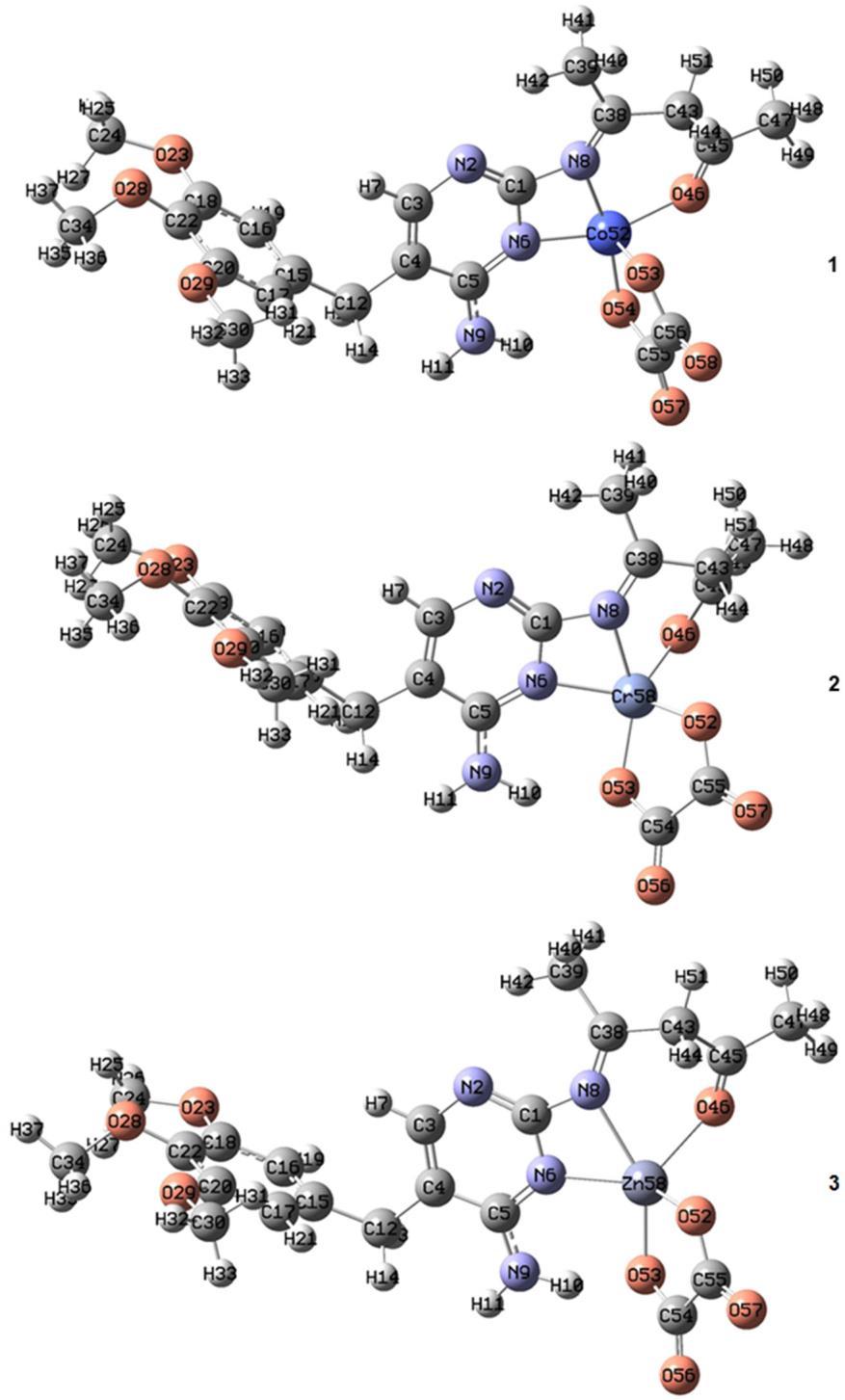
1.Thesuggested structureofthecomplexes
benzene ringprotonsdisplayed in 6.75 8.01ppm[23 24]. The chemical shift of the proton of the pyrimidine ring is observed at 5.966 ppm. The protons of the amine group (NH2) showed two peaks at 6.545 and 6.252 ppm. The reported values for the pyrimidine ring proton and the protons of the amine group are 5.8 and 6.9, respectively [25]. The chemical shifts of the methyl protons of the
andligands


N=C CH3) and ((CO) CH3) groups were observed at 1.880 and 2.490 ppm, respectively [26].
13C-NMR Results
The 13C NMR spectrum of the ligand L1 in DMSO d6 solvent is shown in S2. The spectrum of the ligand, S2, exhibits achemicalshiftat18.53ppm,whichisassigned
to the carbon atom of the methyl group, which is attached to the azomethine group ( N=C CH3). It was reported that the chemical shifts of the carbon atom in the methyl group of C CH3 in the range 19.09 19.87 [22]. The chemical shift at 21.3 ppmis attributed tothe carbon atom of the methyl group, which is attached to the carbonyl group ( (CO) CH3). It was reported that the carbon atom ofthemethylgroupdisplayedchemicalshiftsat 21.06and 21.4 ppm [27]. The signals appeared in the range 40.0 32.9 ppm concerning the DMSO solvent. The chemical shift at 55.8 ppm is assigned to the carbon atom of the O CH3 group. This assignment is based on the previously reported chemical shifts of the carbon atom of this group [21 22]. The chemical shift at 59.9 ppm is due to the carbon atom of the CH2 group. The reported values of the carbon atom of the CH2 group of the mono and diphenyl tin(IV) complexes were 59.9 60.46 ppm [28]. The chemical shift of the carbon atoms of the aromatic ring appeared in the range of 105.6 135.8 ppm [26]. The carbons of the pyrimidine ring showed three signals at 152.7, 154.3, and 161.6 ppm [26]. The peak that appeared at 162.3 ppm can be assigned to the carbon atom of the azomethine group ( C=N). In a previous work reported by Saheb et al., the carbon atom of the azomethine group showed a chemical shift at 162.79 ppm [21]. The signal
appeared at 172.5 ppm concerning the carbon atom of the carbonyl group. The carbon atom of the carbonyl group showed chemical shifts in the reported value of 167.3 170.0 ppm [22].
Vibration Frequencies Results
Table 2 displays the selective vibrational frequencies of infrared spectra of the synthesized complexes and ligands. The assignments for the observed infrared bands were made primarily based on the vibration modes as calculated (theoretically) and on the literature data [27 32]. The infrared spectrum of the free ligand L1 exhibits a sharp band at 1658 cm−1 , which is assigned to ν(−C=N−). This vibrational band appears at alower frequency,intherange 1642 1645cm−1 , in the infrared spectra of all complexes. The shifting of the ν(−C=N−) to the lower frequency after complexation indicates coordination with the metal ion through the nitrogen atom of the −C=N− group. The vibrational frequency of the L1 ligand at 1682 cm−1, which is assigned to the ν(C=O) of the acetyl group, is shifted to lower frequencies intheinfraredspectra ofcomplexes1,2,and 3 and appears in the range of 1670−1674 cm−1 indicating thecoordination isthrough the oxygen atomof the acetyl groupoftheL1 ligand.Inthecasesofcomplexes4and5,
Table 2. Experimental and calculated infrared absorption (cm−1) data of the free ligand and complexes Compounds ν(C=O)b ν(−C=N−) ν(C O)c ν(M N) ν(M O)c ν(M O)b
L1 1682 1658 (1693)a (1665)a
L2 1277 (1310)a
1 1670 1645 1238 524 480 459 (1689)a (1643)a (1273)a (565)a (488)a (443)a
2 1671 1643 1235 526 475 448 (1680)a (1651)a (1272)a (536)a (470)a (422)a
3 1674 1643 1242 542 484 438 (1677)a (1651)a (1270)a (551)a (473)a (401)a
4 1681 1642 1240 513 479 (1694)a (1638)a (1265)a (553)a (447)a
5 1680 1643 1245 532 472 (1694)a (1650)a (1270)a (533)a (463)a
: Calculated value at the B3LYP level of theory with the 6 311G* basis set for C, H, O, and N atoms and LANL2DZ basis set for metal atom., ν: stretching, M: the corresponding metal, b: oxygen of the carbonyl of L1 ligand, c: Oxygen of L2 ligand
the ν(C=O) of the acetyl group appeared at 1681 and 1680 cm−1 , respectively. These peaks are similar to that of the free ligand L1, indicating that the acetyl group in L1 does not participate in the coordination with the metal in complexes 4 and 5 It is interesting to note that the frequencyoftheν(C O) bandofthefreeL2 ligandappears at 1277cm−1 , while intheinfrared spectra of complexes1, 2,3,4,and5,thisbandisdownshiftedandlocatedat1238, 1235,1242,1240,and1245cm−1 ,respectively.Thisresult suggested the coordination of the oxygen atoms of the hydroxyl group, the L2 ligand, with the metal ion. The downshifting of the frequency values of the ν(−C=N−), ν(C=O), and ν(C O) bands of the complexes as compared to the free ligands are further supported by the calculated vibrational frequencies (Table2).In thelower frequencies region of the infrared spectra of all complexes, new bands observed in the range 513 542 cm−1 and 472 480 cm−1 were ascribed to the ν(M N) and ν(M O) vibrations, respectively. Also, in the infrared spectra of complexes 1, 2, and 3, another new bandwas observed at 459, 448, and 438 cm−1 , respectively, and assigned to the ν(M O). Accordingly, the primary ligand L1 binds to the metal ion in a tridentate fashion through two nitrogen atoms and
one oxygen atom in complexes 1, 2, and 3 and as a bidentate ligand fashion through two nitrogen atoms in the case of complexes 4 and 5 (Fig. 1). In the case of the L2 ligand, it binds the metal ion as bidentate donors via an oxygen atom in all complexes.
Geometry Optimization
The optimized geometry around the central metal ion in complex 1 is trigonal bipyramid, where 2 and 3 adopted a distorted square pyramid configuration. These results are based on the observation obtained from the calculated geometries according to the B3LYP level of the theory of these complexes (Fig. 2, Table 3). The distorted square pyramid of the optimized geometry around the metal ion in these complexes is due to the tensioninN6 M N8andN8 M O46angles.Accordingly, we can deduce that the Schiff base ligand (L1) binds to the metal ion as tridentate fashion (NNO) donors, Fig. 2, and as bidentate ligand fashion (NN) donors, Fig. 3, while the (oxalate anion) binds to the metal ion as bidentate donors through oxygen atoms. The optimized geometries for complexes 4 and 5 (Fig. 3, Table 4) showed aslight N6 M N8 angle andindicated adistorted square planar geometry.
Table 3. Selected bond angles (°) and bond distances (Å) of the optimized structures of 1, 2, and 3 at the B3LYP level of theory with the 6 311G* basis set for C, H, O, and N atoms and LANL2DZ basis set for metal atom
Bond lengths (Å) of complex 1 Bond lengths (Å) of complex 2 Bond lengths (Å) ofcomplex 3 Co(52) O(46) 1.927 Cr(58) O(46) 1.968 O(46) Zn(58) 2.196 Co(52) N(8) 1.926 Cr(58) O(52) 1.921 N(6) Zn(58) 2.125 Co(52) N(6) 1.910 Cr(58) O(53) 1.940 N(8) Zn(58) 2.275 O(54) Co(52) 1.849 N(6) Cr(58) 2.071 Zn(58) O(52) 1.949 O(53) Co(52) 1.836 N(8) Cr(58) 2.067 Zn(58) O(53) 2.005 Angles(°)ofcomplex1 Angles(°)ofcomplex2 Angles(°)ofcomplex3 N(6) Co(52) N(8) 69.3 O(52) Cr(58) O(46) 101.9 O(52) Zn(58) N(6) 138.9 N(6) Co(52) O(46) 159.8 O(52) Cr(58) N(6) 131.3 O(52) Zn(58) O(46) 94.8 N(6) Co(52) O(54) 99.4 O(52) Cr(58) N(8) 98.9 O(52) Zn(58) N(8) 114.6 N(6) Co(52) O(53) 99.7 O(52) Cr(58) O(53) 83.6 O(52) Zn(58) O(53) 85.8 N(8) Co(52) O(46) 93.0 O(46) Cr(58) N(6) 116.8 N(6) Zn(58) O(46) 120.2 N(8) Co(52) O(54) 168.7 O(46) Cr(58) N(8) 79.1 N(6) Zn(58) N(8) 60.3 N(8) Co(52) O(53) 93.6 O(46) Cr(58) O(53) 132.6 N(6) Zn(58) O(53) 89.3 O(46) Co(52) O(54) 98.2 N(6) Cr(58) N(8) 63.5 O(46) Zn(58) N(8) 76.4 O(46) Co(52) O(53) 90.8 N(6) Cr(58) O(53) 90.2 O(46) Zn(58) O(53) 126.5 O(54) Co(52) O(53) 87.9 N(8) Cr(58) O(53) 147.2 N(8) Zn(58) O(53) 149.5
The optimized structures of 1, 2,and3 complexesat theB3LYP level oftheorywiththe 6 311G* basisset for C, H, O, and N atoms and LANL2DZ basis set for metal atom
The μeff value for complex 1 is 4.86, complex 2 is 3.52, and complex 4 is 1.98 BM, respectively, while the μeff values for complex 3 and 5 are approximately zero (diamagnetic).
The electronic spectral studies of compounds were
carried out in DMSO (10−3 M) solution [33 36]. Oxalic acid in the DMSO solvent showed two high intensive bands at 262 nm (38167 cm−1) attributed to π→π* and at 310 nm (28490 cm−1) attributed to n→π*, respectively.
Thespectrumofthefreeligand(L1)showedastrongband
Fig3.Theoptimizedstructuresof 4and5complexes at the B3LYPlevel oftheorywiththe6 311G* basis set for C, H, O, and N atoms and LANL2DZ basis set for metal atom
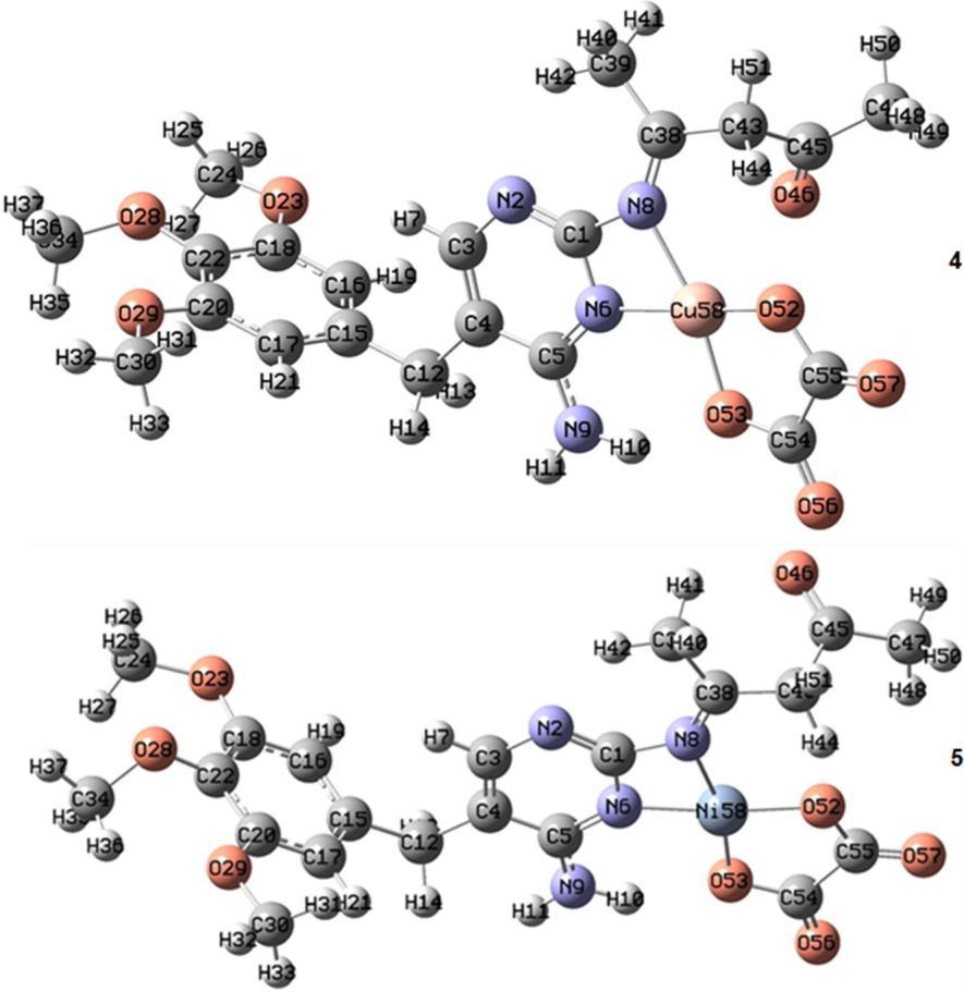
Table 4. Selected bond angles and bond distances (Å) of the optimized structure of 4 and 5 at the B3LYP level of theory with the 6 311G* basis set for C, H, O, and N atoms and LANL2DZ basis set for metal atom Bond lengths (Å) of complex 4 Bond lengths (Å) of complex 5 Cu(58) O(52) 1.877 Ni(58) O(52) 1.844 Cu(58) N(8) 2.026 Ni(58) N(8) 1.987 Cu(58) O(53) 1.876 Ni(58) O(53) 1.846 Cu(58) N(6) 1.949 Ni(58) N(6) 1.886 Angles(°)ofcomplex4 Angles(°)ofcomplex5
N(6) Cu(58) N(8) 67.0 N(6) Ni(58) N(8) 68.5 N(6) Cu(58) O(52) 168.3 N(6) Ni(58) O(52) 173.8 N(6) Cu(58) O(53) 98.6 N(6) Ni(58) O(53) 97.7 N(8) Cu(58) O(52) 107.6 N(8) Ni(58) O(52) 105.4 N(8) Cu(58) O(53) 165.6 N(8) Ni(58) O(53) 166.2 O(52) Cu(58) O(53) 86.3 O(52) Ni(58) O(53) 88.3
at 281 nm (35587 cm−1), which may be ascribed to π→π* electronic transition within the organic ligand.
Complex 1 is considered distorted trigonal bipyramidal, and the electronic spectrum might be
assigned in the “regular” geometry. In a trigonal bipyramidal ligand field, the ground termis split into the states 4A2', 4A1'', 4A2'', 4E", and 4E'; the 4Ptermissplit into 4A2 ' and 4E". The electronic spectrum of complex 1 (d7 ,
1357Indones. J. Chem., 2022, 22 (5), 1348 1364
reported for a distorted square planar of the Ni(II) complexes [38].
Electronic Structures
Term symbol of 4F) exhibits four bands, the first band at v1⁄v2 = 80 (or v2⁄v1 = 1.23 are in the usual range 279 nm (35842 cm−1) assigned to the charge transfer transition. The other three bands are due to d d transitions at 526 nm (19011 cm−1) ,672 nm (14880 cm−1), and 826 nm (12106 cm−1), which are assigned to 4A2'(F)→4A1"(F), 4A2'(F)→4A2"(F), 4A2'(F)→4E"(F) transitions, respectively [36 40].
The electronic spectral data of the chromium complex (complex 2) reveal four bands at 604 nm (16556 cm−1), 509 nm (19646 cm−1), 425 nm (23529 cm−1), and 277 nm (36101 cm−1), (Table 3 and 4) [29 30]. The intense high band appearing at 277 nm is assigned to the π→π* transition of the benzene ring. Furthermore, the band at 425 nm is assigned to the n→π* transitions of the azomethine group. The electronic spectrum of the Cr(III) complex showed two bands at 509 and 604 nm. The spectral band is consistent with that of five coordinated Cr(III) complexes (Cr(III), d3, Term symbol of 4F), so a “regular” square pyramidal geometry may be assigned for this complex [34 35,41]. However, distorted geometry is observed in this instance. The zinc(II) complex (complex 3) displays multiple intense absorption bands in the UV region at 281 nm (35587cm−1) that we assume result from metal ligand charge transfer and ligand internal transitions, which is compatible with this complex having distorted square pyramid structures [35 36].
The electronic spectrum of complex 4 displays a broad single d d band that appeared at 859 nm (11641 cm−1) which implies the three allowed spin transitions, 2B1g→2A1g(ν1), 2B1→2B2g(ν2) and 2B1→2Eg, may be due to square planar (slightly distorted) geometry around the copper(II) ion and 281 nm (35587 cm−1) which assigned to ligand to metal charge transfer (LMCT) [37].
The (UV Vis) spectrum of complex 5 exhibits four bands; the first band at 279 nm (35842 cm−1) is assigned to the π→π∗ transition of the aromatic ring. The second band appeared at 419 nm (23866 cm−1), which is attributed to the charge transfer from ligand, azomethine, to metal. From the electronic spectrum, the observed peak at (628 nm) 15923 cm−1 is assigned to the 1A1→1B1 band and 778 nm (12853 cm−1), which is attributed to the 1A1→1A2 band. These transitions suggest the square planar geometry around Ni(II). The 10 Dq = 2853 cm−1 , and
The calculated electron densities and energies of the frontiers' molecular orbitals (HOMO and LUMO) were investigated to explain the electronic properties of the prepared complexes. Fig. 4 and 5 summarize the calculated absolute energies of the HOMO and LUMO orbitals together with the HOMO LUMO energy gap values. Complex 5 has a more significant HOMO LUMO energy gap. The calculated orbital energy levels and percent composition of particular chosen frontier inhabited and virtual molecular orbitals of the prepared compounds, expressed in terms of composing fragments (L1, L2,andmetal ion),arelisted inTable5.Asshownin Table 5, the HOMO of complex 1 is wholly localized on the L1 ligand, and its LUMO complex is mainly localized on the metal ion. The HOMO and LUMO of complex 2 are mainly localized on the metal ion, as shown in Fig. 4 and presented in Table 5. The percent composition of frontier occupied and virtual molecular orbitals of complex 3 differ from those of complexes 1 and 2, as shown in Table 5. The ligands L1 and L2 of complex 3 contribute 100% of LUMO and 99% of HOMO, respectively. The LUMO orbital of complex 4 is distributed over metal ions,L1 and L2, with almost equal percent, while its HOMO orbital is almost localized on the metal ion (Fig. 5, Table 5). The percent composition of HOMO and LUMO molecular orbitals of complex 5 are verysimilar tothose of complex 3 (Fig. 5,Table 5).
Molecular Electrostatic Potential
The generated molecular electrostatic potential in the space around a molecule, including the charge distribution, is very helpful in understanding the sites for electrophilicattacksand nucleophilic reactions. Also, exploring the charge distribution around the molecule is essential for studying biological recognition processes and hydrogen bonding interactions [42 45]. Hence, the molecular electrostatic potentials for complexes 2 and 5 werecalculatedfromtheoptimizedgeometry(Fig.6).The
Fig 4. The frontier orbitals, HOMO, and LUMO of the complexes 1, 2, and 3 were calculated at the B3LYP level of theory with the 6 311G* basis set for C, H, O, and N atoms and LANL2DZ basis set for metal atom, with the surface isovalue of 0.02
Fig 5. The frontier orbitals, HOMO, and LUMO of complexes 4 and 5 were calculated at the B3LYP level of theory withthe6 311G* basisset for C,H,O, and Natoms andLANL2DZbasisset for metal atoms, withthesurfaceisovalue is 0.02
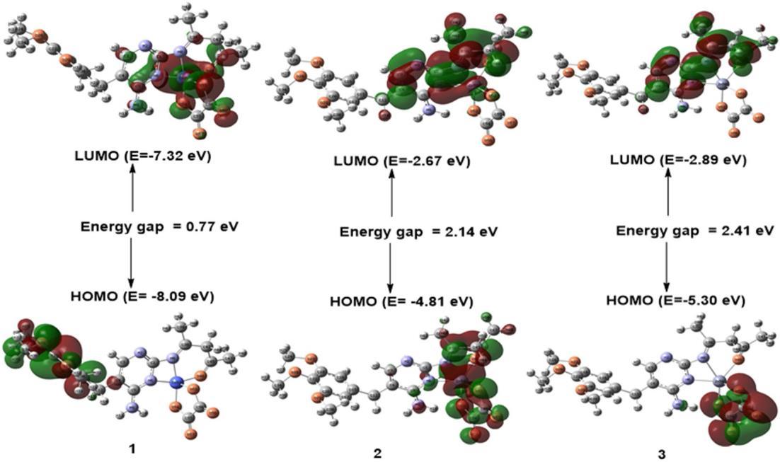
Table 5. Energies andpercent composition of HOMOand LUMO molecular orbitals ofcomplexes, expressedin terms of composing fragments, calculated at the B3LYP level of theory with the 6 311G* basis set for C, H, O, and N atoms andLANL2DZbasisset formetal atom
Compound Molecular orbital Orbital energy (eV)
Fragment
Metal L1 ligand L2 ligand
LUMO 7.32 62 12 26
HOMO 8.09 0 100 0
LUMO 2.67 63 17 20
HOMO 4.81 83 8 9
LUMO 2.89 0 100 0
HOMO 5.30 0 1 99
LUMO 8.12 30 31 39
HOMO 8.52 5 88 7
LUMO 2.88 1 99 0
HOMO 6.30 3 1 96
electrophilic reactive sites of the molecule are concerned with the negative (red) regions of molecular electrostatic potential (Fig. 6). The nucleophilic reactive sites are regarded by the positive (blue) regions of molecular electrostatic potential. For complex 2, the negative electrostatic potential regions are mainly localized over the O57, O56, O23, O28, and N2 atoms (Fig. 6). The V(r) values are 0.092, 0.085, 0.032, 0.037 and 0.022 a.u. for O57, O56, O23, O28 and N2 atoms of complex 2, respectively. According to these results, complex 2 has five possible sites for the electrophilic attack. The most positive electrostatic potential is localized on the C47−H50 (V(r) = 0.045 a.u.) region of complex 2 Therefore, the C47−H50 region represents the most reactive site toward nucleophilic attack within complex 2. For complexes 1 and 3, the most positive and negative electrostatic potentials are similar to those observed for complex 2. For the complex 5, the negative electrostatic potential regions are mainly localized over the O57 (V(r) = 0.080 a.u.), O56 (V(r) = 0.079 a.u.), O46 (V(r) = 0.028 a.u.), and O29 (V(r) = 0.036 a.u.) atoms (Fig. 6). Accordingly, the O57, O56, O46, and O29 atoms represent the possible sites for the electrophilic attack of the complex 5. The most positive electrostatic potential region is localized on the N9−H11 (V(r) = 0.049 a.u.) of thecomplex5 Therefore,theregionN9−H11,incomplex 5, is an electrophilic site and represents the most reactive site toward nucleophilic attack. For complex4 themost
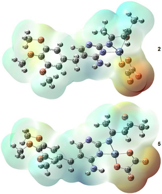
Fig 6. The Molecular electrostatic potential map of complexes 2 and 5
positive and negative electrostatic potentials are similar to those observed for complex 5
Antibacterial Activities
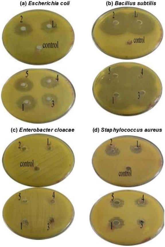
The comparative study of the biological effects of the ligands and their complexes is listed in Fig. 7. The synthesized complexes have been tested against the
Fig7.Thebiological effect of compounds
> complex 5 > complex 4 ≈ complex 1 > complex 3 > L2 = DMSO. This finding means that complexes significantly affect the antimicrobial activity of the organic ligand. Fig. 8. The antibacterial activity (ZI) of all the complexes >> Oxalic acid ≈ DMSO. The DMSO was used as a solvent and negative control. It did not show any activity against bacteria. All tests lacked antibacterial activity against Enterobacter cloacae. All complex tests had anti bacterial activity against test bacteria (Escherichia coli, Staphylococcus aureus, and Bacillus subtilis). The results showed that the nature of the metal ion (M(II), Cr(III)) in complexes plays a significant role in the ZIactivity [46]. Also, the structure of L1 due to the presence of the (C=N) group, which is significant in the mechanism reactions in biological reactions, and ligands with (N) and (O) donor systems might inhibit enzyme production and possibly (π e) delocalization through the whole (chelate ring) system thus include through coordination [46].
DNA Binding Properties
Fig 8. Photograph of the zone of inhibition in mm antibacterial activity of compounds
growth of Escherichia coli, Enterobacter cloacae, Staphylococcus aureus, and Bacillus subtilis. Generally, the antibacterial activities were in the order: Complex 2 > L1
The electrophoresis diagram of compounds is in Fig. 9. In comparison to control samples 1 and 2, there was a complete breakage in samples 3 and 10, which represented CrA and DMSOA, respectively, and the number 5 (lane 5), which represented NiA, was not found to be broken, and in samples 4 (lane 4), 6 (lane 6),
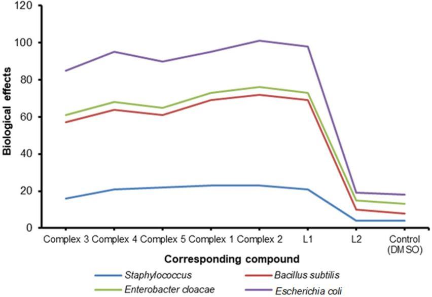
Fig 9. Agarose gel electrophoresis pattern for the DNA binding studies of oxalic acid and Schiff base trimethoprim with various metal ions
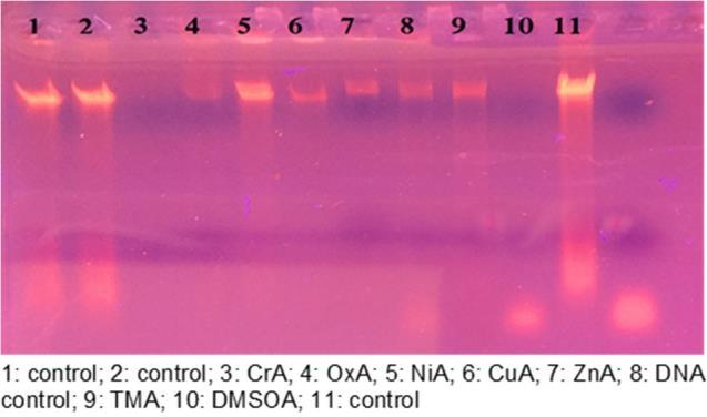
7 (lane 7), 8 (lane 8), and 9 (lane 9), representing OxA, CuA, ZnA, DNA control 3, and TMA, respectively. The most influential sample on DNA was sample 8 (lane 8), which represents DNA control, where a long haze streak showed DNA breaking along the gel transfer line. In the DNA cracking that was detected by the weak radiation of the sample and the use of Red safe stain at the examination under the source of ultraviolet compared to the control sample.The intensity of the metal DNA bands is also reduced when compared to the DNA control [39,46].
■ CONCLUSION
structuresofthesecomplexes,whicharecalculatedatthe B3LYP level of theory with the 6 311G* basis set for C, H, O, and N atoms and LANL2DZ basis set for metal atom. The Schiff base, L1, coordinated to the metal ion in a tridentate fashion through two nitrogen atoms and one oxygen atom in the case of complexes 1, 2, and 3, which adopted highly distorted trigonal bipyramid for complex 1, and distorted square pyramid configuration for 2 and 3, and as bidentate ligand fashion through two nitrogen atoms in the case of complexes 4 and 5 which adopted a highly distorted square planar geometry. The molecular electrostatic potential of the complexes is explored. The results show that the negative electrostatic potential regions are mainly localized over the oxygen atoms in most cases. The electronic structures of the complexesareinvestigatedintermsoftheenergiesofthe HOMO and LUMO orbitals and the electron density distribution of these orbitals over the composing fragments(L1, L2, and metal ions) ofthe complexes.The antibacterial activities of the prepared compounds were in the order: complex 2 > L1 > complex 5 > complex 4 ≈ complex 1 > complex 3 >L2. In the DNA cracking that was detected by the weakradiation of thesample and the use of a Red safe stain, it is observed that the intensity decreased for the metal DNA bands compared to DNA control.
■ ACKNOWLEDGMENTS
The authors wish to thank the technical assistance provided by the Departments of Chemistry in Baghdad and Al Mustansiriya university in the preparation of the samples forantimicrobialactivities are also acknowledged.
■ AUTHOR CONTRIBUTIONS
2
The new mixed ligand complexes are synthesized from the reaction of Schiff base ligand, L1, and oxalate anion, L2, with metal salts. The synthesized Schiff base ligand,L1,waspreparedfromthereactionoftrimethoprim and acetylacetone.Thegeneral formula ofthesynthesized complexes is [M(L1)(L2)], where M represents one of the followingmetals Co(II), Ni(II), Cu(II), Zn(II), and Cr(III), L1 represents the trimethoprim ((Z) 4 ((4 amino 5 (3,4,5 trimethoxybenzyl)pyrimidine 2 yl)imino)pentane 2 one) as the first ligand and L2 represent the oxalate anion (C2O2 ) as a second ligand. The analytical and conductivity measurements support the proposed formulae and indicate that the complexes are non electrolytes except complex 2, which is a 1:1 electrolyte. The results obtained from measurements of UV, IR, and NMR are in good agreement with the optimized
All authors contributed to the study's conception and design. Material preparation, data collection, and analysis were performed by Eid Abdalrazaq, Abdel Aziz Qasem Jbarah, Tagreed H. Al Noor, G.T. Shinain, and M.M.Jawad. Tagreed H. Al Noor wrote the first draft of the manuscript, and all authors commented on previous versions. All authors read and approved the final manuscript.
■ REFERENCES
[1] El Sawaf, A.K., El Essawy, F., Nassar, A.A., and El Samanody, E.S.A., 2018, Synthesis, spectral,thermal and antimicrobial studies on cobalt(II), nickel(II), copper(II), zinc(II) and palladium(II) complexes containing thiosemicarbazone ligand, J. Mol. Struct., 1157, 381 394.
[2] Muralisankar, M., Haribabu, J., Bhuvanesh, N.S.P., Karvembu, R., and Sreekanth, A., 2016, Synthesis, X ray crystal structure, DNA/protein binding, DNA cleavage and cytotoxicity studies of N(4) substituted thiosemicarbazone based copper(II)/nickel(II) complexes, Inorg. Chim. Acta, 449, 82 95.
[3] Mathan Kumar, S., Rajesh, J., Anitha, K., Dhahagani, K., Marappan, M., Indra Gandhi, N., and Rajagopal, G., 2015, Synthesis, characterization, crystal structure and cytotoxic properties of thiosemicarbazide Ni(II) and Zn(II) complexes, Spectrochim. Acta, Part A, 142, 292 302.
[4] Pahonţu, E., Julea, F., Chumakov, Y., Petrenco, P., Roşu, T., and Gulea, A., 2017, Synthesis, characterization, crystal structure and antiproliferative activitystudiesofCu(II), Ni(II) and Co(II) complexes with 4 benzoyl 5 pyrazolones derived compounds, J. Organomet. Chem., 836 837, 44 55.
[5] Pu, L.M., Zhao, Q., Liu, L.Z., Zhang, H., Long, H.T., and Dong, W.K., 2018, Synthesis and fluorescence properties of a new heterotrinuclear Co(II) Ce(III)complex constructed from a bis(salamo) type tetraoxime ligand, Molecules, 23 (4), 804.
[6] Chen, Q.L., 2016, Synthesis and structural characterizationofapyridineoxalatomolybdenum(V) complex, Int. J. New Technol. Res.,2(1),40 43.
[7] Frisch, M.J., Trucks, G.W., Schlegel, H.B., Scuseria, G.E., Robb, M.A., Cheeseman, J.R., Scalmani, G., Barone, V., Mennucci, B., Petersson, G.A., Nakatsuji, H., Caricato, M., Li, X., Hratchian, H.P., Izmaylov, A.F., Bloino, J., Zheng, G., Sonnenberg, J.L., Hada, M., Ehara, M., Toyota, K., Fukuda, R., Hasegawa, J., Ishida, M., Nakajima, T., Honda, Y., Kitao, O., Nakai, H., Vreven, T., Montgomery, J.A., Jr., Peralta, J.E., Ogliaro, F., Bearpark, M., Heyd, J.J., Brothers, E., Kudin, K.N., Staroverov, V.N., Kobayashi, R.,
Normand, J., Raghavachari, K., Rendell, A., Burant, J.C., Iyengar, S.S., Tomasi, J., Cossi, M., Rega, N., Millam, J.M., Klene, M., Knox, J.E., Cross, J.B., Bakken, V., Adamo, C., Jaramillo, J., Gomperts, R., Stratmann, R.E., Yazyev, O., Austin, A.J., Cammi, R., Pomelli, C., Ochterski, J.W., Martin, R.L., Morokuma, K., Zakrzewski, V.G., Voth, G.A., Salvador, P., Dannenberg, J.J., Dapprich, S., Daniels, A.D., Farkas, Ö., Foresman, J.B., Ortiz, J.V., Cioslowski, J., and Fox, D.J., 2009, Gaussian 09 Revision E.01,Gaussian,Inc., Wallingford,CT.
[8] Makkonen, I., Ervasti, M.M., Kauppila, V.J., and Harju, A., 2012, Exchange correlation potentials for inhomogeneous electron systems in two dimensionsfromexact diagonalization: Comparison with the local spin density approximation, Phys. Rev. B, 85, 205140.
[9] Becke, A.D., 2019, Dependence of the virial exciton model on basis set and exact exchange fraction, J. Chem. Phys., 150, 241101.
[10] Chen, H., Nusspickel, M., Tilly, J., and Booth, G.H., 2021, Variational quantum eigensolver for dynamic correlation functions, Phys. Rev. A, 104 (3), 032405.
[11] Wang, G., Annaberdiyev, A., Melton, C.A., Bennett, M.C., Shulenburger, L., and Mitas, L., 2019, A new generationofeffectivecorepotentialsfromcorrelated calculations: 4s and 4p Main group elements and firstrowadditions, J. Chem. Phys.,151,144110.
[12] Hill, J.G., and Shaw, R.A., 2021, Correlation consistent basis sets for explicitly correlated wavefunctions: Pseudopotential based basis sets for the group 11 (Cu, Ag, Au) and 12 (Zn, Cd, Hg) elements, J. Chem. Phys., 155, 174113.
[13] Hassan, S.S., Shoukry, M.M., and Jbarah, A.A.Q., 2020, Cordination compound of dimethyltin(IV) with N,N,N’,N’ tetraethylethylenediamine: speciation and theoretical approach, J. Mex. Chem. Soc., 64 (2), 24 43.
[14] Dennington, R., Keith, T.A., and Millam, J.M., 2016, GaussView, Version 6, Semichem Inc., Shawnee Mission, KS.
[15] Abu Yamin, A.A., Jbarah, A.A.Q.M., AlKhalyfeh, K., Matar, S., Alqasaimeh, M., Rüffer, T., and Lang,
1363Indones. J. Chem., 2022, 22 (5), 1348 1364
H., 2022, Crystal structure, spectroscopic studies, DFT calculations, and biological activity of 5 bromosalicylaldehyde based Schiff bases, J. Mol. Struct., 1262, 132976.
[16] O’boyle, N.M., Tenderholt, A.L., and Langner, K.M., 2008, Cclib: A library for package‐independent computational chemistry algorithms, J. Comput. Chem., 29 (5), 839 845.
[17] Mary Juliet, B.M., and Amaladasan, M., 2014, Preparation and properties of macrocyclic ligand, Int. J. Recent Innovation Trends Comput. Commun., 2 (8), 2102 2105.
[18] Jeffery, G.H., Bassett, J., Mendham, J., and Denney, R.C., 1989, Vogel’s Textbook of Quantitative Chemical Analysis, 5th Ed., John Wiley & Sons Inc., New York, US.
[19] Orekhov, M.A., 2021, Coordination numbers of bivalent ions in organic solvents, Russ. J. Phys. Chem. A, 95 (10), 2059 2064.
[20] Marcus, R.A., 1964, Chemical and electrochemical electron transfer theory, Annu. Rev. Phys. Chem., 15 (1), 155 196.
[21] Saheb, V., Sheikhshoaie, I., and Stoeckli Evans, H., 2012, A novel tridentate Schiff base dioxo molybdenum(VI) complex: Synthesis, experimental and theoretical studies on its crystal structure, FTIR, UV visible, 1H NMR and 13C NMR spectra, Spectrochim. Acta, Part A,95, 29 36.
[22] Agrwal, A., Verma, A., Chantola, N., Verma, S., and Kasana, V., 2022, Synthesis, molecular docking and extensive structure activity relationship of substituted DHP derivatives: A new class of herbicides, J. Environ. Sci. Health, Part B, 57(5), 379 420.
[23] Odion, E.E., Enadeghe, D.O., and Usifoh, C.O., 2021, Synthesis, characterization and antibacterial assessment of 3,4,5 trimethoxy 3’,4’ dimethoxychal cone and 2,4,6 trimethoxy 3’,4’ dimethoxychalcone, Niger. J. Pharm. Appl. Sci. Res.,10(2),1 5.
[24] Jin, R.Y., Sun, X.H., Liu, Y.F., Wong, W., Lu, W.T., and Ma, H.X., 2014, Synthesis, crystal structure, IR, 1H NMR and theoretical calculations of 1,2,4 triazole Schiff base, J. Mol. Struct.,1062, 13 20.
[25]
Aparna, E.P., and Devaky, K.S., 2018, Microwave assisted solid phase synthesis of trisubstituted pyrimidines, J. Chem. Pharm. Res., 10 (8), 67 72.
[26] Jacobsen, N.E., 2017, NMR Data Interpretation Explained: Understanding 1D and 2D NMR Spectra of Organic Compounds and Natural Products, John Wiley&Sons, Inc.,Hoboken,NewJersey,US.
[27] Ebrahimi, H.P., Hadi, J.S., Abdulnabi, Z.A., and Bolandnazar, Z., 2014, Spectroscopic, thermal analysis and DFT computational studies of salen type Schiff base complexes, Spectrochim. Acta, Part A, 117, 485 492.
[28] Adeyemi, J.O., Olasunkanmi, L.O., Fadaka, A.O., Sibuyi, N.R.S., Oyedeji, A.O., and Onwudiwe, D.C., 2022, Synthesis, theoretical calculation, and biological studies of mono and diphenyltin(IV) complexes of N methyl N hydroxyethyldithio carbamate, Molecules, 27 (9), 2947.
[29] Al Noor, T.H., Karam, N.H., Ghanim, F.H., Kindeel, A.S., and Al Dujaili, A.H., 2017, Synthesis, characterization and liquid crystalline properties of novel benzimidazol 8 hydroxyquinoline complexes, Inorg. Chim. Acta, 466, 612 617.
[30] Nakamoto, K., 2009, Infrared and Raman Spectra of Inorganic and Coordination Compounds, Part B: Applications in Coordination, Organometallic, and Bioinorganic Chemistry, 6th Ed., John Wiley & Sons, Inc., Hoboken, New Jersey, US.
[31] Mezey, R.Ș.,Máthé, I., Shova, S., Grecu, M.N., and Roșu, T., 2015, Synthesis, characterization and antimicrobial activity of copper(II) complexes with hydrazone derived from 3 hydroxy 5 (hydroxymethyl) 2 methylpyridine 4 carbaldehyde, Polyhedron, 102, 684 692.
[32] Bellamy,L.J.,1975, The Infra Red Spectra of Complex Molecules, Springer, Dordrecht, Netherlands.
[33] Housecroft, C.E., and Sharpe, A.G., 2018, Inorganic Chemistry, 5th Ed., Pearson Education Limited, Harlow, UK.
[34] Jamil, Y.M.S., Al Qadasy, J.M.K., Al Azab, F.M., and Al Maqtari, M.A., 2018, Synthesis, characterization andantibacterialstudyofsome3d metalcomplexes
Eid Abdalrazaq
1364 Indones. J. Chem., 2022, 22 (5), 1348 1364
of paracetamol and 1,10 phenanthroline, Jordan J. Chem., 13 (4), 203 212.
[35] Fleming, G.R., Lewis, N.H.C., Arsenault, E.A., Wu, E.C., and Oldemeyer, S., 2019, "Two Dimensional Electronic Vibrational Spectroscopy" in Coherent Multidimensional Spectroscopy, Eds. Cho, M., Springer, Singapore, 35 49.
[36] Galić, N., Dijanošić, A., Kontrec, D., and Miljanić, S., 2012, Structural investigation of aroylhydrazones in dimethylsulphoxide/water mixtures, Spectrochim. Acta, Part A, 95, 347 353.
[37] Abbas, S.H., 2017, Synthesis, characterization and biological activity of some nickel(II) mixed ligands complexes of dithiocarbamate and 1,10 phenanthroline, Eur. J. Chem., 8 (4), 367 370.
[38] Yallur, B.C., Krishna, P.M., and Challa, M., 2021, Bivalent Ni(II), Co(II) and Cu(II) complexes of [(E) [(2 methyl 1,3 thiazol 5 yl)methylidene]amino]thio urea: Synthesis, spectral characterization, DNA and in vitro anti bacterialstudies, Heliyon,7(4),e06838.
[39] Rajendran, N., Periyasamy, A., Kamatchi, N., and Solomon, V.,2020, Synthesis and efficacy ofcopper(II) complexes bearing N(4) substituted thiosemicarbazide and diimine co ligands on plasmid DNA and HeLa cell lines, J. Serb. Chem. Soc., 85 (3), 321 334.
[40] Dance, I.G., Gerloch, M., Lewis, J., Stephens, F.S., and Lions, F., 1966, High spin five coordinate cobalt (II), Nature, 210 (5033), 298.
[41] Kafi Ahmadi, L., and Shirmohammadzadeh, L., 2017, Synthesis of Co(II) and Cr(III) salicylidenic
Schiff base complexes derived from thiourea as precursors for nano sized Co3O4 and Cr2O3 and their catalytic, antibacterial properties, J. Nanostruct. Chem., 7 (2), 179 190.
[42] Bendjeddou, A., Abbaz, T., Gouasmia, A., and Villemin, D., 2017, Determination of reactive properties of a series of mono functionalized bis tetrathiafulvalene employing DFT calculations, ASRJETS, 29 (1), 308 326.
[43] Politzer, P., and Murray, J.S., 2021, "Molecular Electrostatic Potentials: Significance and Applications" in Chemical Reactivity in Confined Systems: Theory, Modelling and Applications, Eds. Chattaraj, P.K., and Chakraborty, D., John Wiley & Sons Ltd, Hoboken, New Jersey, US.
[44] Gadre, S.R., Suresh, C.H., and Mohan, N., 2021, Electrostatic potential topology for probing molecular structure, bonding and reactivity, Molecules, 26 (11), 3289.
[45] da Silva, G.C.Q., Cardozo, T.M., Amarante, G.W., Abreu, C.R.A., and Horta, B.A.C., 2018, Solvent effects on the decarboxylation of trichloroacetic acid: Insights from ab initio molecular dynamics simulations, Phys. Chem. Chem. Phys., 20 (34), 21988 21998.
[46] Muthukkumar, M., Malathy, M., and Rajavel, R., 2015, Antimicrobial and DNA cleavage activities of macrocyclic Cu(II), Ni(II), Co(II), and Zn(II) Schiff base complexes, Chem. Sci. Rev. Lett., 4 (16), 1227 1236.
Eid Abdalrazaq et al.