STAWA Year 12 General Human Biology Units 3 & 4 - Digital Version
Year 12 General Human Biology
Workbooks Unit 3 & Unit 4
Student name:
Class: Teacher: Human Reproduction & Infectious Disease
Editor
Annabel Kanakis
Annabel Kanakis
Rebecca Cooke
Michelle Edwards
Shannon Fletcher
Zac Gomes
Fiona May Writers
Acknowledgements
The authors would like to acknowledge the work of the previous writers of the STAWA Human Biology resources, from which several activities have been adapted for this publication. We would also like to acknowledge Shenton College Science staff for providing us with the resources that they developed and use for teaching Human Biology. Some of the activities and diagrams from those resources have been used due to their quality and appropriate level of difficulty for this course. We would like to thank Lorraine McKenzie for editing the documents and making constructive suggestions.
STAWA Year 12 General Human Biology Workbook ISBN: 978-0-9925739-2-8
All rights reserved. Except under the conditions described in the Copyright Act 1968 of Australia and subsequent amendments, no part of this publication may be reproduced, stored in a retrieval system or transmitted in any form or by any means, electronic, mechanical, photocopying, recording or otherwise, without prior permission of the copyright owner.
Schools Curriculum and Standards Authority Disclaimer: The inclusion of Schools Curriculum and Standards Authority material in this publication does not constitute Schools Curriculum endorsement of the publication.
Copying for Educational Purposes: Where copies of parts of this book are made by an educational institution under the Copyright Act, and an agreement exists between the Copyright Agency Limited and the relevant educational authority to pay the fee for such copying, no further fee is due. In the absence of such an agreement, the law requires that the institution keep records of such copying. In such cases, the copyright owner is entitled to claim payment.
Printed in Australia by: Daniels Printing Craftsmen - 10 Walters Drive, Osborne Park, WA 6017. Tel: (08) 9204 6800
Graphic Design by: Kattie Muir - Digital Crayon digitalcrayon@hotmail.com
Illustrations by: David Keigwin
12 GENERAL HUMAN
CONTENTS
Structure and use of the general Human Biology resources
Glossary of key words used in the formulation of questions
CHAPTER 1 Cell Reproduction
CHAPTER 2 Reproductive Systems
CHAPTER 3 Pregnancy
CHAPTER 4 Reproductive Technologies
CHAPTER 5 Disease
CHAPTER 6 Immune System
CHAPTER 7 Community and Global Health
Structure and use of general Human Biology resources
General information for teachers and students
This resource has been produced to support teachers and students in the absence of a textbook for this course. Each chapter corresponds to the topics outlined in the Science Understanding strand of the syllabus, with Science Inquiry and Science as a Human Endeavour incorporated where appropriate. The Syllabus Dot Points are included, and Learning Intentions and Success Criteria are provided so that teachers and students have a clear understanding of exactly what is expected to learn. Please note that these are suggestions from the writers only. Teachers and students are encouraged to formulate their own success criteria as part of the learning process.
Teachers should not use this book as their sole source of information and resources. This is a guide and provides some further resources to explore such as websites and educational films. Once again these are suggestions only. Practical activities are included in each chapter. These may include experiments, dissections, and interpretation of second-hand data. Safety issues have been highlighted where applicable. Teachers do not have to use all of the activities. These are suggestions but should be able to be completed even with somewhat limited resources. Students are encouraged to formulate their own tables for data collection and presentation, as well as practicing their graphing skills. There are several opportunities for students to draw labelled scientific diagrams.
Students should use this book as a source of essential information covering the syllabus dot points, but should seek other resources for greater depth of understanding. A glossary at the beginning of each chapter provides a list of key terms that students should define as they progress through the text. Students are encouraged to write their own notes using the ‘Checkpoints’ as a guide. Some Checkpoint questions enable students to write answers in this book, but they are encouraged to write their own notes for revision. These have been included following each section of text information to enable students to consolidate their understanding of the key concepts and summarise the key points. Chapter Review Questions are found at the end of each chapter and should be answered by students in their notebooks as revision for each topic. There are ‘Extras for Experts’ for students who want to check their depth of understanding of some concepts outlined in the chapter.
Glossary of key words used in the formulation of questions
Note – definitions in the glossary available from SCSA website syllabus documents are generic and applicable across all courses. Students should be aware of the meaning of the terms so as to be able to understand the questions asked in the book.
Word key Definition
Account
Advise
Analyse
Apply
Assess
Calculate
Choose (multiple-choice)
Clarify
Classify
Comment on
Compare
Complete
Consider
Account for: state reasons for, report on.
Give an account of: narrate a series of events or transactions
Recommend or inform
Identify components and the relationship between them; draw out and relate implications
Use, utilise, employ in a particular situation
Make a judgement of value, quality, outcomes, results or size
Ascertain/determine from given facts, figures or information
Decide or select the most suitable from a number of different options
Make clear or plain
Arrange or include in classes/categories
Make reference to and expand upon
Show how things are similar and different
Finish an outlined task
Reflect on and make a judgement/evaluation
Construct Make; build; put together items or arguments
Contrast
Correlate
Show how things are different or opposite
Demonstrate a mutual or complementary relationship
Create Make, invent something
Deduce
Define
Demonstrate
Draw conclusions
State meaning and identify essential qualities
Show by example
Describe Provide characteristics and features
Determine
Decide, find out
Discuss Identify issues and provide points for and/or against Distinguish
Draw (diagrams etc.)
Evaluate
Recognise or note/indicate as being distinct or different from; note differences between
An instruction, as in draw a circle
To ascertain the value or amount of; appraise carefully
Examine Inquire into
Word key Definition
Explain
Explore
Relate cause and effect; make the relationships between things evident; provide why and/or how
Similar to ‘explain’ (see above), but requires the quoting of specific examples or statistics or possibly the drawing of maps, graphs, sketches, etc.
Draw meaning from
To plan, search or inquire into; examine in order to obtain the true facts
Support an argument or conclusion; give reasons for your statements or comments
Identify by placing a name or word used to describe the object or thing
Provide a series of related words, names, numbers or items that are arranged in order, one after the other
Provide a word or term used to identify an object, person, thing, place etc. (something that is known and distinguished from other people or things)
Sketch in general terms; indicate the main features of
Suggest what may happen based on available information
Put forward (for example, a point of view, idea, argument, suggestion) for consideration or action
Present remembered ideas, facts or experiences
Recount Retell a series of events
Respond to…
Provide an answer; reply
Select Choose somebody or something from among several
Show Give information; illustrate
Sketch
A picture or diagram that is done quickly, roughly; a brief outline
State Express the main points of an idea or topic, perhaps in the manner of 'describe' (see above)
Summarise
Express, concisely, the relevant details
Unit 2
Unit description
The focus for this unit is on the reproductive choices that people make for personal reproductive health and the delivery of a healthy baby.
Offspring show features of both parents which result from new chromosomal combinations. Reproductive systems are specialised to produce differentiated gametes and ensure the chances of successful fertilisation and implantation. The healthy development of the embryo and foetus can be monitored and options are available for the safe delivery of the baby. Lifestyle choices can impact an individual’s sexual health and their fertility may require the use of reproductive technologies.
Students apply their knowledge to construct a DNA model and demonstrate cell division processes. They analyse and evaluate the various contraceptive methods, assisted reproductive technologies and delivery methods in terms of risks, effectiveness and personal circumstances. Students are encouraged to use information and communication technology to interpret data and communicate their findings in a variety of ways.
Unit content
Each unit includes the knowledge, understandings and skills described below.
Scientific Method
Identify a topic for investigation; research and construct questions for investigation.
Determine the appropriate methodology for investigations.
Design scientific investigations, including the formulation of investigable questions and/or hypotheses, materials required, procedure to be followed to collect valid and reliable data, and identification of safety and ethical considerations.
Conduct risk assessments to identify potential hazards and prevent potential incidents and injuries.
Select appropriate equipment and techniques to safely, competently and methodically collect valid and reliable data, and use equipment with precision, accuracy and consistency.
Represent qualitative and quantitative data in meaningful and useful ways, including the construction of appropriately labelled tables, process quantitative data using appropriate mathematical relationships and units, and draw appropriate graphs.
Analyse data to identify and describe trends, patterns and relationships, including the use of appropriate mathematical techniques, and recognise errors and limitations in data.
Draw conclusions consistent with the evidence and relevant to the question being investigated, identify further evidence that may be required, and recognise the limitations of conclusions
Evaluate the investigative procedure, including the relevance, accuracy, validity and reliability of data, and suggest improvements.
Communicate information and ideas in a variety of ways using scientific conventions and terminology, including the selection and presentation of data and ideas to convey meaning to selected audiences in written, oral and multimedia formats.
Scientific Literacy
Distinguish between opinion, anecdote and evidence, and scientific and non-scientific ideas.
Use reasoning to construct scientific arguments, and to draw and justify conclusions consistent with the evidence and relevant to the question under investigation.
Identify examples of where the application of scientific knowledge may have beneficial, harmful and/or unintended consequences.
CHAPTER 1
Syllabus dot points
Science Understanding:
Chromosomes are made up of large molecules of DNA found in the cell nucleus.
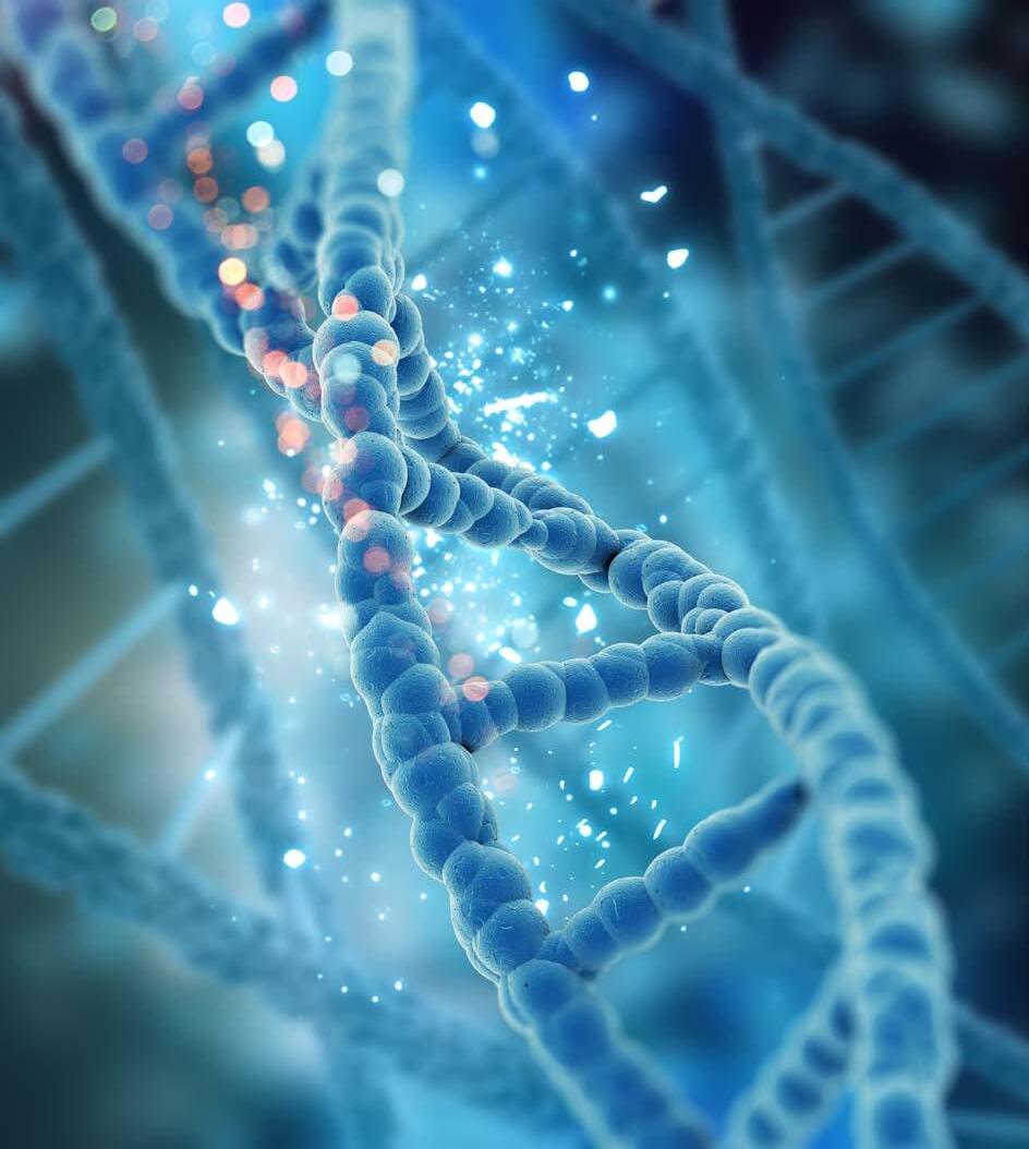
DNA has a double helix structure that is made up of nucleotides with complementary base pairing.
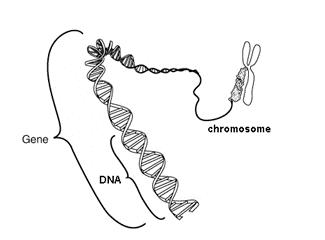
Genes are units of inheritance and are responsible for carrying genetic information from one generation to the next.
Mitosis produces diploid cells for the purpose of growth and repair and meiosis produces haploid cells for the purpose of gamete production (names and specific details of stages not required).
The Learning Intentions and Success Criteria are included as a guide to understanding expectations of students as outlined in the syllabus. Students could use them to review their understanding of the syllabus prior to assessments.
Learning intentions
1. Understand that DNA controls the characteristics of an organism and that these characteristics are inherited.
2. Understand that genes and chromosomes are comprised of DNA and carry the genetic information from generation to generation.
3. Understand that cells must divide for growth, repair and the production of gametes.
Success criteria
Be able to:
Describe the make-up of chromosomes.
identify the location of chromosomes in the human cell.
Describe the structure of DNA.
Describe the structure of a nucleotide.
Identify the base pairings G with C, and A with T in DNA.
Explain the reason for complimentary base pairing in the double helix of DNA.
Describe the structure and function of genes.
Describe why the body needs to be capable of making new cells.
Describe 3 purposes of making new cells.
Label simple diagram of a parent cell with (2N) to produce 2 daughter cells with (2N) each.
Name the process of cell division that occurs in somatic or body cells.
Describe and summarise the process of mitosis.
Explain why there are 2 stages of cell division in meiosis.
Name the process of cell division that occurs in gametes or sex cells.
State why the number of chromosomes is reduced in a gamete/sex cells to (N) haploid
Explain why the process of fertilization requires that the gametes are (N) haploid.
Key terms
Identify and fill in the definitions for the following key terms:
Key term
Cell cycle
Centromere
Chromatid
Chromosome
Complimentary
Daughter cell
Diploid
DNA
Double helix
Gene
Haploid
Meiosis
Mitosis
Nucleotide
Parent cell
Definition
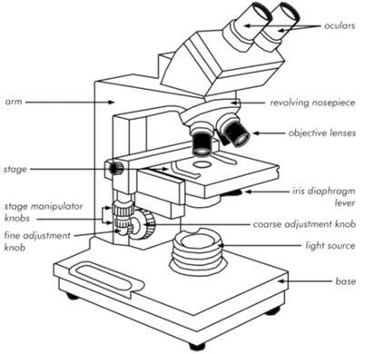
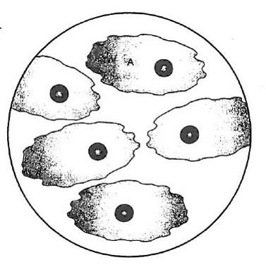
Structure of chromosomes
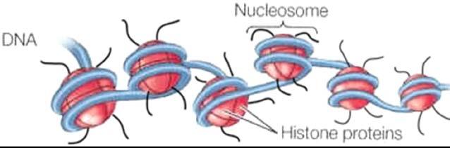
The DNA molecules are very long and need to be wrapped around a group of special proteins called histones. The histones form a nucleosome, and these enable the DNA to be tightly packed. The DNA can unwind when it is required to be copied.
Part of a coiled chromosome
When a cell is not dividing, the DNA exists in a mass of long thin fibres called chromatin. In chromatin the DNA is packed into a smaller volume.
Chromosomes are formed when the DNA is very tightly coiled and form a structure that is large enough to been seen by a light microscope. Chromosomes become visible during cell division.
In a chromosome - two parallel chromatids can be seen, and these are joined at a central point called a centromere.
One chromatid from each chromosome will be separated into the new daughter cells.
In human body cells, there are 46 chromosomes, half of which come from each parent. The gameteseggs and sperm have only HALF the number of chromosomes as in the body cells.
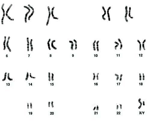
Genetic information can be taken from cells, usually by a blood test. Chromosomes can be seen in the process known as a karyotype. This is the arrangement of all the chromosomes from largest to smallest. Chromosomes displayed this way can be viewed to look at the size, shape and number of chromosomes. Geneticists use this information for the detection of differences in individual’s chromosomes.
Karyotype
Homologous chromosomes
Of the forty-six (46) chromosomes in each cell, forty-four (44) are chromosomes for the inheritance of all characteristics other than the sex chromosomes. These 44 are called autosomes. These chromosomes occur in homologous pairs. These are chromosomes of the same length, have the centromere in the same position, and have genes at the same location. Genes are the small, coded sections or unique coding for a particular characteristic on each chromatid.
Sex chromosomes
The two chromosomes that determine gender or sex, are called sex chromosomes. Sex determination is biological, and you inherit one sex chromosome from each biological parent.
Females inherit two X chromosomes. One X from their mother and one X from their father. Females are (XX).
Males inherit one X chromosome from their mother and one Y from their father. Males are (XY)
Females have 2 copies of X chromosomes – one from each parent.
Males have on copy of each sex chromosome X and Y – one from each parent.
The sex determination of a child at fertilization depends on whether the male sperm as an X or a Y sex chromosome.
Checkpoint
Describe how chromosomes are tightly packed forms of DNA.
State how many chromosomes are in human body cells.
Describe where chromosomes are found in the cell.
Describe what a karyotype display can tell about the about the genetic makeup of the individuals. Why is this useful?
Draw and label a chromosome including chromatid, centromere, genes, alleles.
Describe homologous chromosomes.
Describe an autosome.
How many autosomes chromosomes are there in each cell?
How many sex chromosomes are there in each cell?
Genetic material
Deoxyribonucleic acid (DNA) is the chemical that determines the characteristic you inherit. It is located in the nucleus of the cells and in the organelle - mitochondria. The DNA is a coded format that the body can use to produce new cells- replication. It can code for proteins to be made by the cells and it controls the characteristics that are inherited. Variations may occur due to changes in the DNA codes. This is known as a mutation.
The genome is the entire genetic code for an organism. The human genome has been mapped as 3,117,275,501 base pairs.
Checkpoint
Complete these sentences:
DNA is the abbreviation for:
The two locations where DNA can be found is:
DNA can code for:
The structure of DNA
There are two types of DNA; nuclear DNA, located in the nucleus and mitochondrial DNA located in the mitochondria.
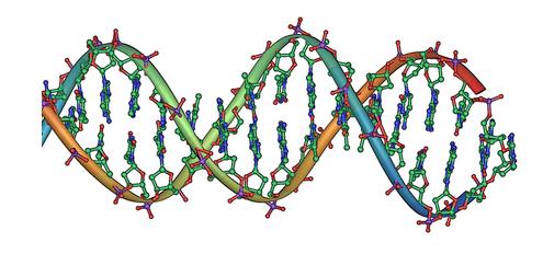
DNA molecule that we refer to is the Watson-Crick model. This winding ladder-like model forms a double helix.
DNA is made up of nucleotides. These are the basic structural component.
A nucleotide consists of three parts: a sugar, a phosphate and one of four nitrogenous bases.
The bases are either Adenine (A), Guanine (G), Cytosine (C) and Thymine (T).
These bases pair in a complimentary way. Adenine with Thymine, (A-T) and Cytosine with Guanine, (C-G).
Weak hydrogen bonds hold the bases together. Adenine and Thymine share two hydrogen bonds and Cytosine and Guanine share three hydrogen bonds.
The sides of the double helix or ‘ladder’ are a linked chain of ribose sugar and phosphate molecules.
The ‘rungs’ of the ladder or steps are attached to the sugar molecules and are called bases.
The order of the bases in the DNA molecule forms the genetic code.
Structure of DNA nucleotides and complimentary base pairs
Activity: Build a DNA molecule
Activity purpose
To create a molecule of DNA
Safety
It is not safe to eat food in a science laboratory.
Materials
Confectionery listed below (any suitable and available lollies can be used)
Marshmallow
Jelly babies
Long Toothpicks
Licorice strands
Tray to create DNA strands on
Gloves
Procedure
1. Wearing gloves and using a clean tray sort all the confectionery into types and colours.
2. Four jelly baby colours need to be allocated a nitrogenous base. Write down the colour allocation.
3. Assemble Jelly baby pairs A with T and C with G.
4. Licorice strand separate. Write down that licorice is the deoxyribose sugar ‘backbone’.
5. White marshmallow. Write down that this is phosphate.
6. Toothpicks are the ladder rungs.
7. Referring to a diagram of DNA recreate a double helix.
8. Peirce the jelly babies in nitrogenous base pairs. Centre these on the toothpick.
9. Peirce the licorice strands through.
10. Add the marshmallow to the ends.
11. Repeat this following the DNA strand.
12. Pick up and the strands and twist to make it a double helix.
Results
Take a photo of your DNA molecule and paste below.
Questions
Describe why the numbers of the nitrogenous base pairs always equal?
Explain how the complimentary base pairs ensure exact replication of DNA.
Activity: Extraction of DNA
Activity purpose
To isolate the DNA from a sample of fruit.
Extraction of DNA from fruit.
Observe the structure of DNA from plant cells.
Materials
Fruit (strawberries and kiwi fruit work well)
1 heavy duty zip-lock bag
Gauze cloth
Wooden popstick
Funnel
Test tube
DNA Extraction buffer solution (made of 50mL liquid dishwashing detergent, 15 g of NaCl and 950 mL water)
Ice-cold 95% alcohol (collect just before it is needed so it doesn’t evaporate)
Pipette
DNA ‘hook’
Time
Approximately 45 minutes
Safety
It is not safe to eat food in a science laboratory.
Wear safety glasses at all times
Procedure
1. Place your piece of fruit into a zip-lock bag, squeeze out as much air as possible and seal.
2. Squash the fruit for approximately 2 minutes.
3. Add 10 mL of extraction solution into the bag.
NOTE : The detergent dissolves the lipids (fats) that hold the membranes together and this releases the DNA into the solution. The extraction solution contains a type of salt which enables the DNA strands to aggregate (come together).
4. Mash again for approximately 1 minute.
5. Put two layers of gauze into the funnel. Pour the fruit mixture into the funnel and, collect the
filtrate in a test tube. Throw away the gauze with the remains of the fruit pulp.
6. Slowly pipette 8 mL of the ice-cold ethanol down the side of the test tube creating a layer of ethanol above the ‘fruit juice’. DO NOT SHAKE THE TEST TUBE.
7. Allow the solution to sit for 2 – 3 minutes. You should see DNA stands form at the interface where the two liquids meet.
8. The DNA can be removed and examined using the DNA hook.
Questions
What did the sample you extracted look like? Is this what you expected?
What is the purpose of the extraction solution?
Write a few sentences to describe the function of this material from your fruit sample.
Checkpoint
Complete these sentences:
DNA forms a... DNA is made up of...
The three main parts of a nucleotide are...
The complimentary pairing of bases is specific in...
Draw a nucleotide in the space below:
List the pairings of bases.
In the space below, draw and label a double helix including the base pairs, the hydrogen bonds, the sugar and the phosphate.
Genes
Located on each chromatid of the chromosome are small, coded sections of DNA. These are called genes. Genes are the unique coding for a particular functional protein. These can be a characteristic or a trait.
Genes can vary in number of base pairs and the order of the base pairs. This makes each gene different. The coding of the base pairs is for a particular protein. These proteins are what we need to survive.
Genes are located on the chromatids. You have two copies of each chromatid on the chromosome.
The genes are inherited from each parent and each chromatid has the copy from each parent. You have two copies of the gene. Offspring inherit all their genetic information from their parents.
Checkpoint
Describe what a gene is.
Describe the location of genes.
Cell division
All organisms which reproduce sexually begin life as a single fertilized egg cell. Cells need to divide to replicate and grow. Cells need to be replaced as they die or become damaged. Organisms grow when cells divide to produce new cells. There are two types of cell division.
Mitosis is the process of cell division for growth and cell replacement. This produces new cells that have 46 chromosomes. This is called the diploid number and is given the notation 2N (46).
Meiosis is the process of cell division for reproduction. It halves the chromosome number so that the gametes have half the number of chromosomes found in other body cells. This is called the haploid number and is given the notation N (23).
Checkpoint
Describe the purpose of mitosis.
Describe the purpose of meiosis.
Explain the purpose of halving the number of chromosomes in cells produced in meiosis.
State the number of chromosomes in a human diploid cell. ______________________________________________
State the number of chromosomes in a human haploid cell. _____________________________________________
Mitosis
Mitosis produces two new ‘daughter’ cells from a ‘parent’ cell. These daughter cells are genetically identical to the parent cell.
The doubling of the cell’s chromosome number during replication, followed by a single division at the end of mitosis ensures that the two daughter cells, both exactly the same as the original parent, are produced. Mitosis is a continuous process.
The stages of mitosis are outlined in the diagram below.
Two identical daughter cells are formed during the process. They have the same number of chromosomes.
Checkpoint
Name the process of cell division that occurs in somatic or body cells.
Summarise the process of mitosis.
Meiosis
Meiosis is the process of cell division for reproduction. This occurs in the testes and ovaries. This process happens in two stages and produces gametes that have half the number of chromosomes, 23. Four haploid (N) daughter cells are produced, and they are not identical.
Meiosis produces 4 non identical daughter cells. During the first part of the process ‘variation’ can occur. Chromosome pairs can ‘cross over’ genetic information between the two adjacent chromatids. The point of contact is called a chiasmata. This process provides new genetic variations and combinations. The daughter cells each have 23 chromosomes, the haploid number.
Meiosis has two divisions compared with mitosis that only has one division.
Differences in the products of meiosis in males and females
Males produce four equal haploid cells, called sperm cells.
Females produce one large egg cell that contains most of the cytoplasm and three smaller polar bodies. This will be further discussed in Chapter 3.
Checkpoint
Name the daughter cells of meiosis in males and females.
How many of each type of gamete in males and females are produced from the meiosis of one parent cell?
Why do gametes require just one set of chromosomes?
Name the process of cell division that occurs to produce gametes or sex cells.
Explain why there are two stages of cell division in meiosis.
How many chromosomes are there in a normal human body cell?
How many chromosomes are there in a human gamete?
Describe why the number is haploid.
Explain why the process of fertilization requires that the gametes are (N) haploid.
Chapter review
Describe the structure of a chromosome.
Draw and label a nucleotide.
Describe the function of DNA?
Draw and label a DNA molecule.
Describe the relationship between chromosomes, genes and DNA
Where does mitosis occur?
How many daughter cells are produced from mitosis?
State how many chromosomes are in the daughter cells from mitosis.
Where does meiosis occur?
How many daughter cells are produced from meiosis?
State how many chromosomes are in the daughter cells following meiosis.
Describe the purpose of mitosis and meiosis.
Extras for experts
Describe the relationship between chromatin and chromosomes.
Describe how scientists can use a karyotype to determine the gender of a foetus.
Explain why the number of chromosomes is different in a body cell and a gamete cell.
Create a table to compare and contrast mitosis and meiosis.
Describe why mitosis has one stage and meiosis has two stages.
CHAPTER 2
Reproductive Systems
Syllabus dot points
Reproductive systems
The production and delivery of gametes is facilitated by the structures of the male and female reproductive systems; females have additional structures that support the development of the unborn baby.
The male reproductive hormones follicle stimulating hormone (FSH), luteinising hormone (LH) and testosterone have a role in the production and maturation of sperm.
The female reproductive hormones follicle stimulating hormone (FSH) and luteinising hormone (LH) have a role in the production, maturation and release of ova; ooestrogen and progesterone have a role in preparing the uterus for implantation after fertilisation (detailed menstrual and ovarian cycle not required).
Sexually transmitted infections (STIs) can be prevented through safe sex methods and, if left untreated, can lead to serious health consequences.
The Learning intentions and Success criteria are included as a guide to understanding expectations of students as outlined in the syllabus. Students could use them to review their understanding of the syllabus prior to assessments.
Learning intentions
1. Understand the structure and function of the parts of the male and female reproductive systems.
2. Understand that hormones play a vital role in the production of sperm and ova, and to prepare the uterus for pregnancy.
3. Understand that fertilization is the union of gametes and that conditions must be optimal for this to take place.
4. Understand that sexually transmitted infections can be prevented.
Success criteria
Label and provide the function of the main parts of the female and male reproductive systems.
Describe the female reproductive organs that support the development of a growing foetus.
Describe the process of sperm production.
Describe the functions of FSH, LH and testosterone in the production and maturation of sperm.
Describe the functions of FSH and LH in the production, maturation and release of ova.
Describe the functions of ooestrogen and progesterone in the preparation of the uterus for possible fertilization and pregnancy.
Describe the cause, mode of transmission, symptoms, and treatment of common STIs: bacterial – Chlamydia, Gonorrhoea, Syphilis & viral – Genital herpes, HIV.
Define notifiable diseases.
Identify STIs that are notifiable to the Department of Health.
Key terms
Identify and fill in the definitions for the following key terms:
Key term Definition
Cervix
Ejaculation
Erection
Fertilisation
Gamete
Menopause
Menstruation
Ovaries
Ovulation
Ovum/ova
Penis
Prostate gland
Semen
Seminal vesicle
Sperm
Spermatozoa
Key term Definition
Testes
Uterine tubes
Uterus
Vagina
Female reproductive system
The purpose of sexual reproduction is to provide for the continuation of the species, to produce offspring. The structure of the reproductive systems in both male and female are designed for this function. The reproductive system also has an influence on behaviour, the way people grow, appearance and how the body works, through the production of hormones.
Male and females produce sex cells or gametes which combine during fertilization forming a zygote from which a new unique individual will grow. However, male, and female reproductive systems are quite different.
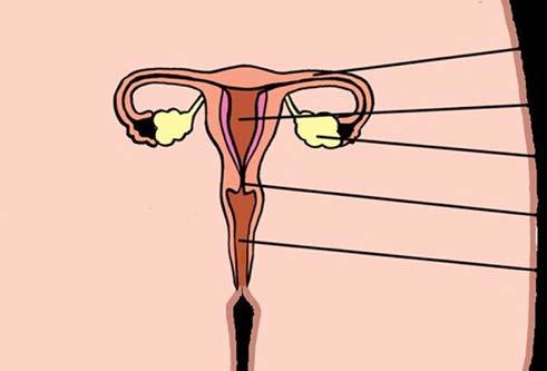
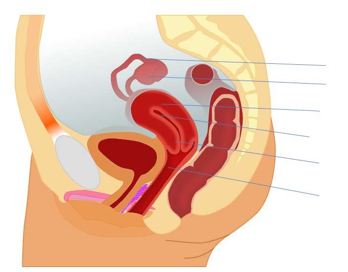
The main function of the female reproductive system is to produce eggs and to grow, nurture and produce a baby.
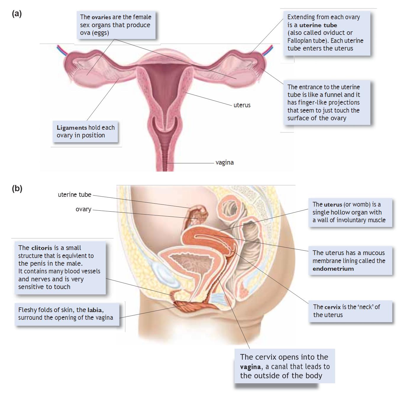
The female reproductive system as viewed: (a) from the front; (b) from the side showing the position of the internal organs.
The ovaries are the female sex organs and produce the female gamete, the ovum or egg. The ovaries are about the size of an olive, found in the abdominal cavity, on either side of the uterus, held in place by ligaments. Extending from each ovary are the uterine tubes (also known as Fallopian tubes), and
Same image on page 65. Confirm this is correct.
these catch the ova as it is released from the ovary during ovulation. Small projections lining the tubes waft the ova towards the uterus. It is here, in the uterine tube, that fertilisation occurs if the ova meets a sperm.
The uterus is the ultimate destination for a fertilized or unfertilized ovum as both uterine tubes enter at the top of the uterus. The uterus is a muscular hollow organ which is lined by a membrane called the endometrium. The endometrium has a rich blood supply and is where the fertilised ova (embryo) will embed and grow.
At the base of the uterus is the cervix, which opens into the vagina (also known as the birth canal). There are folds of skin which protect and surround the opening of the vagina called the labia and the clitoris which is very sensitive to touch containing many blood vessels and nerve endings.
Checkpoint
State the function of the ovaries.
Identify where the ovaries are located in the female.
Name the tubes that allow the ova to move from the ovaries to the uterus.
Describe the function of the lining of the uterus, the endometrium.
Why is the vagina also called the birth canal?
Label the following parts of the female reproductive system on both diagrams above and provide their function:
Name Function
Ovary
Uterine Tube
Uterus
Endometrium
Cervix
Vagina
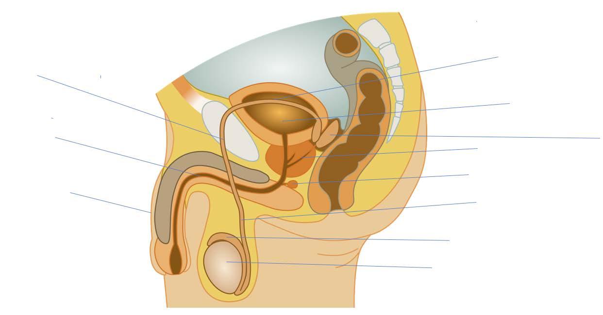
Male reproductive system
The main role of the male reproductive system is to produce the male gamete, sperm and to transfer the to the body of the female.
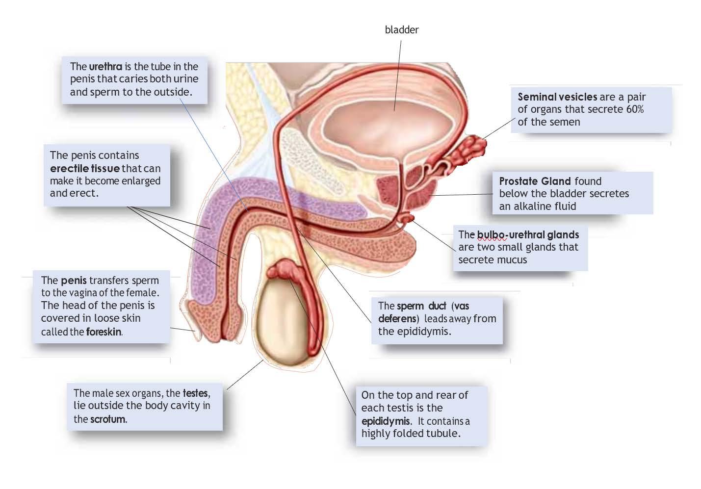
The male sex cells or gametes are sperm (spermatozoa) and these are produced in the testes. Males have two testes, each about the size of a large grape surrounded by a sack of skin called the scrotum. These are located outside the body behind the penis. This is because production and development of sperm requires temperatures lower than normal body temperature so they cannot be located internally. Males produce sperm each day from puberty until death.
Sperm are produced in the seminiferous tubules inside the testes but move to the epididymis to mature. The epididymis is found on the top of each testes and the sperm mature there for up to a month. Once mature, the sperm will be able to move from the epididymis through the vas deferens, a tube which lead from each teste, to the penis.
The sperm have tails (or flagella) which enable them to move and the fluid or semen through which they move is produced and secreted by a number of glands which form part of the male reproductive system.
The seminal vesicles produce a thick sugar rich fluid which provides energy for the sperm (remember they are using their tails to move!). There is one seminal vesicle found on each vas deferens. Most of the seminal fluid comes from these glands.
Just below the bladder is the doughnut shaped prostate gland. It is here both vas deferens join the urethra (the tube from the bladder to the outside world) and the prostate gland contributes a thin alkali fluid to the semen. This fluid neutralizes the acids normally present in the vagina.
Just underneath the prostate gland are two pea sized organs called the bulbo-urethral glands. These secrete thick mucus which acts as a lubricant to aid the insertion of the penis into the vagina during intercourse.
Finally, the semen containing the sperm leaves the body through the urethra via the penis.
Checkpoint
State where the male gamete is produced.
State where the sperm is matured.
Name the tubes that carry the matured sperm away from the testes.
Name the three glands that contribute fluid to the seminal fluid or semen.
Describe the location of the prostate gland.
Describe the pathway of the sperm from testes to penis (hint: look at the diagrams).
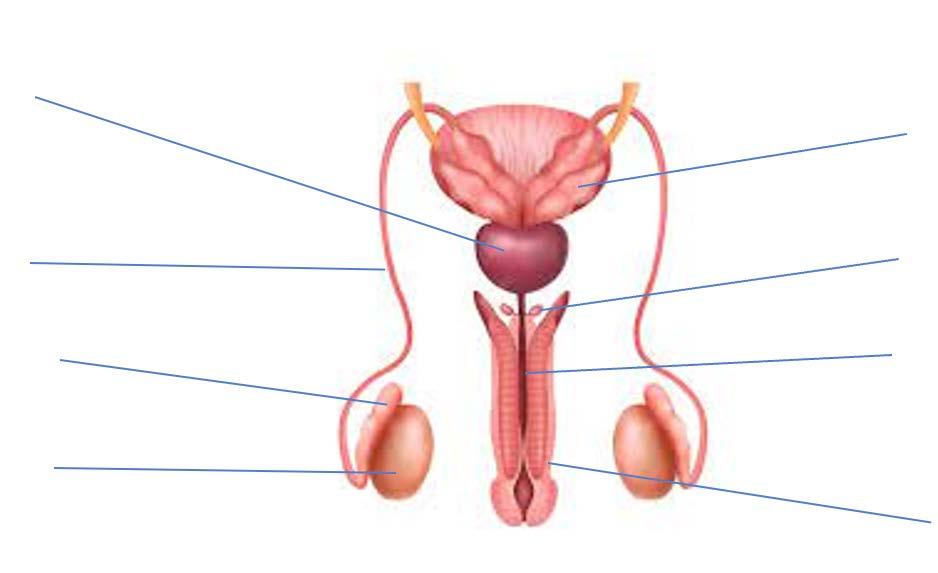
Label the following parts of the male reproductive system on both diagrams on the following page and provide their function:
Name Function
Testes
Vas Deferens
Epididymis
Seminal Vesicles
Urethra
Prostate Gland
Bulbo-urethral Glands
Penis
Male reproductive hormones
The primary role of the male reproductive system is to produce the male gamete, sperm.
The male sex cells or gametes are sperm (spermatozoa) and these are produced in the testes under the influence of hormones secreted from the pituitary gland in the brain and from the testes.
The male reproductive hormones are follicle-stimulating hormone (FSH), luteinizing hormone (LH) from the pituitary gland, and testosterone produced and secreted by the testes. These chemicals play crucial roles in the production and maturation of sperm, a process known as spermatogenesis.
Follicle-Stimulating Hormone (FSH):
In males, FSH stimulates specialised cells called Sertoli cells within the testes to stimulate the production of sperm and to nurture developing sperm cells through the process of spermatogenesis.
It promotes the growth and development of the seminiferous tubules in the testes, where spermatogenesis occurs.
Luteinizing Hormone (LH):
In males, LH acts on specialised cells called the Leydig cells in the testes, stimulating them to produce testosterone.
Testosterone is essential for various aspects of male reproductive function, including the development of secondary sexual characteristics, libido, and most importantly, the initiation and maintenance of spermatogenesis.
LH is particularly active during puberty.
Testosterone:
It is the primary hormone involved in the development and maintenance of male reproductive tissues, including the testes and prostate.
Testosterone also plays a critical role in stimulating spermatogenesis, particularly in the early stages of sperm cell development.
It is crucial for the maturation of sperm cells.
Overall, the coordinated action of FSH, LH, and testosterone is essential for the proper functioning of the male reproductive system, particularly in the production and maturation of sperm. Any disruptions in the levels or function of these hormones can lead to infertility or other reproductive health issues in men.
Female reproductive hormones
Follicle-stimulating hormone (FSH), luteinizing hormone (LH), oestrogen, and progesterone are the primary hormones that control the maturation of eggs or ova, ovulation and the menstrual cycle.
Follicle-Stimulating Hormone (FSH) and Luteinizing Hormone (LH) in Ovulation:
FSH and LH are both produced by the pituitary gland in the brain.
During the menstrual cycle, FSH stimulates the growth and development of ovarian follicles in the ovaries.
These ovarian follicles contain immature eggs (oocytes). As the follicles grow, they produce increasing amounts of oestrogen.
Rising oestrogen levels lead to a surge in LH levels. This surge in LH triggers ovulation.
Ovulation is the process where a mature egg is released from the ovary into the uterine tube, where it can be fertilised by sperm. This is caused by the action of LH on the mature follicle, causing it to rupture and release the egg.
Oestrogen:
Oestrogen is primarily produced by the developing ovarian follicles.
Oestrogen plays multiple roles in the menstrual cycle and reproductive system:
- It stimulates the thickening of the endometrium (the lining of the uterus) during the menstrual cycle.
- Oestrogen also helps in the development of secondary sexual characteristics in females, such as breast development and the widening of the hips.
- Additionally, oestrogen is involved in maintaining the health of the lining of the vagina and promoting cervical mucus production, which facilitates sperm transport.
Progesterone:
Progesterone is primarily produced by the corpus luteum, which forms from the remains of the ovarian follicle after ovulation.
Progesterone prepares the endometrium for implantation of a fertilised egg by promoting its further thickening and the development of glandular structures that will support early pregnancy.
It also helps maintain the uterine lining throughout the early stages of pregnancy.
Together, FSH, LH, oestrogen, and progesterone control the 28 day menstrual cycle and ensure the proper functioning of the female reproductive system. These hormones regulate the production, maturation, and release of eggs (ova), as well as prepare the uterus for potential implantation
and support early pregnancy if fertilisation occurs. Disruptions in the balance or function of these hormones can lead to menstrual irregularities, infertility, or difficulties in maintaining a pregnancy.
Checkpoint
State the name of the hormone responsible for the stimulation of sperm growth.
State the name of the hormone responsible for the development of secondary sex characteristics in males.
Describe the function of Luteinising hormone in the male.
State the names of the hormones produced in the ovaries.
Describe the function of Luteinising hormone in the female.
Describe two functions of oestrogen.
Sexually transmitted infections (STIs)
Key term Definition
Abstinence
Antibiotics
Infertility
Inflammation
Safe sex
Sexually transmitted infections
Symptoms
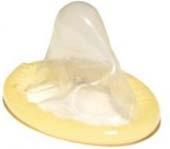
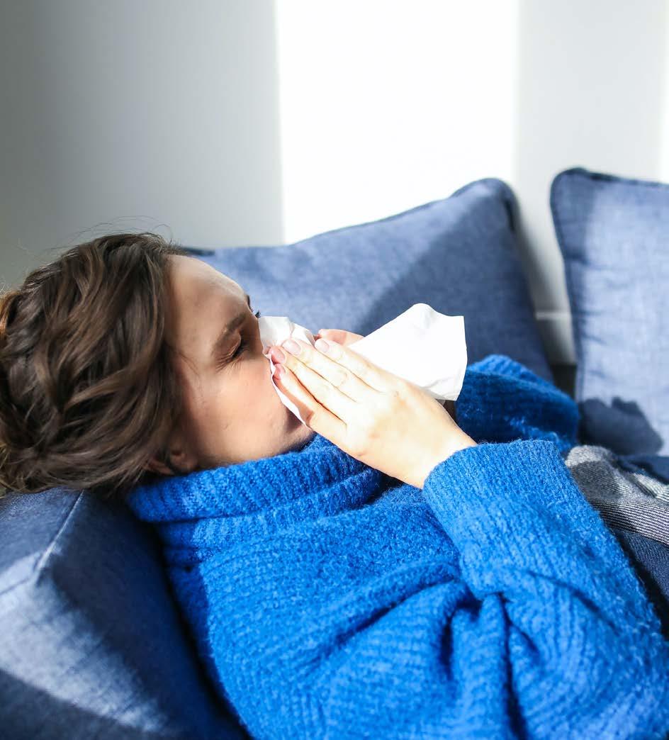
A Sexually Transmitted Infection (STI) is an infection or disease that spread during unprotected sexual intercourse with an infected partner. STIs can be caused by bacteria, viruses, fungi, or parasites. Infection occurs during the exchange of bodily fluids so can occur during vaginal, anal, and oral sex. Using barrier protection during intercourse, such as a condom, will prevent the spread of many STIs. Abstaining from sexual intercourse and having one sexual partner will greatly reduce the risk of contracting most STIs. However, some STIs can also be spread via skin-to-skin contact and through blood.
Recognising symptoms of various STIs is useful. However, many people who contract an STI do not display any symptoms. Thus, knowing:
How to practice safe sex,
Where and when to get tested for STIs, and
How to set and maintain personal boundaries using assertive communication skills, are important life skills.
This section will present information about some of the more common diseases so that students can develop an understanding about how the diseases are caused and how they are spread so that they develop a clear comprehension of the prevention of STIs.
Activity: Handshake transmitted infections
A handshake transmitted infection (HTI) is a fictional infection that, as the name suggests, is passed on through shaking hands with an infected person. It is a symptomless and short-lived infection, but it has a 100% infection rate if you shake hands with an infected person.
Materials
Pieces of paper equal to the number of students (one piece labelled “HTI”, two pieces labelled “glove”)
Procedure
1. Your teacher will give you a piece of paper. Do not read it. Put it aside until instructed.
2. When instructed by your teacher, shake hands with two people. Record their names in the table under “Person One” and “Person Two” for Round One.
3. Repeat Step 2 until you have completed four rounds, recording the names of those people you shook hands with in the relevant round number.
4. After Round 4, your teacher will instruct you to read your piece of paper. If it says “HTI” then you were infected at the start of the activity. Notify your teacher and those with whom you shook hands. If your piece of paper says “G”, you were wearing gloves and thus were protected from the HTI!
Results
Determine who was infected by completing the tree diagram below.
Predict the number of rounds needed to infect everyone who was not wearing gloves.
In terms of STIs, what does the glove represent?
Teacher Notes: Variations
Adjust the number of people who start with the HTI, the number of handshakes per round, or the number of rounds depending on class sizes.
The activity could continue until everyone is infected to test students’ prediction.
Students could abstain from shaking hands for some or all rounds. Discuss what this represents in terms of STIs.
Bacterial infections
Many STIs are caused by bacteria. This means that they are infectious but can be treated. The most prevalent STIs are caused by bacteria.
Chlamydia
Cause and transmission
Chlamydia is caused by a bacterium called Chlamydia trachomatis. The bacteria lives in semen and vaginal fluid, so it can be transmitted through vaginal, oral, or anal sex.
Symptoms
An infected person often has little to no symptoms but is still capable of infecting sexual partners. This is one of the primary reasons chlamydia is so common today. When chlamydia does display symptoms, in females it causes:
abnormal vaginal discharge.
bleeding or spotting between periods.
pain when urinating.
pain during sexual intercourse.
If left untreated, chlamydia can cause Pelvic Inflammatory Disease (PID), PID causes inflammation and scar tissue to develop along the walls of the uterus and uterine tubes, which in turn leads to fertility problems.
Long-term chlamydia can also cause eye problems and arthritis. It can also infect unborn babies, causing eye or lung problems at birth.
In males it causes:
clear or milky discharge from the penis.
pain when urinating.
redness near the urethra.
If left untreated, chlamydia causes swelling of the testicles and epididymis, resulting in fertility problems.
Treatment
If detected early, chlamydia is easily treated with antibiotics. However, the infected person does not become immune to chlamydia and can be reinfected. Early detection and treatment of chlamydia is vital to a full recovery, as the damage caused by PID or to the testicles, eye problems and arthritis is irreversible.
Checkpoint
The Western Australian Department of Health keep records on the occurrence of notifiable sexually transmitted infections. The table below shows the number of cases of chlamydia from 2011 to 2020.
Graph this data on a sheet of graph paper using a column graph.
Suggest a reason for the decline in notified cases in 2020.
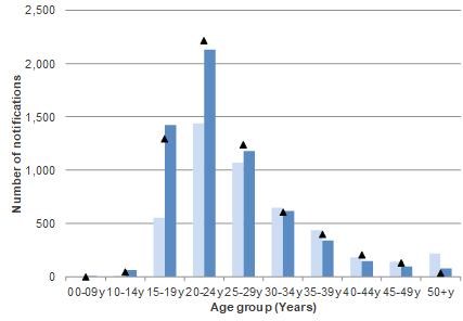
Figure 1 below shows the number of chlamydia notifications by sex and age for Western Australia in 2020. (Ignore the black triangles)
Figure 1
Describe the trend evident in the number of notifications of chlamydia by age in:
Females:
Males:
Suggest two possible reasons why there is the difference in the number of notifications between males and females aged less than 24 years.
Gonorrhoea
Cause and transmission
Gonorrhoea, commonly known as ‘the clap’, is also caused by bacteria. The bacteria live in semen and vaginal fluid, so it can be transmitted through vaginal, oral, or anal sex.
Symptoms
Like chlamydia, gonorrhoea often has no symptoms. When symptoms do appear, in females:
unusual vaginal discharge.
pain when urinating.
irregular bleeding, especially between periods or after sex.
dry, sore throat.
anal discharge and discomfort.
Like chlamydia, if left untreated, gonorrhoea can cause PID, and can infect newborn babies as they pass through the birth canal.
In males it causes:
yellow discharge from penis.
pain when urinating.
pain in the testicles.
dry, sore throat.
anal discharge and discomfort.
Left untreated, it damages the tubes that carry sperm, resulting in infertility. In males and females, untreated gonorrhoea also damages the eyes, heart, and brain.
Treatment
Gonorrhoea can also be treated with antibiotics, however it is developing a resistance to many antibiotics. Still, it is important to see a doctor early if an infection is suspected as the long-term damage to the reproductive organs, eyes, heart, and brain is irreversible, even after the infection is cured.
Syphilis
Cause and transmission
Syphilis is another bacterial infection and is commonly called ‘the pox’. Like the previous infections, the bacteria live in semen and vaginal fluid, so it is spread through vaginal, oral, and anal sex.
Symptoms
Some people do not exhibit any symptoms. However, the symptoms of syphilis go through four stages.
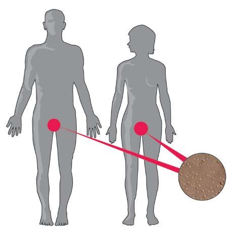
The primary stage begins after three or four weeks with one or more small sores in or around the penis, vagina, mouth, or anus, but can appear anywhere on the body and is usually painless. The sores go away by themselves, which can lead infected people to believe they are healed. They are so wrong.
The second stage begins a few months after the first stage. There is a broad range of symptoms, such as:
flu-like symptoms.
skin rash that lasts for weeks.
swollen glands in the armpit or groin.
ulcers in the mouth, nasal cavity, or genitals.
pain in bones, muscles, and joints.
The person is highly infectious during this stage.
The symptoms will go away without treatment and progress to the next stage.
During the latent stage, the person does not display any symptoms. Only a blood test will reveal the presence of syphilitic bacteria. The latent stage can last for years.
The tertiary stage is the final stage of syphilis. It appears between five to 20 years after the initial infection. The symptoms associated with this stage are widespread and devastating. It causes:
syphilitic heart disease,
weakened blood vessels,
blindness,
deafness,
dementia and,
insanity.
Pregnant women can pass the bacteria to their foetus. This can lead to miscarriages, stillbirths, or babies born with tertiary stage syphilis.
Treatment
Syphilis is treated with antibiotics. If detected and treated during the first stage, there are no lasting complications. If treated during the later stages, it is still curable, the damage over the later stages can be irreversible.
Checkpoint
Name the bacterial STI that most often shows symptoms.
Identify how bacterial STIs can be treated.
List the symptoms of chlamydia that a female would notice.
List the symptoms of gonorrhoea that a male would notice.
Describe Pelvic Inflammatory Disease and explain how it can lead to infertility.
Viral infections
A number of serious STIs are caused by viruses. They often mutate so viral infections are very difficult to treat.
Genital Herpes
Cause and transmission
Genital herpes is caused by a virus. It is present in semen and vaginal fluids as well as blisters on the skin around the genital area of an infected person and so can be transmitted via vaginal, anal, or oral sex. Since genital herpes lives on the skin, barrier methods of contraception reduce the chance of infection, but they do not eliminate it.
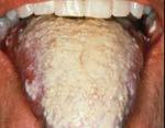
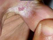
Symptoms
The symptoms for genital herpes are similar for males and females. Blisters develop around the genitals (penis and vulva) and anus. The infected person also experiences flu-like symptoms upon first infection. Other symptoms include stinging in the genital area and a burning feeling when urinating. The blisters last for a few weeks before going away, but they can reappear at any time. Even when blisters are not present, there is still a risk of infecting another sexual partner. Genital herpes can also infect a baby during childbirth if delivered vaginally.
Treatment
The blisters are treated with pain medication, ointment for the affected areas, and bathing the area with salt solution. However, there is no cure. The virus migrates to and multiplies in the spinal cord, where it remains for life.
Human
immunodeficiency virus (HIV)
Cause and transmission
Human immunodeficiency virus (HIV) is caused by a virus. It is present in semen and vaginal fluid, so can be transmitted through vaginal, oral, and anal sex. The virus is present in blood, so can be spread through contact with open wounds and sharing needles. HIV can also be passed on to a baby during pregnancy, childbirth, and breastfeeding.
Symptoms
Roughly two weeks after infection, the person develops normal flu-like symptoms, so an infected person may not suspect HIV. These symptoms resolve without treatment and no other symptoms appear, but the virus can remain latent in the body for many years.
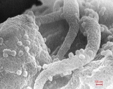
Left untreated, in five to 10 years the virus infects and destroys white blood cells – cells that are necessary for healthy immune function. When this happens, the body can no longer fight infections from other pathogens, nor prevent some cancers from developing – even a common cold infection can lead to pneumonia. At this point, the person is said to have developed Acquired Immune Deficiency Syndrome (AIDS).
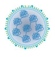
The image below shows a scanning electron micrograph of HIV budding from infected white blood cell (lower left) and other surrounding structures.
Treatment
There is no cure for HIV. However, if detected early, an infected person can take antiretroviral drugs that slow the replication of HIV. These drugs, if taken exactly as prescribed, can prevent a person developing AIDS.
Checkpoint
Outline the similarities and differences between genital herpes and Human Immunodeficiency Virus in terms of their symptoms and treatments.
Similarities
Differences
Suggest why viral infections that infect a person for life.
Preventing the spread of STIs
Abstinence, that is, refraining from sexual intercourse, is the only method of preventing the transfer of most STIs. However, the risk of infection can be reduced by having one uninfected sexual partner.
Practicing safe sex to ensure semen or vaginal fluids do not enter the partner’s body also reduces the risk of infection. Safe sex involves the use of condoms and dental dams, covering open wounds and covering other infectious body parts (for example, blisters from genital herpes).
Having regular STI checks, especially after a new sexual partner, can also detect STIs early and make early treatment possible for the infected person and limiting infectious period.
Notifiable STIs
Notifiable diseases are those illnesses about which public health authorities must be informed. Many STIs are notifiable diseases. The reason is to know where significant diseases are spreading to take steps and to take steps to limit the spread (to avoid epidemics) and increase public education. The identity of the infected individual is kept anonymous to protect their privacy. However, the infected individual may want to – or need to – notify current and previous sexual partners after a positive diagnosis of an STI.
At the time of publication, the notifiable STIs in WA are:
chlamydia,
gonorrhoea,
syphilis,
donovanosis,
chancroid,
HIV and
hepatitis A, B and C.
Checkpoint
Name and describe two ways people can reduce their chances of contracting STIs.
Suggest two actions an infected person can take to reduce the spread of infection.
‘notifiable disease’.
Identify two things public health authorities can do with statistics about STI infections.
Chapter review
Complete the summary table below for the sexually transmitted infections. Name of
Chlamydia
Syphilis
Gonorrhoea
Genital herpes
Explain how HIV can lead to AIDS, and why AIDS can be life-threatening.
Circle the correct option for each statement:
STIs are transmitted through vaginal, anal, and oral sex.
Chlamydia, gonorrhoea and syphilis are caused by bacteria
STIs often display symptoms.
Genital herpes can be cured with antibiotics.
A person with HIV will always develop AIDS
True/False
True/False
True/False
True/False
True/False
Abstinence is the only way to prevent contracting an STI True/False
Notifications to the Department of Health keep the infected True/False person anonymous.
Chapter review
Describe the function of the uterine tubes?
Describe the function of the ovaries.
Describe the pathway that an egg (ovum) would take following ovulation from the ovary to the cervix.
Explain why the testes are located in the scrotum.
Besides sperm, what else is makes up the semen?
Describe the function of the vas deferens.
Describe the function of the seminal vesicles.
Name the hormones that have an effect on the male reproductive system.
Describe the function of Luteinising hormone and testosterone in the male reproductive system.
State which hormones have an effect on the ovaries.
Describe the functions of oestrogen.
Describe the functions of progesterone.
List the STIs caused by bacteria.
State the symptoms of chlamydia in males and females.
Describe the main methods people can adopt to prevent the spread of STIs.
Explain why it is important for females to prevent STI infection and describe the possible consequences of not doing so.
Explain why it is important for people to notify medical professionals if they suspect they have an STI.
Extras for experts
Explain why the semen should be alkaline.
Describe the differences between the pituitary hormones on the male and female reproductive systems.
Explain how the functions of oestrogen and testosterone are similar.
Discuss with a partner or in small groups the following questions:
Why is it important that the identity of the infected individual is kept anonymous when a doctor notifies the public health authorities?
Are there any STIs, if not all STIs, that an infected person should be legally required to inform past, present and future sexual partners?
CHAPTER 3
Syllabus dot points
Fertilisation combines the male and female gametes producing a zygote with genes from both parents and pregnancy will be established if implantation occurs.
Embryonic and foetal development have a known and predictable sequence of events (details of specific milestone events not required).
The placenta has an important role in the provision of nutrients to and removal of wastes from the developing baby.
The unborn baby can be monitored utilising a variety of techniques, including ultrasound and blood tests.
Parental, embryonic and foetal testing can be done to detect a range of genetic and chromosomal abnormalities through the examination of karyotypes and dna profiles.
Maternal lifestyle choices, including the use of drugs, alcohol and smoking, will affect the developing baby and ongoing health of the child.
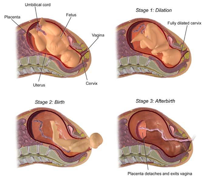
The sequence of events in the birth process prepare the baby and mother for delivery.
Various methods of delivery of the baby are available.
The Learning intentions and Success criteria are included as a guide to understanding expectations of students as outlined in the syllabus. Students could use them to review their understanding of the syllabus prior to assessments.
Learning intentions
1. Understand that an embryo is produced from the successful unity of an egg and sperm, and must be maintained for an approximate 40 week period by the mother before the baby is delivered.
2. Understand that the embryo and foetus progress through predictable stages of development unless there is a genetic or environmental issue that affects those processes.
3. Understand that lifestyle choices and accessibility to health services will affect the outcome of the development and growth of the baby and the developing infant. Unborn babies can be monitored throughout the pregnancy to detect growth and any abnormalities.
Success criteria
Define the term gamete.
Define fertilisation as the restoration of the 2N number of chromosomes by combining gametes.
Describe the formation of the embryo as cells produced from the fertilisation of the egg by the sperm, to combine the genes from both parents.
Define the term zygote.
Describe the events leading to the production of an embryo.
Label a diagram outlining the succession of the embryo’s growth as it moves from the oviduct into the uterus.
Define implantation.
Describe how implantation occurs.
Describe how the placenta is formed and maintained.
Explain the purpose of the placenta.
Describe the known and predictable sequence of development from the zygote through embryonic stages to foetal development.
Distinguish between an embryo and a foetus.
Describe the use of ultrasound technology to determine the health of the baby.
List some environmental factors that may affect the mother and foetus during pregnancy.
Describe how both the mother and foetus are affected by environmental factors.
Explain how maternal lifestyle choices will affect foetal development and ongoing health of the baby.
Describe the possible consequences of an unhealthy lifestyle during pregnancy, such as drug taking, malnutrition and alcohol.
List and describe tests that detect foetal development and abnormalities.
- Identify and describe tests that can be done to check foetal development.
- Identify and explain tests that can show genetic make-up of foetus and how this is used to check for genetic disorders.
Describe the sequence of events in the birth process that prepare the offspring and mother for delivery.
Recognise and label the events that occur during labour.
Describe the complications that can arise due to the positioning of the placenta and umbilical cord.
Key terms
Identify and fill in the definitions for the following key terms:
Key term Definition
Implantation
Infant
Labour
Oviduct
Placenta
Pregnancy
Ultrasound
Umbilical cord
Uterus
Zygote
The reproductive system
Successful reproduction is essential for the survival of the species. In the previous chapter, you learned about the reproductive system and where the sperm and eggs are produced. You also learned about the major organs and their functions, including the oviduct, uterus, vagina and penis. In this chapter you will learn about the fertilisation of the egg by the sperm and the resulting pregnancy. Successful pregnancy, including the optimal health of both mother and baby, results in birth and the continuing development of the infant.
The pictures below show the structure and function of the female reproductive system which is the focus of this chapter.
Same image on page 34. Confirm this is correct.
Checkpoint
The female reproductive system is responsible for the receiving of the sperm from the male and the production and ovulation of the egg to be fertilised. It is also responsible for the implantation of the fertilised egg and subsequent pregnancy.
Describe the function of the ovary.
Name the organ in which fertilisation takes place.
Describe the functions of the uterus.
Describe the functions of the vagina.
Fertilisation
The ejaculated semen contains some 500 million sperm.
The seminal vesicles contain sugar to provide the sperm with energy. The sperm has to move approximately 18cm to reach the egg. The prostate gland produces a fluid which is alkaline (high pH) which helps the sperm to survive the rather acidic condition found in the cervix and uterus.
Once in the vagina the sperm start to swim, vaginal contractions during intercourse help push the sperm through the cervix. The death rate of sperm is very high. Many sperm die through contact with the walls of the uterus, whilst others may go in the wrong direction taking the uterine tube without an egg. Of the 500 million sperm ejaculated only about 1000 will reach the egg.
The egg secretes chemical signals to the sperm so it knows where the egg is so that it can be fertilized. On finding the egg, the sperm breaks through the protective outer layers, and this will often take many sperm. Once one sperm has entered the egg a chemical change occurs in the eggs membrane which prevents any further sperm form entering. Once the head of the sperm joins with the nucleus of the egg fertilization has occurred and a zygote has been formed.
Checkpoint
Describe the route of sperm from Seminiferous Tubules in the Testes to the point of fertilisation in the uterine tube.
Fertilisation and implantation
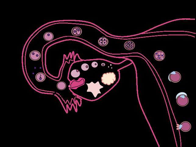
Fertilisation is also known as conception and marks the beginning of pregnancy. It occurs when an egg, or ovum, and sperm fuse together. This enables the total number of chromosomes in the resulting nucleus to be restored to 46 (2N), as 23 chromosomes from the sperm and 23 chromosomes from the egg join as a nucleus is formed within the egg. The resulting zygote thus has half of its genes from the female and half from the male.
Fertilisation occurs in the oviduct. The resultant zygote then passes along the oviduct, dividing repeatedly to form a hollow ball of cells called a blastocyst. The blastocyst continues to grow, the cells reproducing by mitosis very rapidly. It reaches the uterus within 4-6 days and consists of about 150200 cells. It then buries itself into the lining of the uterus. This is called implantation. The developing embryo can then start to receive nutrients for its growth and development from the blood vessels and glands in the uterine lining.
Checkpoint
Fertilisation occurs when:
The number of chromosomes in a zygote is: ______________________________________________________________
Fertilisation occurs in the:
It takes ________________________________________ days for the developing blastocyst to arrive at the uterus.
The term that describes the burying of the forming embryo into the lining of the uterus is:
First trimester
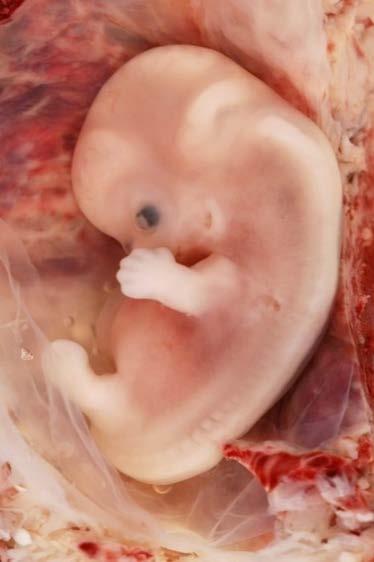
Cell division occurs very quickly, and some of the cells within the hollow ball start to form the embryo
After about 16 days the brain, spinal cord and heart have started to form. By the end of the eighth week the embryo can be recognised as human, and limb buds can be clearly seen. All organs have are present, but they are not functional. Miscarriages are most common during the first trimester as the developing embryo is mostly easily affected by drugs, such as alcohol and nicotine, and pathogens such as rubella and other viruses.Some of the cells that make up the hollow ball of cells (blastocyst) develop into the placenta. The embryo is called a foetus from 8 weeks onwards.
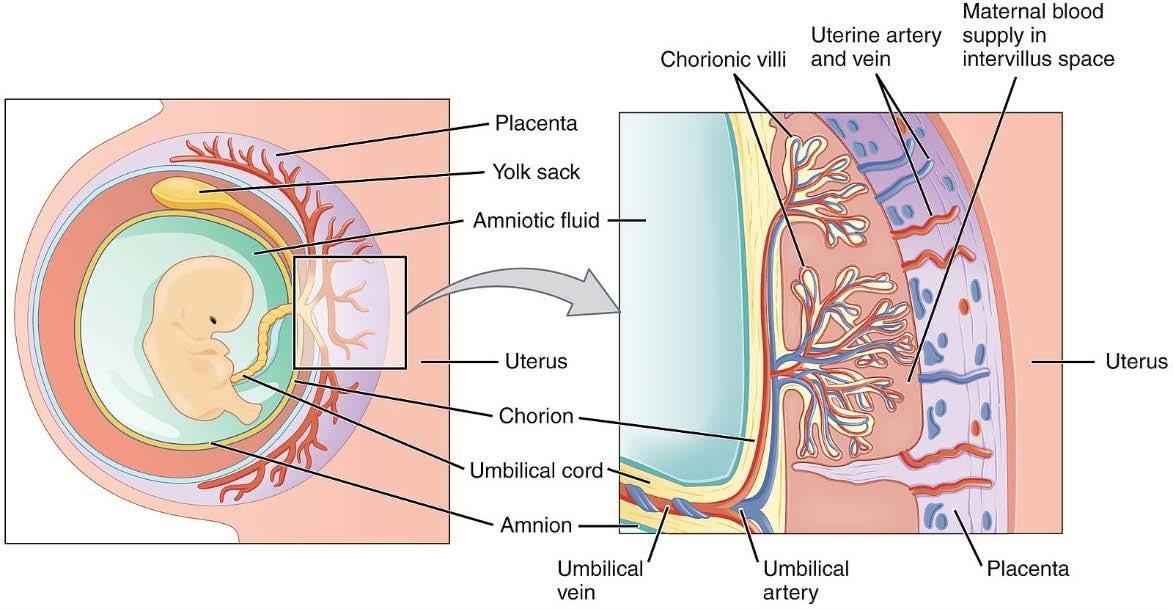
The placenta
The placenta is an organ that allows oxygen and nutrients to be passed from the mother into the embryonic and foetal blood. Even though the placental blood and the blood of the baby are very close, they never mix. The placenta attaches to the baby via the umbilical cord that carries wastes from the baby to the mother and oxygen and nutrients to the baby. By the end of the eighth week the placenta is fully formed.
Checkpoint
Define the term embryo.
Summarise what happens during pregnancy from Weeks 5-12 to end the first trimester.
Describe the roles of the placenta.
For how many weeks during pregnancy is the baby called an embryo?
Describe what happens during the embryonic period?
State the name of the developing baby from Week 10 of pregnancy?
The second and third trimester
By the end of the first trimester all of the major organ systems are established, but during the second trimester the organ systems develop further and become well established. If a baby is born prematurely at 24 weeks has approximately a 60% survival. Once the organs have developed the foetus continues to grow.
Label the diagram of the foetus in the uterus:
The embryonic period is characterised by the development of all of the body systems. The embryonic period lasts from Week 3 to Week 10 of pregnancy (8 weeks).
The foetal period is characterised by rapid growth, weight gain and development of complex structures. The foetal period is from Week 11 to Week 40 of pregnancy.
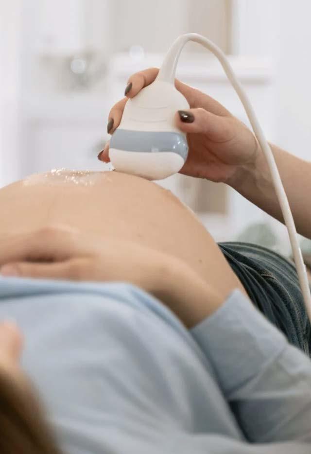
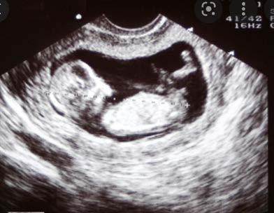
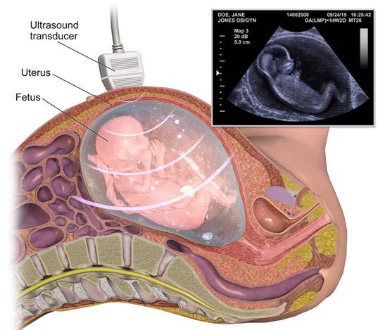
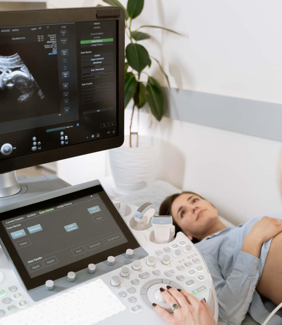
Regular medical checks during pregnancy ensure that the growth and development of the foetus can be monitored and any problems can be diagnosed quickly. The growth and development of the embryo and foetus can be monitored using ultrasound techniques. A trained technician called a sonographer presses a small, hand-held device (transducer) against the skin of the abdomen and moves it to capture the images. The transducer sends sound waves into the uterus, collects the ones that bounce back and sends them to a computer, which creates the images.
Pregnant woman having ultrasound
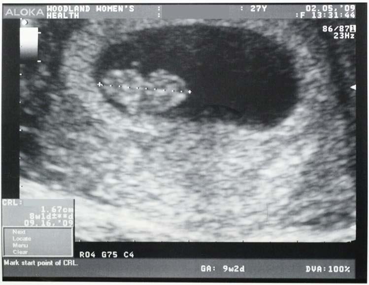
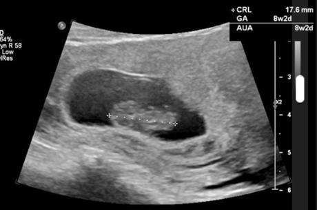
Ultrasound picture of 8 week old embryo
Activity: Pregnancy
During gestation, the period between conception and birth, a foetus grows in size. This growth is accompanied by changes in form and function or development.
Activity purpose
To describe the structural and physiological changes happening during embryonic and foetal development.
To name the techniques presently available for studying the foetus in utero.
Materials
graph paper
Procedure
1. Graph the data on separate pieces of graph paper for embryo and foetal length and mass that appear in the table below.
2. Indicate the trimesters on the graph.
Timetable of Foetal Growth and Development
Note: These data have been obtained by combining several sources. Figures are rounded for simplicity. Developmental stage details are not required for assessment in this course.
0 0 0 Fertilisation.
1 0 0 Embryo reaches uterus. Implantation.
2 0 A flat, 2-layered disc i.e. only ectoderm and endoderm. Saclike digestive tract with no mouth or anus. Umbilical cord forming.
3 2.5 3 layers present; ectoderm, mesoderm and endoderm. Beginnings of skeletal and nervous systems.
4 6 Simple 2-chambered heart, beating 60 beats/min. Tail, gill pouches, limb buds. Muscular system forming. Neural tube closing to form spinal cord and brain.
5 12 Mouth, eyes, webbed fingers and toes, lungs and regions of digestive canal form.
All major systems formed. Now called a foetus. Ossification (replacing cartilage by bone) begins. Makes small movements, but not yet felt by mother.
‘Quickening’ (movement) felt by mother. Heart can be heard.
Heart rate 140 beats/min. Head hair appears. Skin glands produce vernix caseosa a white creamy paste to protect delicate new skin. Sleeps and wakes.
Vigorous movements.
Testes descend. Fat deposited. Fine hair (lanugo) covers head and body.
Lanugo drops away. Takes up birth position, head down usually. 38 500 3250
Full term. Skin covered with cheese-like vernix caseosa. Foetus has moved down in pelvis. Foetus’ pituitary signals for birth to begin.
Questions
In which of the following intervals does the baby form the major body systems?
months 0-3
months 4-6
months 7-9
During which of the following time intervals is increase in length most rapid?
months 0-3
months 4-6
months 7-9
Using the graph, justify your answer to the previous question.
During which of the following intervals is increase in mass most rapid?
months 0-3
months 4-6
months 7-9
What developmental changes could cause this increase in mass?
Name the process that increases cell numbers as the baby grows?
The data supplied came from several sources, some pre-dating modern techniques for examining the foetus in utero. How do you think these older data were obtained?
Identify some new techniques are available for studying the foetus in utero?
Babies born before 25 weeks have a very small chance of survival without serious problems arising. State the main problems that affect the survival of very premature babies.
The pregnant mother must maintain a healthy lifestyle so that the development of the baby is not affected adversely. A healthy balanced diet, plenty of water and regular exercise help the mother and baby to remain healthy throughout the three trimesters.
Research the nutrients a pregnant woman should consume that are essential for optimal embryonic and foetal growth.
If the pregnant mother is in contact with some types of chemicals, including drugs such as alcohol and nicotine in cigarettes, there may be serious consequences for the developing baby.
Use reference materials to find out the effects of the following drugs on embryonic and foetal development. Where possible explain how the drug acts on the developing baby.
Alcohol
Cigarette smoke, particularly nicotine and carbon monoxide.
Select another drug of your own choice to research: _____________________________________________________
Methamphetamine
Diagnosis of foetal health
Parental, embryonic, and foetal testing are crucial tools in identifying a range of genetic and chromosomal abnormalities.
Parental testing involves examining the genetic makeup of prospective parents to assess the risk of passing on genetic disorders to their offspring. This type of testing often includes carrier screening, where individuals are tested for specific genetic mutations that may not affect them but could cause genetic diseases if inherited from both parents. For example, prospective parents might undergo screening for conditions like cystic fibrosis, sickle cell anaemia, or muscular dystrophy. Parental testing helps individuals make informed reproductive decisions and may involve genetic counselling to discuss the implications of the results.
Prenatal screening
Prenatal screening can give parents valuable information about the baby’s health. The screening can identify if the baby has birth defects and genetic disorders. These tests include blood tests, ultrasounds and DNA screening.
Types of screening
First trimester the mother is offered a blood test and an ultrasound to measure the size of the clear space in the tissue at the back of the baby’s neck. This can identify if the foetus is developing Down Syndrome and other conditions.
Second trimester the mother is offered another blood test that looks for chromosomal disorders such as Down Syndrome and other serious abnormalities.
The ultrasound images below show the difference in the first trimester and the second trimester.
The image below shows how an ultrasound is used to monitor a baby’s development and look for abnormalities.
First trimester ultrasound Second trimester ultrasound
Prenatal screening for genetic disorders
Screening tests can identify if the baby has any abnormalities, these tests are optional. Some of these tests are invasive. These tests are offered to people who have had:
Previous pregnancy with a genetic disorder.
The parents have an increased risk due to a previous scan done in early stages of pregnancy.
If there is a family history of a genetic condition.
Embryonic testing, also known as preimplantation genetic testing (PGT), is performed on embryos conceived through in vitro fertilization (IVF) before they are implanted in the uterus. This testing is particularly valuable for couples at risk of passing on genetic disorders or for those who have experienced recurrent pregnancy losses or failed IVF cycles.
Foetal testing involves assessing the genetic and chromosomal status of a developing foetus during pregnancy. Techniques such as amniocentesis and chorionic villus sampling (CVS) are used to obtain foetal cells for analysis.
Amniocentesis can be carried out between 16-20 weeks of pregnancy and is invasive, involving the removal of about 130mL of amniotic fluid from the amniotic sac. Chorionic villus sampling requires the removal and testing of cells from the chorion, a foetal membrane. These tests can detect a wide range of genetic and chromosomal abnormalities, including Down syndrome, trisomy 18, and neural tube defects.
Types of screening includes:
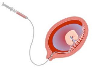
Chorionic villus sampling (CVS)
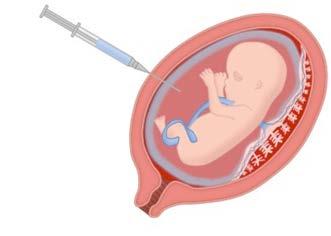
Amniocentesis
Duration of procedure
When is the test done?
What is the procedure?
Short procedure no need for anaes-thetic.
Short procedure no need for anaesthetic.
What is the test looking for?
The test is done between 12-14 weeks of pregnancy.
A needle is inserted through the abdomen of the female into the uterus taking a sample of the pla-centa cells. The procedure is done using an ultrasound to avoid dam-age to the foetus.
The sample is then analysed, and the DNA retrieved.
Image of procedure Suction tube removes foetal cells from placenta.
Checkpoint
The test is done between 15-20 weeks of pregnancy.
A needle is inserted through the abdomen to take a sample of the amniotic fluid. The pro-cedure is guided by ultrasound to avoid dam-age to the foetus
The sample is then analysed, and DNA is retrieved.
Needle draws out amniotic fluid.
Rye and Dave have been trying to have a baby for two years. Identify factors that may affect a male and female’s fertility.
Rye and Dave visited a reproductive technology clinic, and they were advised to undergo fertility tests. List and describe the tests they could use to check both Rye’s fertility and Dave’s fertility.
After a few weeks Rye and Dave have managed to conceive their first baby. Rye is delighted and wants to make sure that the baby is forming well. List and describe the test that can be carried to determine foetal development and disorders like Down syndrome.
Rye and Dave’s baby was developing well but they are concerned that the genetic disorder in Rye’s family could affect their baby. Identify and explain tests that can be done to check the foetus for genetic disorders.
Blood samples from the mother can also be used to detect abnormalities as some foetal cells can be isolated from the mother’s own blood cells.
Foetal testing is typically offered to pregnant individuals who are at increased risk of having a baby with a genetic disorder, such as women over the age of 35 or those with a family history of genetic conditions.
Results from foetal testing can help parents make decisions about the management of the pregnancy, including preparation for the birth of a child with special needs or considering options such as termination of pregnancy in cases of severe abnormalities.
Both parental, embryonic, and foetal testing rely on the examination of karyotypes (the number and appearance of chromosomes) and DNA profiles to identify genetic and chromosomal abnormalities. These tests provide valuable information to individuals and couples, allowing them to make informed decisions about family planning and pregnancy management.
Karyotype showing the chromosomes of a normal male
Checkpoint
Explain why it is important to check foetal health during pregnancy.
Name two foetal tests that involve the removal of foetal cells for DNA testing.
Describe what a karyotype shows.
Explain the purpose of testing parents before pregnancy.
Childbirth
The process of childbirth, also called labour, is divided into three stages. Approximately two to three weeks before labour begins, the foetus rotates in the uterus and moves lower down. The head can then settle into the mothers’ pelvis ready for birth.
The first stage of labour is dilation. The mother experiences contractions of the muscles of the uterine wall that become progressively stronger and more frequent. This is the longest stage of labour and can last for many hours. During this stage the cervix dilates allowing the baby’s head to move further into the vagina or birth canal. It is during this stage that the membranes surrounding the foetus that contain the amniotic fluid break. This is often referred to as the “waters breaking”.
The second stage is the expulsion stage. This is when the actual delivery of the baby occurs. The cervix must be fully dilated (about 10 cm) and the baby makes its descent through the birth canal. After the baby is born the uterine muscles continue to contract pushing out the placenta and the membranes that surrounded the developing foetus. This is referred to as the ‘afterbirth’. This final stage of labour, the placental stage, culminates in the umbilical cord being clamped and cut.
The best position for the placenta during pregnancy is for it to develop on the rear wall of the uterus, towards the mothers’ spine. This is because it is best for the baby as it allows the baby to grow and descend to the right position and align in the birth canal for a vaginal birth.
Stages of childbirth
If the placenta stays low in the uterus, near to or covering the cervix, it may block the opening. This is called low-lying placenta or placenta praevia. It affects about 1 in every 200 births, but in most cases the cervix is not completely covered.
Having an anterior placenta increases the chances of having the baby’s head being down but the back being against the mother’s spine. This is called the OP position (occiput posterior). If this happens the baby’s face is turned upward. This can cause longer labour and more pain in the mother’s back. The longer the labour, the more risk there is of complications for both the mother and baby.
The umbilical cord also needs to be in the correct position during labour to limit any complications. Some problems include umbilical cord prolapse. This is when the umbilical cord slips into the vagina (birth canal) ahead of the baby during the first and second stages of labour. The cord can get pinched, so the baby may not get enough oxygen. This does not happen very often. Vasa previa is when one or more blood vessels from the umbilical cord or placenta cross the cervix. These blood vessels aren’t protected by the umbilical cord or the placenta, so they can tear during labour. This can cause lifethreatening haemorrhaging of the baby. This can cause death and, even if the blood vessels don’t tear, pressure on them during labour can cause problems for the baby. A nuchal cord is an umbilical cord that gets wrapped around a baby’s neck. Babies with a nuchal cord usually are born healthy, but it sometimes can affect their heart rate. The midwife or doctor can see a nuchal cord on an ultrasound and usually can slip the cord off the baby’s neck during labour and birth.
Checkpoint
Describe each of the stages of labour.
Explain why the position of the placenta being at the rear of the uterus is so important.
Assisted delivery and interventions
There are several methods of delivering a baby besides a normal vaginal delivery, each with its own considerations based on factors such as the health of the mother and baby, the stage of labour, and any existing medical conditions. Vaginal delivery is preferred when there are no complications during pregnancy or labour, and the baby is in the proper position for birth.
In some cases, assistance may be needed to facilitate vaginal delivery. This can include the use of instruments such as forceps or vacuum extractors to help guide the baby through the birth canal. Assisted vaginal delivery is typically considered when there are concerns about the progress of labour or foetal distress, but the baby is still in a position suitable for vaginal birth.
Forceps can be used to assist the birth of the baby.
A caesarean section is a surgical procedure in which the baby is delivered through an incision made in the mother’s abdomen and uterus. C-sections may be planned in advance (elective) or performed as an emergency procedure if complications arise during labour. Common reasons for a C-section include foetal distress, breech presentation (when the baby’s feet or buttocks are positioned to come out first), placenta previa (when the placenta partially or completely covers the cervix), or previous C-sections.
These are just a few of the methods of delivery available to women, each with its own benefits, risks, and considerations. The choice of delivery method should be made in consultation with healthcare providers and based on individual circumstances, preferences, and medical needs.
Chapter review
Define the term gamete.
Define fertilisation as the restoration of the 2N number of chromosomes by combining gametes.
Describe the formation of the zygote.
Describe the events leading to the production of an embryo.
Describe the succession of the embryo’s growth as it moves from the oviduct into the uterus, and then implants.
Describe how the placenta is formed and maintained.
Explain the purpose of the placenta.
Describe the known and predictable sequence of development from the zygote through embryonic stages to foetal development.
Distinguish between an embryo and a foetus.
Describe the how the health of the baby in the uterus can be monitored.
List some environmental factors that may affect the mother and foetus during pregnancy.
Describe how both the mother and foetus are affected by environmental factors and the mother’s lifestyle choices.
Describe the possible consequences of an unhealthy lifestyle during pregnancy, such as drug taking, malnutrition and alcohol.
Describe three methods used to test for foetal abnormalities.
Recognise and describe the three stages of labour.
Explain why a mother would require a caesarian section as a method of birthing a baby.
Extras for experts
Find out about the “hygiene hypothesis” and develop an argument for and against the use of cleaning agents in the home.
Describe what a caesarean section is and explain why this may be used instead of a “natural” vaginal birth.
Explain why pregnant mothers are advised to supplement their diets with folate.
CHAPTER 4
Reproductive Technologies
Syllabus dot points
Contraceptive methods or devices are used to prevent fertilisation or implantation.
There are a variety of infertility treatments which help overcome infertility problems; each has its limitations, risks and benefits.
The Learning intentions and Success criteria are included as a guide to understanding expectations of students as outlined in the syllabus. Students could use them to review their understanding of the syllabus prior to assessments.
Learning intentions
1. Understand that there are many methods available to control reproductive cycles and to prevent pregnancy.
2. Understand that people who are not able to become pregnant have several methods available to assist them to achieve pregnancy.
Success criteria
List and classify methods of contraception used for males and females.
Describe mechanical methods for pregnancy prevention.
Explain the function of female hormones to assist with mature egg production and prepare the female body for pregnancy.
Describe the methods used for pregnancy prevention using hormonal control.
Explain how hormonal methods work to prevent conception.
Explain how the female ovarian and menstrual cycles can be used to prevent pregnancy.
Compare the effectiveness of different methods of pregnancy prevention techniques for women and men.
List and describe the different fertility treatments.
List and explain tests used to identify conditions that affect fertility.
List and describe the tests that check the level of fertility in males and females.
List and describe the reasons why a person may choose to undergo a reproductive technology.
List the steps associated with IVF-ET.
Outline the risks associated with IVF-ET.
Describe the advantages and disadvantages of undergoing IVF-ET treatment.
List the steps associated with gamete intrafallopian transfer GIFT.
Describe the advantages and disadvantages of undergoing GIFT treatment.
Describe the steps associated with zygote intrafallopian transfer ZIFT.
Describe the advantages and disadvantages of undergoing ZIFT treatment.
List the steps associated with frozen embryo transfer FET.
Describe the advantages and disadvantages of undergoing FET treatment.
Compare the difference between IVF, GIFT, ZIFT and FET.
Key terms
Identify and fill in the definitions for the following key terms:
Key term Definition
Conception
Contraception
Embryo
Fertility
Foetus
Infertility
Contraception
Contraception prevents pregnancy. There are many different types of contraception, and some types of contraception are more effective than others. Contraception is the responsibility of both sexual partners. There are factors to consider when choosing a contraception for example:
The effectiveness of the type of contraceptive method for pregnancy prevention.
The cost and availability of the contraception.
Does the contraceptive method offer an effective protection against sexually transmissible infections (STI’s)?
Is the contraceptive method easy to use?
Male contraception
Types of contraception for males include:
Condom
Male Sterilisation (Vasectomy)
A male contraception. It is a sheath made of latex or polyurethane that is rolled around the penis. This type of contraception is a barrier that prevents semen from entering the uterus.
An operation on the vas deference to prevent sperm from joining ejaculation fluid.
Withdrawal Method The penis is removed before ejaculation happens.
Can be an effective method of contraception if used correctly.
Can be a protection from STI’s. It can be used in conjunction with other methods of contraception to increase effectiveness. It can be purchased accessibility.
Female partner does not need to take contraception.
Male has complete control over personal fertility.
May on occasions break.
A new condom needs to be used every time intercourse happens. Some people may be allergic to the latex.
A permanent change to pregnancy prevention. Can be reversed but not always possible.
Not very effective form of contraception.
Female contraception - hormonal
Types of contraception for females using hormonal control include:
Type of contraception
The Combination Pill
Vaginal Ring
Progesterone Pill
Hormonal Intrauterine Device (IUD)
- Mirena – larger and has higher amount of hormone
- Kyleena – smaller and has lower amount of hormone
How it is used
An oral contraceptive that is taken daily. Contains hormones oestrogen and progesterone. These the hormones to prevent the ovaries from releasing eggs and thickens mucus at the entrance of uterus (cervix) to stop sperm (semen) from moving in the uterus. Menstruation still occurs.
A soft plastic ring that releases hormones, oestrogen and progesterone. The ring is self-inserted and remains in the vagina for three weeks. It stops ovaries from releasing eggs (ovum). It also thickens the mucus at the opening of the uterus (cervix) limiting sperm (semen) movement.
A pack of twenty-eight pills taken orally every day. It contains the hormone progesterone. Does not stop ovulation. Thickens the mucus at the entrance of the uterus, to reduce movement of sperm (semen) in the uterus.
This device is small and T-shaped. It is placed in the uterus by a medical practitioner. It slowly releases a low dose of hormone. This thickens the mucus at the opening of the uterus (cervix) limiting sperm (semen) movement. It also prevents a fertilised egg from attaching to the wall of the uterus.
Advantages
May assist to reduce heavy menstrual flow.
Considered mostly effective if correctly taken every day.
Disadvantages
If not taken every day as prescribed, it will lose its effectiveness.
Some people experience unwanted side effects.
Not a protection for STI’s. Has to be prescribed by a medical practitioner.
Not effective if you are vomiting.
May not be effective if taking other medication.
Considered mostly effective if used correctly.
Can be fitted by yourself.
May not be effective if inserted incorrectly.
Not a protection for STI’s. You have to remember to remove ring after three weeks and apply a new one a week later.
There may be unwanted side effects.
Considered mostly effective if correctly used every day.
Safe to use when breastfeeding.
Not a protection from STI’s
If not taken as prescribed it is not effective.
Must be taken at the same time every day.
One device can be used for many years.
The IUD is considered an effective form of contraception when used correctly.
Safe to use when breastfeeding. Not expensive.
Not a prevention for STI’s. Involves a procedure to be inserted.
May have unwanted side effects.
Contraceptive Injection
Emergency Contraception Pill (ECP) (Morning After Pill)
- Levonorgestrel-ECP (LNGECP)
- Ulipristal acetate (UPA)
An injection of (DMPA) Depo Provera or Depo Ravovera. It is given as an injection every twelve weeks. It stops ovulation. It thickens the mucus at the opening of the uterus (cervix) limiting sperm (semen) movement.
Advantages
It is considered very effective. Can be used when a female is breastfeeding.
Can be useful if a monthly menstruation is not wanted.
Disadvantages
Not a prevention for STI’s. Has to be taken regularly. Can have side effects.
Need to remember to take it every twelve weeks.
It may take a long time for fertility to return once the injections have been stopped.
Not recommended for people under 18 as it influences bone density.
Used when a female has had intercourse without contraception. The pill is taken within 72 hours after unprotected intercourse. It stops the ovaries from releasing an egg (ovum)
LNG-ECP is effective. Safe to use more than once a month if necessary. UPA is effective up to five days after intercourse.
Can buy at a chemist, easily available.
Not a method for regular contraception.
Most effective if taken soon after unprotected intercourse.
Not a prevention for STI’s.
Contraceptive Implant A small device inserted under the skin on the inside of the arm above the elbow. It releases the hormone progesterone, preventing ovulation. Menstruation may stop.
Female contraception - non-hormonal
One device can be used for many years. Considered mostly affective if used correctly. You don’t have to remember to take this contraception. Safe to use when breastfeeding. Inexpensive form of contraception.
Types of contraception for females using hormonal control include:
Intrauterine Device (IUD)Copper This is a small device made from plastic surrounded by copper. It is fitted into the uterus. It thickens the mucus at the opening of the uterus (cervix) limiting sperm (semen) movement. It also prevents a fertilised egg from attaching to the wall of the uterus
Can change menstrual cycle pattern, may stop all together or become irregular. Can have unwanted side effects.
Not a prevention for STI’s. Can only be inserted or removed by a medical practitioner.
One device can be used for many years. It has no effect on the menstrual cycle.
Does not contain hormones. Safe to use when breastfeeding.
Disadvantages
Not a prevention for STI’s. Has to be inserted and removed by a medical practitioner, insertion may be uncomfortable.
Diaphragm
Female Condom
Female Sterilisation (Hysterectomy)
A soft dome shaped silicone cap with a flexible rim. It is worn inside the vagina at the cervix. Providing a barrier to stop sperm entering the uterus. After intercourse it must be left in for at least six hours.
A sheath that has two flexible rings to keep it in place when inserted into the vagina. It collects the sperm (semen)
Advantages
Considered an effective form of contraception when used correctly.
Disadvantages
Needs to be fitted correctly to be effective.
Considered effective when inserted correctly. Helps protect against STI’s. Can be used with other methods of contraception like the pill.
May break if used with a male condom. A new condom needs to be used every time intercourse happens. May not be readily available in shops.
An operation on the fallopian tubes to stop the egg (ovum) from reaching the uterus.
Female contraception - natural methods
Male partner does not need to take contraception.
Female has complete control over personal fertility.
A permanent change to pregnancy prevention.
Types of natural contraception for females using menstruation cycle and the ovarian cycle: Type of contraception
Fertility Awareness
Billings Ovulation or Rhythm Method
This method relies on not having intercourse while the female is most fertile.
To identify this: period, changes in body temperature, cervical mucus and changes in the cervix are measured.
This is a natural form of pregnancy prevention without using hormones or purchasing contraception.
May not be accurate in preventing pregnancy.
Not effective form of pregnancy prevention.
Not a prevention for STI’s. Need expertise and specific education to identify times in the menstrual cycle.
Daily tracking required.
The cervix secretions change before the female body releases an egg, it creates an environment that will help the sperm to travel through the cervix, uterus and fallopian tubes to reach the egg.
A type of pregnancy prevention is to identify changes in cervix mucus and identify when the female is more likely to release an egg (ovulate). This method needs expert training to track the changes.
Visit https://www.naturalcycles.com/ to learn more about Bluetooth Thermometer natural cycle tracking applications.
Type of contraception How it is used
Ovulation Temperature Method
Advantages
Disadvantages
When the female body releases an egg the body temperature rises. By tracking the females normal body temperature every day, she can work out when she is releasing an egg by her temperature rise.
This method of contraception may have inaccurate results if the female is ill and has a fever, if she drinks alcohol, and some medications may affect her normal body temperature.
It is important to note that no method of contraception is 100% effective, and choosing the correct contraceptive method for the individual is essential to avoid unintended pregnancy.
Checkpoint
Male contraception can include using a condom and male sterilisation. Describe the method that would be best for an older male who already has children and does not intend to have any more? Give a reason for your answer.
There are different types of female hormonal contraception methods. Using the table below to compare the combination pill and the progesterone pill, outlining the advantages and disadvantages of these two pills.
Type of hormonal contraception
Compare the two types of contraception
What are the disadvantages for each type of contraception?
Progesterone Pill
There are two types of IUD’s. Identify the two types and describe the differences between them.
Assisted reproductive technologies
As a female goes through her menstrual cycle her hormones oestrogen and progesterone fluctuate before and after ovulation. These hormones influence the thickness of the uterus lining (endometrium). For an embryo to attach to the uterus the lining of the uterus needs to be thick enough for the embryo to embed into it. Hormone therapy can assist with preparing the females body for pregnancy. Hormone therapy can have unwanted side effects, for example an increased risk of developing certain cancers, premenstrual symptoms like bloating, mood swings, cravings and irritability. High oestrogen levels can also lead to life threatening blood clots.
Below are some types of fertilisation treatments couples can undergo when trying to become pregnant.
In-vitro-fertilisation
(IVF-ET) (IVF-ET) in-vitro-fertilisation (ET – embryo transfer) is a series of procedures to assist the female with her fertility, prevent genetic problems and conceive a child.
Steps for IVF
Female undergoes hormone treatment to mature the eggs in her ovaries.
Mature eggs are collected from the female ovary.
Mature sperm is collected from the male’s testes.
The egg is fertilised by the sperm and transferred to the uterus.
Frozen embryo transfer (FET)
This is a process of transferring stored frozen embryos into the female uterus. The female would have undergone a treatment to harvest mature eggs and fertilise them with sperm. The fertilised eggs that were not used were frozen after they had divided into a few cells called a blastocyst. Some fertilised eggs may not survive the thawing process, if they are not viable another frozen egg is thawed and checked for viability.
Steps for FET
The female needs to be at the right stage of her menstrual cycle to allow the fertilised egg to attach to a thick lining of her uterus (endometrium).
The fertilised egg is warmed and analysed under a microscope to determine viability.
The fertilised egg is transferred into the female uterus.
The female’s hormonal levels are monitored to ensure the internal environment remains ideal for the fertilised egg to grow and implant into the uterus.
Gamete intrafallopian transfer (GIFT)
The GIFT procedure is more like normal pregnancy as it resembles unassisted reproduction. GIFT is usually more expensive and more invasive than IVF treatment as GIFT requires a surgical procedure. It is also the least selected technique for fertility treatment. A problem with the GIFT fertility treatment is that a few mature eggs are collected from the female at the same time and inserted into the fallopian tubes with the male sperm, this can lead to multiple pregnancies and complications for birth and labour.
Steps for GIF
The female undergoes hormonal treatment to stimulate the eggs in her ovaries to mature.
Female mature eggs are collected, and mature male sperm is collected. These procedures are done at the same time.
The egg and sperm are immediately inserted into the female fallopian tube by a surgical procedure ready to be fertilised.
Zygote intrafallopian transfer (ZIFT)
The ZIFT is a reproductive technology that has derived from a combination of IVF fertility treatment and GIFT fertility treatment. The ZIFT has a relatively high success rate in assisting pregnancy.
Steps for ZIFT
The female undergoes hormonal treatment to stimulate the eggs in her ovaries to mature.
Female mature eggs are collected, and mature male sperm is collected.
The mature egg is fertilised by the mature sperm in a petri dish. The cells start to divide before the fertilised egg is placed into the female.
The fertilised egg is inserted into the female fallopian tube by a surgical procedure ready to be fertilised.
Checkpoint
Research the difference between frozen embryo transfer and fresh embryo transfer, which one would be better or have less risks?
Advantages
Disadvantages
IVF-ET
GIFT
ZIFT
FET
Associated risks with this technique
IVF-ET
Egg-retrieval procedure complications
GIFT ZIFT FET
Multiple births – give an explanation
Factors that may prevent this treatment from being effective
Fallopian tube damage or blockage
Ectopic pregnancygive an explanation
Laparoscopy may puncture internal organs – give an explanation
Uterine fibroids
Endometriosis
General anaesthesia–give an explanation
Ethics of reproductive technologies
To understand the ethics of using reproductive technologies it is important to understand when a human life becomes a person, this is debated as people have different views on when a human becomes a person with rights. Some people also object to artificially creating life that would not be possible naturally.
There are many ethical issues that have been raised, they include:
Do parents understand what they are consenting to?
The use of reproductive technologies used to choose the sex of the unborn child.
The storage and fate of frozen embryos.
Activity: Debate and brochures
The following represents a choice of class activities that broaden the understanding of the topic and promotes discussion.
Prepare a class debate
Stimulus – A high proportion of the world’s population is living in poverty, yet people in first world countries have the enough money and opportunity to undergo reproductive technique treatments and have children that they could not naturally conceive.
Issue: You can’t deny people the opportunity to have children.
Question: Is it better to spend money on having children in a world that has children without parents already, or should we be giving money to people who are struggling without money and living in poverty.
Create an informative brochure on reproductive technologies for couples
Create a brochure to explain the different choices of reproductive technologies. The brochure must include:
Name and explanation of reproductive technology.
Advantages and disadvantages for each type of reproductive technology.
What the risks are for each reproductive technology.
Your brochure must be informative and colourful.
Create an informative brochure for teens to inform them about methods of contraception
Create a brochure for teenagers to inform them about the risks of unprotected intercourse and different contraception methods that are available. Your brochure should include:
Name and explanation of contraception method.
Advantages and disadvantages for each contraception method.
Risks for each contraception method.
Identify if the contraception method can give protection against STI’s.
Your brochure must be colourful and informative.
Testing for fertility
Below is a comparison of fertility testing available for males and females.
Fertility in males
Some of the causes of male infertility, include, a problem with the male sperm count or a problem blocking the sperm from getting ejaculated. To determine a males fertility requires an analysis of the sperm (semen).
Tests to analyse male fertility
Semen analysis looks at the number of sperm the male is producing and how many sperm have the correct shape enabling them to move correctly. A more detailed analysis can be done on the DNA carried by the sperm’s head, if the DNA is damaged it may result in the fertilised egg being miscarried.
Blood tests are done if the female has recurring miscarriages, this test looks for family genetic conditions and chromosomal abnormalities.
Ultrasound are done to look for obstructions in the vas deference that block the sperm from joining ejaculation fluid. An ultrasound can also detect precancerous changes in the testes.
Fertility in females
Some causes of female infertility include, long term illness or surgery. The use of birth controls and sexually transmitted diseases.
Tests to analyse female fertility
Pap smear can detect abnormal cells around the cervix. This may be a form of pre-cancer.
Urine sample can be analysed to test to measure the female’s hormone levels, such as LH luteinizing hormone that is elevated just before ovulation.
Blood test can check the female’s progesterone hormone level.
Ultrasound may be done to check the health and structure of the fallopian tubes, ovaries and uterus.
Chapter review
Compare and contrast the different mechanical methods of contraception.
Compare mechanical contraceptive methods with hormonal methods.
Explain what couples can do if they are unable to use either mechanical or hormonal contraceptive methods due to medical issues or beliefs/religion.
Explain the different types of morning after pills and identify how they can be used safely.
Jen is in a long-term relationship but does not want to have any children yet. She can be forgetful when taking medication; and has mentioned that having a menstrual cycle is difficult for the work she does. She is considering taking the contraceptive injection or the contraceptive implant. What could you recommend for Jen, and give reasons for your answer?
Mel and Jia would like to start a family and want to use a natural way to identify when Mel is most fertile. Explain the steps to identify when Mel is most likely to fall pregnant.
Freda is at a religious school and her family are also religious. She has a long-term boyfriend and wants to have an intimate relationship with him. She does not want to disrespect her parents. She also does not have a job or money of her own.
- If Freda decides to have an intimate relationship with her boyfriend which contraception method, would you recommend, give a reason for your answer.
- What pressures would Freda have from her family, community, and peers about her choice in having an intimate relationship with her boyfriend and using contraception.
Mia and Ed have had an intimate relationship for eight months; Mia is taking the pill but is afraid to continue to use it as she remembered that her Mum used the pill and she put on a lot of weight. Mia has not told her parents that she is on the pill and is worried that they will find out. Mia wants to change her contraception method.
- What contraceptive method do you recommend for Mia, give a reason for your answer.
- What pressures would Mia have from her family, peers and community?
Chen and Sia met at a party, they had unprotected intercourse at the party. The next day Chen is worried that she may get pregnant.
- What short term contraceptive method would you recommend for Chen? Why?
- What long term contraceptive method would you recommend for Chen? Why?
- What pressures would Chen and Sia have from their peers before they had intercourse?
Compare and contrast the different methods of fertility treatment available. Use a table and include how the method works and the advantages and disadvantages.
Syllabus dot points
Science understanding:
Infectious disease is caused by the invasion of a pathogen, including bacteria, viruses, fungi, protozoa and parasites
Transmission of a pathogen from one host to another occurs by various mechanisms, including direct and indirect contact
The Learning intentions and Success criteria are included as a guide to understanding expectations of students as outlined in the syllabus. Students could use them to review their understanding of the syllabus prior to assessments.
Learning intentions
1. Understand that not all diseases are transmissible but can be caused by other genetic or lifestyle factors.
2. Understand that diseases that are transmitted between persons are caused by pathogens.
3. Understand there are different types of pathogens that have their own mechanism of causing disease.
4. Understand methods of transmission of disease between hosts can be both direct and indirect.
Success criteria
Define the term disease.
Distinguish between the cause of infectious and infectious disease.
Recall types of pathogens; bacteria, viruses, fungi, protazoa and parasites.
Recall examples of diseases caused by bacteria, fungi, viruses and parasites and describe how they cause disease.
Describe indirect and direct methods of disease transmission.
Key terms
Identify and fill in the definitions for the following key terms:
Key term Definition
Fission
Key term Definition
Disease
DNA
Ectoparasite
Endoparasite
Flagella
Fungi
Fungicide
Host
Incubation period
Infectious
Micro-organism
Parasite
Pathogen
Protein Coat
Protozoan
RNA
Spores
Key
Describing disease
The term disease is used to describe any condition that prevents the body from functioning normally. In ancient times disease was thought to be caused by evil spirits or gods. Religious ceremonies and rituals were practiced in the hope of pleasing the gods and releasing any evil spirits from the afflicted. Around 400BC Hippocrates, a Greek physician, became known as the ‘father of modern medicine’ as he believed disease was caused by natural causes and therefore could be treated and cured. He separated the idea that disease was caused by the supernatural and witchcraft, he studied symptoms of disease and offered suggestions for treatment. It wasn’t until much later in the early 1600’s that an understanding of human anatomy and how the human body worked allowed disease to be better understood. The discovery of the microscope around the same time led to the detection of microorganisms which uncovered the world of pathogens and infectious disease.
Types of disease
Disease can be classified as infectious or non-infectious.
Non-infectious diseases are not able to be spread from one person to another, they are not transmissible. Non-infectious diseases are inherited, such as haemophilia or cystic fibrosis, whilst others are caused by lifestyle related issues, such as anemia, heart disease, asthma or rickets.
Infectious diseases or communicable diseases are caused by pathogens. Pathogens are microorganisms that cause disease by invading the body. Any disease caused by a pathogen can be transmitted, infecting others when they come into contact with an infected person.
Check point
Fill in the missing words.
Any condition that prevents the body from functioning normally is called a _____________________________ . Diseases they can be transmitted from one person to another are called _______________________ and are caused by a ______________________ that invades the body. _____________________ and ______________________ are examples of these types of diseases. _______________________ ________________________ are caused by either genetic factors or lifestyle conditions. They are not able to be _________________________ from one person to another. ___________________________ and ________________________________ are examples of these types of diseases.
Activity: Myth or truth?
Science inquiry skills
Interpret a range of scientific and media texts, and evaluate models, processes, claims and conclusions by considering the quality of available evidence, including interpreting confidence intervals in secondary data; and use reasoning to construct scientific arguments
What do you already know about diseases, their causes and how we get sick?
1. You might like to work with a partner to complete this activity.
2. For each of the beliefs below choose whether you think the statement is true or a myth.
3. Research to find out if science says it is the truth or a myth.
Belief
Cold or wet weather makes you sick.
Drinking too much milk makes you phlegmy.
If you pick up food within seconds of it hitting the ground, it’s safe.
It’s safe to double dip.
Flu shots can cause the flu.
Green mucus indicates a sinus infection.
Myth or truth?
What does the science say?
Vitamin C will keep you from getting a cold.
Importance of the microscope
Infectious disease refers to disease caused by a pathogen. In 1861, Louis Pasteur demonstrated experimentally that micro-organisms can be present in non-living matter and can cause disease. The development of the microscope was important in linking specific pathogens to specific diseases.
The invention of the microscope has opened up a whole new dimension in science and medicine. By using microscopes scientists were able to discover the existence of microorganisms, study the structure of cells, and see the smallest parts of plants, animals, and fungi. Today, the microscope is still a commonly used tool to diagnose illness in hospitals and clinics all over the world.
Scientists call the study of the structure of cells, cytology. Since cells are so small, your naked eye is not capable of observing cells and you must use a microscope to study cells. The instrument you use in the school laboratory is called a compound light microscope. The magnification range of most of these microscopes is from around x40 to x600, depending on the combination of lenses the microscope has. To study any living tissue with this type of microscope the material must be only a few thousands of a millimetre thick to allow light to pass through.
The scientist will then put the thin section on a glass slide and can stain it with special chemicals to highlight various features. Finally, the scientist covers the thin section with a thin, glass coverslip. This process is called mounting the specimen. The slide is then ready to use.
When you place a slide on the stage of your microscope, light passes through the thin section on the slide, then through a series of lenses. The lenses produce a magnified image of the thin section for you to see.
Remember that microscopes are expensive and delicate instruments, so treat them carefully!
Care of the microscope:
Try not to wet the objectives lenses and the stage.
Carry the microscope by its arm, with your other hand under the base. Hold it close to your body and place it down carefully on the desk.
Always look from the side when moving the tube downwards with the coarse focus, otherwise you may smash the slide and the objective lens.
Always change to LP before removing a slide from the stage, otherwise you may chip the objective lens.
When you finish with the microscope leave the LP in position and return it as you found it!
Parts of the microscope
Activity: Microscopes
Please note: microscopes and calculations (Part C) are not specific syllabus dot points. This activity is included for teachers who think students would benefit from learning these skills.
Science inquiry skills
Conduct investigations safely, competently and methodically for the collection of valid and reliable data
Part A: Preparing the compound microscope for use.
Procedure
1. Place the microscope on a flat surface, with the arm pointing towards you.
2. Use the diagram on the previous page to identify all the parts of the microscope.
3. Turn on your microscope.
4. The eye piece contains the ocular lens that magnifies objects. It has its magnification written on its side.
What is the magnification on the ocular lens? __________________________________
5. Carefully observe the objective lenses. Note that you can find the magnification etched on each lens casing, for example 5X.
6. Complete the following table to calculate the total magnification for each objective lens.
TOTAL MAGNIFICATION = Objective Lens x Ocular Lens
Ocular lens
Objective lens Total magnification
7. Use the coarse adjustment knob to lower the stage so that the objective lens does not hit the stage when the revolving nosepiece is rotated.
8. Rotate the nosepiece until the lowest power objective lens (the shortest lens) is in line with the barrel.
9. Make sure that the iris diaphragm on your microscope is open. It’s a good idea to gently move the lever backwards and forwards while looking down through the ocular lenses to see what happens when the diaphragm is adjusted.
10. Do not move the microscope and you are now set up ready to go.
Part B: Focusing on low power and high power.
Materials
Compound light microscope
One prepared slide e.g., daphnia
Procedure
1. Set-up the microscope (see part A).
2. Use the nosepiece to turn the objective lenses so that the smallest lens is in position. You are now at low power. What is the total magnification at low power? ____________________________________
3. Use the coarse focus to make sure that you move the stage down, or as far away as possible from the objective lens.
4. Now place your prepared slide on the stage in the correct position using the stage clip. Remember to hold the slide on the side – you don’t want any fingerprints on your glass slide! What you are looking at is written on the slide.
5. Now use the stage manipulator knobs to centre the slide in the middle of the field of view (the field of view is the circle of light you look down at).
6. While looking at the microscope from the side, turn the coarse adjustment to bring the low power objective within about 2mm of the slide.
7. Now, looking through the ocular lens, slowly turn the coarse adjustment to increase the distance between the objective lens and the slide until the specimen is in focus.
8. Use the fine focus to adjust if required to bring the image into sharp focus. Try to keep both eyes open when making observations. If you cannot, try to alternate between your left and right eye to reduce fatigue.
9. In the space provided on the next page, draw what you see at LP (low power).
10. Turn the nosepiece to the next largest objective lens. DO NOT TOUCH THE COARSE ADJUSTMENT. The specimen should be in focus still but if need be, you can use the fine focus knob. You are now focused on high power.
11. Draw what you see at HP (high power) in the space provided on the next page.
Total magnification: ______________________________
Checkpoint
Name the lens system closest to your eye when looking down a microscope.
Name the lens system closest to the specimen.
Of the low and high-power objective lenses, the shortest is the _________________________ and the longest is the ______________________________________.
Which objective lens is always used first when focusing on a specimen?
Why do you always look from the side when using the coarse focus to bring a slide and objective lens closer together?
Which adjustment knob is used with the high-power lens?
When we increase the magnification, we are actually observing smaller / larger (circle the answer) pieces of the specimen.
The section that we are observing just looks smaller / larger (circle the answer).
This circle is called the field of view. The higher the magnification, the more detail is seen and vice versa.
Complete the following table:
Microscope part
Ocular lens
Objective lens
Coarse adjustment knob
Fine adjustment knob
Stage
Stage clip
Stage manipulator knob
Nosepiece
Iris diaphragm
Function (job)
Part C: The field of view and size of objects viewed.
Materials
Compound light microscope
One prepared slide of the letter “e”
Mini-grid
One prepared slide
Procedure
1. Set up your microscope at low power.
2. Hold the letter “e” slide up to the light so you can read the letter.
3. Now, place the prepared letter “e” slide on the stage so it is in the same position (i.e., you can read it) and following the correct procedures, focus the letter “e” at low power.
4. In the space below, draw what you see. Hint: The letter “e” should be inverted (upside-down and back-to-front).
5. Remove the letter “e” slide from your microscope and leave your microscope at low power.
6. Scientists call the circle of light you see when you look through your microscope the field of view (FOV). It is important to know the diameter of the field of view so you can estimate the size of structures you are examining.
7. Collect a mini-grid slide and place it in position on the stage of your microscope. Move the mini-grid slide so that one of the grid lines is on the very left of the field of view (see diagram below).
8. As the grid lines are 1mm apart, estimate how many millimetre squares there are across the diameter of the field of view at low power. Convert the diameter length into micrometres (don’t forget that 1mm = 1000 µm).
_________________ mm = ______________________ µm
Enter this number into the table below.
9. If you increase your magnification, you will proportionally reduce your field of view because you will be looking at a smaller area in greater detail. If you double your magnification, you will halve the field of view; but, if you reduce your magnification, you will increase your field of view by a similar factor. Complete Table below by calculating the fields of view of the magnifications on your microscope starting from the lowest magnification, the field of view of which you have already measured.
Microscope Measurements
*If you are not sure how to calculate the numbers to complete this table, see your teacher for help!
10. Once you have determined the diameter of the field of view, you can now estimate the size of the object you are viewing under the microscope. We do this by judging what proportion of the diameter of the field of view that the object takes up. Before we do the real thing with a microscope practice with the example below:
- If the field diameter is 0.5mm, what is the approximate length of cell A in millimetres and micrometres?
- Draw and measure a small animal from a prepared slide. To measure the animal, estimate the fraction of the field of view it takes up. Multiply the field of view diameter by this fraction.
A student used a microscope with a 10X ocular lens and a 10X objective lens. The diameter of the field of view observed was 1200 µm. The object observed occupies a quarter of the field, calculate the:
1. Total magnification.
2. Size of object under LP.
3. Diameter of HP field, when the objective lens is changed to 40x.
4. Size of the object if it has a length of ¾ of the field and a width of 1/3 of the field.
For each letter below, draw what it would like when viewed under a microscope:
Types of pathogens
Pathogens are organisms that cause disease. The body has several ways of defending itself from these organisms, but sometime pathogens manage to evade these defence systems and enter the body causing illness. Diseases caused by pathogenic organisms are called infectious diseases and may also be referred to as contagious or communicable diseases as they are able to be transferred by direct contact with someone infected with the disease or through indirectly coming into contact with something that the infected person has touched.
There are five main categories of pathogens.
1. Bacteria
2. Viruses
3. Fungi
4. Protazoa
5. Parasites
Bacteria
Only some bacteria cause disease. Most bacteria are harmless (non-pathogenic) and many are considered essential to life on Earth. They play an important role in decomposing and the recycling of organic matter, food, and medicine production.
Bacteria are unicellular (contain only one cell) which can only be seen using a microscope. Their diameter is around one thousandth of a millimetre in diameter or 1μm. A bacterial cell contains a cell membrane like a body cell but also contains a cell wall that protects each single bacterial cell from damage. There is no nucleus and the DNA is found tangled and floating free in the cytoplasm. Some bacteria have flagella that assist in movement.
There are several main shapes of bacteria that can be used by scientists to classify them.
Bacilli are rod shaped, cocci are round or spherical in shape, spirilla are spiral shape. Bacteria regardless of shape that contain flagella are called flagellated bacteria. An example of flagellum containing bacteria is, E coli, that is known to cause food poisoning.
Bacteria reproduce asexually by a process called binary fission where exact copies are reproduced. This allows bacteria multiply quickly and large colonies are able to form over short periods of time if the conditions are right.
Pathogenic bacteria cause disease by injecting a toxin produces inside the bacterial cell that destroys
cells and disrupting their function.
The discovery of penicillin in the early 1900’s and the subsequent development of other antibiotics since has revolutionised the treatment of bacterial infections. They act by breaking down the cell wall of the bacteria thus destroying it.
Examples of diseases caused by bacteria are salmonella, tuberculosis, tonsillitis, chlamydia, gonorrhea, the bubonic plague, and leprosy.
Checkpoint
Are all bacteria pathogenic? _______________________________________________________________________________
State the size of a typical bacteria. ________________________________________________________________________
Draw and label a generalized bacterial cell- include cell wall, cell membrane, DNA, and flagella:
Diagram showing a typical bacterial cell
Explain the purpose of flagellum.
Explain how bacteria reproduce.
Scientists classify bacteria based on their shape. In the table below name the type of bacteria and describe their shape:
Type of bacteria
Diagram
Description
Explain how bacteria cause disease.
the two major ways bacteria cause illness.
State how a bacterial infection can be treated.
Viruses
Viruses are much smaller than bacteria and can only be seen by powerful electron microscopes. Viruses are non-cellular as they do not perform life processes like cells do. They consist of genetic material (either DNA or RNA) and are surrounded by a protein layer.
Virus particles are not able to reproduce on their own they need a host in which they inject their genetic material into. The virus’s genetic material instructs the cells to start producing new virus particles. The cell is destroyed, and hundreds of new virus particles are released to infect other host cells. The process continues destroying more and more of the hosts cells and producing more virus particles, causing the host to become ill.
Unlike bacteria, viruses do not respond to antibiotics. Vaccination is the only way to prevent viral infection.
Diseases caused by viruses include COVID-19, Ross River, chicken pox, measles, mumps, polio, cold sores, influenza, rabies, HIV, rubella and genital herpes.
Virus particle
Virus mechanism of infecting cells
Checkpoint
Compare the size of viruses to bacteria.
Name the scientific instrument needed to see a virus particle.
Explain why a virus is not considered a living organism.
Draw and label a diagram of the general structure of a virus particle:
Fungi
Like bacteria, most fungi do not cause disease and are essential for breaking down organic matter in the environment. Only some fungi are pathogenic to humans mainly causing skin infection, and very few are life threatening. Fungi cells contain a cell wall and nucleus but unlike plant cells do not contain chlorophyll so do not photosynthesis instead relying on absorbing nutrients from other organisms. Fungal disease is spread by spores that need a warm moist place to grow. Most fungal infections are found on and round the skin in the warmest and most sweat prone areas.
Types of fungal infections that affect humans are tinea, thrush, and ringworm. They can be highly contagious in the right environment and are sometimes difficult to treat. A chemical called a fungicide is used to kill the fungus.
Checkpoint
Are all fungi pathogenic? __________________________________________________________________________________
Fungi are not able to produce their own ___________________________ . They live off the nutrients provided by another o______________________________.
Fungal diseases are spread by s______________________ and grow well in m______________________ and d_______________________ areas.
Most fungal diseases in animals are n_________ l_____________ threatening and affect the s______________.
Examples of common fungal diseases are ________________________________________________________________.
Thrush infection in mouth Tinea infection between toes Ringworm infection
Protozoa and Parasites
A parasite is any living organism that lives on or in another organism called the host. Taking in nutrients from that organism and providing nothing in return and in some cases causing great harm. Parasites that live on the outside of the host are called ectoparasites, such as lice, ticks, and fleas. Endoparasites live inside the host, often not detected until the host becomes sick, such as tapeworm and roundworm. Small single-celled organisms called protozoans are also parasites that cause diseases such as malaria, amoebic dysentery, and toxoplasmosis. Parasites are often transmitted via vectors, a third host, that passes the pathogen on between one host to another. For example, malaria, is caused by a protozoan parasite transmitted by mosquitoes infected with the pathogen.
Protozoa can be found in various environments, including freshwater, marine environments, soil, and the bodies of other organisms. While many protozoa are free-living, some are parasitic and can cause diseases in humans, animals, and plants.
Protozoan parasites can cause disease through several mechanisms:
Some protozoa invade and replicate within host tissues, causing direct damage to cells and tissues. For example, the protozoan parasite Trypanosoma brucei, responsible for African trypanosomiasis (sleeping sickness), invades the central nervous system, leading to neurological symptoms.
of protozoans
Certain protozoa produce toxins that can cause damage to host tissues. For example, the protozoan parasite Plasmodium, which causes malaria, releases toxins as part of its life cycle, leading to symptoms such as fever, chills, and organ dysfunction.
Some protozoa compete with the host for nutrients, leading to malnutrition and weakening of the host’s immune response. For example, the protozoan parasite Giardia lamblia, which causes giardiasis, attaches to the intestinal lining and competes with the host for nutrients, leading to symptoms such as diarrhea and weight loss.
Examples
Mosquito carrying the parasite that causes malaria
Checkpoint
Define an ectoparasite.
Name some examples of ectoparasites.
Define an endoparasite.
Name some examples of endoparasites.
Describe three ways that protozoa can cause disease in humans.
How disease is transmitted
The human body, like that of other large animals, is under constant attack from a wide range of potential parasites and pathogens. Pathogens may be transferred, or transmitted, from one individual to another by several methods. It is possible for some diseases to be transmitted in more than one way.
Direct transmission
This occurs when there is direct physical contact with a source of infection. This may involve touching the person directly or through being exposed to their body fluids e.g., touching, kissing, sexual intercourse and sharing needles.
Indirect transmission
This includes touching objects that have been in contact with the source of infection. Examples include eating utensils, drinking cups, bedding, toys, money and used syringes.
Airborne transmission
Occurs when droplets of the pathogen are released when talking, coughing, or sneezing. These droplets may be breathed in by another person in close proximity or land on utensils or surfaces that may then enter the mouth.
Foodborne transmission
Consuming food that is contaminated with the pathogen may cause illness. Common examples are bacteria that cause food poisoning (e coli and salmonella) and some endoparasites (tapeworm).
Waterborne transmission
Disease can be spread while bathing, washing, drinking water, or by eating food exposed to contaminated water. Examples of these are cholera, typhoid fever, giardiasis, and dysentery.
Vector transmission
Vectors are organisms that carry the disease-causing pathogen without being affected by the disease themselves. Mosquitos, houseflies, rats, and mice are all examples of vectors. Malaria is mostly
commonly known to be spread by the female anopheles’ mosquito, while the bubonic plague was spread by fleas on rats.
Methods of disease transmission
Checkpoint
Summarise the various forms of disease transmission in the table below: use the internet to assist you with your research.
Form
Direct contact
Form of transmission Description Example of pathogen / disease
Body fluids
Foodborne transmission
Waterborne transmission
Airborne transmission
Vector transmission
Preventing transmission of pathogens
Many factors can influence the spread of disease, including the social climate, diet, general health, and access to medical care. There are several ways to slow down and even stop the spread of disease.
Behavioural factors
Human intervention and modification of behaviour can reduce the transmission rate of some diseases and inhibit their spread. This can be done by:
The use of personal physical barriers e.g. condoms, protective clothing such as masks, gloves and mosquito nets.
Adoption of good personal hygiene practices e.g. regular handwashing, covering nose and mouth when coughing and sneezing or using tissues.
Isolation of people infected by the illness.
Quarantine for those people that may be infected but have not yet fallen ill.
Public sanitation
Cleaning up the environment also lowers the incidence of disease by reducing the likelihood that pathogens or their vectors will survive. The effective control of infectious disease depends on knowing the origin of the outbreak (its natural reservoir), its mode of transmission within the population, and the methods that can be feasibly employed to contain it.
This can be done ensuring effective management of sewerage, garbage disposal and treatment of drinking water.
Environmental management
The environment can be made less suitable for the survival of growth of some pathogens. Reduction of open drainage areas and draining swamps can reduce stagnant suitable breeding environments for mosquitos carrying disease.
Public vaccination schedules
The use of widespread vaccination programs adopts the herd immunity theory that if most of the public is immune then outbreaks of disease will be limited to sporadic cases and the symptoms are not as severe. Some people cannot be vaccinated for many reasons and herd immunity is essential for the decrease in transmission to everyone.
Public laws
The Australian Quarantine Inspection Services (AQIS) play a critical role in keeping Australia free from many plant and animal pests and disease that affect other countries. Australia has strict quarantine rules. By limiting or prohibiting the entry of certain goods ensures protection of Australian agricultural industries and the entry of unwanted diseases or pests that may affect the plant, animal and human health or our environment.
Chemicals and sterilisation techniques
The use of disinfectants and autoclaves to sterile equipment and destroy pathogenic microbes on surfaces before they have the chance to infect.
Diseases are often classified according to how they behave in a particular population. Any disease that spreads from one host to another, either directly or indirectly, is said to be a communicable disease. Those that are easily spread from one person to another, such chicken pox or measles, are said to be contagious. Such diseases are a threat to public health and many must be notified to health authorities.
Checkpoint
Describe when you should carry out the following behaviours to reduce the spread of disease:
Behaviour When to follow
Cover your mouth
Wear gloves
Stay at home away from school
Wipe surfaces with disinfectant
Use tongs, pliers, or tweezers
Behaviour When to follow
Never share personal items
Define “mechanical barrier” and name some examples.
Describe why surgeons wear surgical masks?
Explain briefly how each of the following assists in the control of a specific disease:
The use of condoms to control HIV infection rates.
Drainage of stagnant water to control malaria.
Thorough handwashing to stop infection with Campylobacter.
Describe how each of the following methods is used to control the growth of disease-causing microbes. You may need to do some research on-line to complete this question.
Method
Disinfectants
Antiseptics
Heat
Ionising radiation (gamma rays)
Desiccation
Cold
How this is used to control disease-causing microbes
Activity: Tinea between my toes
Tinea is a fungus that can grow on the skin, hair, or nails. As it grows, it spreads out in a circle, leaving normal-looking skin in the middle, which makes it look like a ring. Because of the way it looks, tinea infection is often called “ringworm.” When tinea infects the feet, it is called “athlete’s foot”. Fungi are plant-like organisms that grow best on living tissue. Given the right conditions, a fungal infection called tinea can attack the skin of the feet or groin. Tinea is a highly contagious infection.
Activity purpose
To design and conduct an investigation to determine the most favourable conditions for growth of fungi.
Science inquiry skills
Identify a topic for investigation; research and construct questions for investigation.
Determine the appropriate methodology for investigations.
Design scientific investigations, including the formulation of investigable questions and/or hypotheses, materials required, procedure to be followed to collect valid and reliable data, and identification of safety and ethical considerations.
Conduct risk assessments to identify potential hazards and prevent potential incidents and injuries.
Select appropriate equipment and techniques to safely, competently and methodically collect valid and reliable data, and use equipment with precision, accuracy and consistency.
Represent qualitative and quantitative data in meaningful and useful ways, including the construction of appropriately labelled tables, process quantitative data using appropriate mathematical relationships and units, and draw appropriate graphs.
Analyse data to identify and describe trends, patterns and relationships, including the use of appropriate mathematical techniques, and recognise errors and limitations in data.
Draw conclusions consistent with the evidence and relevant to the question being investigated, identify further evidence that may be required, and recognise the limitations of conclusions.
Safety
Students should not open the bags or petri dishes once they are set up.
Students should wash their hands thoroughly before and after setting up the experiment.
Materials
1 slice of 3 day old bread - cut into 6 equal pieces
6 plastic petri dishes or sealable plastic bags
6 labels
sticky tape
water
List the conditions existing around your feet that differ from those of your hands.
Procedure
1. You have 6 pieces of bread, representing your skin’s surface. Decide on the conditions you will test to observe the growth of fungus on the bread.
2. Discuss your ideas with your team members.
3. Once you have decided, label each petri dish and its conditions.
4. Refine your ideas to determine the variables to be tested, measured and controlled in your investigation.
5. At the end of one week, record your results in the table below.
6. Wrap the petri dishes and leave for your lab technician to dispose of.
Describe the changes to the contents of petri dishes that occurred over the week.
Was there more than one type of fungus growing on the bread? How can you tell?
Why was the fungus able to grow on the bread?
Explain why the conditions you chose provided different levels of fungal growth.
State the precautions that can be taken to reduce the chance of transmission of tinea from one person to another.
State the general name given to a chemical that kills fungi.
Explain why does tinea grows between the toes and not the fingers.
Activity: The 5 second rule - myth or truth?
How many times have you dropped food you were about to eat on the floor, shouted “Five seconds!” picked up the food and continued to eat it? This urban myth has unknown origins but is an accepted routine for many people. The stipulated time frame of five seconds can also change from three to 10 seconds. Little thought goes into what could be on the ground where the food dropped or, more importantly, what is transferred from the ground to the food you proceed to eat.
Bacteria are all around us – in the air we breathe, the money we touch, hand rails and computer keyboards and of course on the ground the food was dropped on but many of us do not give this a second thought when counting three, five or 10 seconds. How long does food have to stay on the ground before it becomes contaminated with bacteria?
Activity purpose
To investigate bacterial growth from food that has been dropped on the ground for various amounts of time.
Science inquiry skills
Identify a topic for investigation; research and construct questions for investigation.
Determine the appropriate methodology for investigations.
Design scientific investigations, including the formulation of investigable questions and/or hypotheses, materials required, procedure to be followed to collect valid and reliable data, and identification of safety and ethical considerations.
Conduct risk assessments to identify potential hazards and prevent potential incidents and injuries.
Select appropriate equipment and techniques to safely, competently and methodically collect valid and reliable data, and use equipment with precision, accuracy and consistency.
Represent qualitative and quantitative data in meaningful and useful ways, including the construction of appropriately labelled tables, process quantitative data using appropriate mathematical relationships and units, and draw appropriate graphs.
Analyse data to identify and describe trends, patterns and relationships, including the use of appropriate mathematical techniques, and recognise errors and limitations in data.
Draw conclusions consistent with the evidence and relevant to the question being investigated, identify further evidence that may be required, and recognise the limitations of conclusions.
Materials
5 x Jelly Babies
6 x prepared Agar plates
plastic forceps (sterile)
sticky tape
incubator (or heat lamps)
stopwatch
face mask
gloves
Safety
Students should wear gloves and face masks during the set up of this experiment. This will prevent contamination of agar plates and protect students from transmission of bacteria.
Students should not open petri dishes once they are set up.
Students should wash their hands thoroughly before and after setting up the experiment.
NOTE: There are many different variations of this investigation. This is a suggestion only. Other websites:
The 5 Second Rule | Science project | Education.com
Testing the 5-second rule - ABC Education
Designing your own experiment to debunk the ‘five-second rule’ - Society for Science
Procedure
1. Without opening the prepared Agar plates, label the bottom of the petri dishes with a permanent marker with which test it is e.g., 0 sec, 3 sec, 5 sec, 10 sec, 20 sec and Untouched.
2. Use the sterile forceps to place a Jelly Baby on the floor and time for 3 seconds.
3. Use the forceps to pick up the Jelly Baby and wipe it over the surface of the Agar thoroughly over the entire surface. Ensure that you only open the lid just enough to fit the Jelly Baby and the forceps through. Try not to breathe over the petri dish while you are doing this.
4. Close the lid and sticky tape the petri dish closed.
5. Repeat steps 2 to 4 for the 5 seconds, 10 seconds, and 20 seconds Agar plates.
6. Use the forceps to take one Jelly Baby and wipe it on the surface of the Agar without dropping it on the floor.
7. Close the lid and sticky tape the petri dish closed.
8. Take the remaining petri dish and sticky tape it closed without opening it.
9. Give the petri dishes to your teacher to be incubated.
10. Examine the petri dishes for growth of micro-organisms every 24 hours for 2 – 3 days.
11. Do not remove the sticky tape or open the petri dishes at any time.
12. Record your observations.
Independent variable
Dependent variable
Controlled variables
Results
The following will assist you in your descriptions of the colonies grown
Shape & Size – small, large, round, irregular.
Edge of colony – smooth, curved, wavy, has lobes.
Elevation – flat, raised, concave.
Texture – “hairy”, smooth.
Questions
Draw/insert photo each plate below to show how the bacterial colonies have spread across the Agar surface.
Describe any changes in the appearance of the cultures over the incubation period. Are you able to explain any of these changes? Consult some reference material to assist you in your answer.
What was the purpose of including an untouched petri dish?
What was the purpose of testing a Jelly Baby that had not touched the floor?
Did you expect to find any micro-organisms on the Jelly Babies? Why or why not?
If you were successful in culturing any micro-organisms, do you think they could be harmful? Why or why not?
Were there any aspects of the investigation that could have influenced your results? If so, what could you do next time to ensure your results were as accurate as possible?
What can you conclude from this experiment?
What is a “colony” of bacteria?
List the various defences the body has to protect itself from various bacterial invasions.
If we follow “The 5 Second Rule” why don’t we get sick every time?
Activity: Pathogen case study
Use https://www.healthywa.wa.gov.au/ to research information on the following infectious diseases:
Disease Pathogen type Transmission method Symptoms
Salmonella
Ross river
Malaria
Tinea
Influenza
Who is most at risk?
Activity: Prevention of disease in healthcare
Use the resource Australian Guidelines for the Prevention and Control of Infection in Healthcare (https://www.safetyandquality.gov.au/publications-and-resources/resource-library/australianguidelines-prevention-and-control-infection-healthcare) to answer the following questions.
Describe the steps of routine hand hygiene in hospitals in WA.
Describe each item of PPE and when it must be used.
Describe the term ‘aseptic technique’.
How must sharps be safely handled and disposed of?
List the areas that must be cleaned in workplaces- add detail about how each one should be cleaned (e.g. what with)?
See section ‘Transmission- based precautions’. List 3 areas of transmission are covered by this protocol.
Chapter review
Define the term infectious disease.
Describe how infectious diseases differ from non-infectious diseases.
Define the term pathogen.
How has the development of the microscope aided in our understanding of infectious disease?
Outline the general characteristics of bacteria and describe how bacteria are classified.
Name the method by which bacteria reproduces.
Outline how bacteria cause illness in humans.
Describe why viruses are considered non living and explain how they infect a human to cause disease.
Describe the main types of diseases caused by fungi, and give two examples.
Explain the difference between endoparasites and ectoparasites, provide examples of each. How do they cause disease?
Explain the difference between direct and indirect contact.
Name and describe the ways in which disease can be transmitted
Define the term vector? Give an example of vector transmission of disease.
How do physical barriers prevent spread of disease? Explain by using an example.
Describe some behaviours that people can adopt to prevent the spread of disease?
Present some things that local councils and governments can do to ensure the public are protected from infectious diseases?
Distinguish between isolation and quarantine.
Extras for experts
Do a quick Google search and find out what the ‘Hippocratic oath’ is. What is the significance of the oath and how does it impact the medical profession today?
Until the middle of the 20th Century infectious diseases account for more deaths worldwide than non-infectious diseases. Since around 1930 in developed countries such as Australia, USA, and the UK more people have died of non-infectious diseases. Can you explain some of the reasons for this occurrence?
Investigate the history of the microscope. Create a timeline of events that describe significant developments that lead to the discovery of microbes and diagnosis of disease.
Who was Alexander Fleming? Explain why is he is well known in the area of microbiology.
Research the different types of viruses. State the group of viruses that Covid-19 belongs to?
CHAPTER 6 Immune System
Syllabus dot points
Immune system:
The first line of defence involves external non-specific biological, chemical and physical barriers to prevent the entry of pathogens.
The second line of defence involves phagocytic cells, including neutrophils and macrophages, and other non-specific responses, including inflammation and fever.
The third line of defence involves specific responses to antigens, including the production of antibodies and memory cells for short-term and long-term immunity (details of b and t cells are not required).
Passive and active immunity can be acquired through natural and artificial means.
Antivirals and antibiotics can be used to reduce the rate or severity of infection.
Use and misuse of antibiotics can lead to the development of multidrug-resistant bacteria.
The Learning intentions and Success criteria are included as a guide to understanding expectations of students as outlined in the syllabus. Students could use them to review their understanding of the syllabus prior to assessments.
Learning intentions
1. Understand that the body can target pathogens and uses various responses to overcome their effects.
2. Understand that the body can respond to specific antigens and produce an immune response.
3. Understand that medicines are developed to reduce the rate of infections and reduction of symptoms.
Success criteria
Explain the difference between non-specific and specific in terms of defence mechanism of the body.
Describe the body’s (non-specific) external non-specific biological, chemical and physical defences that acts to defend against pathogens entering the body.
Describe the (non-specific) inflammatory response and how it acts to defend the body against invading pathogens.
Describe the purpose of fever as a non-specific response to pathogens.
Explain the significance of the (specific defence) immune response - the 3rd Line of Defence.
Describe antigens and antibodies.
Explain how immunity to specific diseases is obtained.
Explain the difference between “passive” and “active”, “natural” and “artificial” immunity and provide examples.
Discuss medical interventions to pathogens (use of antibiotics, antivirals and antiseptics).
Describe how the overuse of antibiotics have led to development of multi resistant organisms (superbugs).
Key terms
Identify and fill in the definitions for the following key terms:
Key term Definition
Active immunity
Antibiotic
Antibody
Antibody mediated
Antigens
Antiseptic
Antiviral
artificial immunity
Autoimmune disease
Cell mediated
Histamine
Immune response
Inflammation
Natural immunity
Non-self-antigen
Non- specific immunity
Key term Definition
Passive immunity
Pathogen
Protective reflexes
Self antigen
Specific immunity
Specific and non-specific response
Non-specific immunity is the term used to describe the body’s response to all pathogens. This response is to stop the pathogen from entering the body. Barriers to pathogens are in different parts of the body and can include the skin, mucous membranes, cilia, chemicals and secretions of fluids or oils. We have also developed reflex responses to stop the pathogen entering the body. Coughing and sneezing are example of these.
Specific immunity is the term used to describe the body’s response to specific pathogens. If the pathogen has entered the body, there are mechanisms to target a specific pathogen to reduce the effect it has on the body.
Checkpoint
Describe how non-specific immunity is different to specific immunity.
Complete the table to show the key features of non-specific immunity to specific immunity.
Non-specific response
Specific response
Pathogen
Mechanism body uses on pathogen.
First line of defence
External barriers
Non-specific external responses to pathogens include skin, mucous membranes, chemical barriers.
Skin - Unbroken skin provides a barrier that pathogens cannot enter through. Skin also has sweat and sebaceous glands which produce substances that do not support the growth of pathogens. Skin has two layers: the epidermis and dermis. The surface epidermal layer is waterproof, tough and flexible. It contains a tough protein called keratin which aids in the defence of pathogens.
Mucous membranes – provide barrier to pathogens. Mucous membranes can be found in many organ systems such as the reproductive, respiratory, urinary, and digestive system. These systems are likely pathways for pathogens to enter. Mucous membranes provide a thick covering of mucus to trap pathogens. Some have cilia that will beat to create movement of the fluid and help to remove the pathogen.
Chemical barriers - provide properties that can destroy the pathogen. These can include antiseptic oils in the skin, tears that contain an enzyme called lysosyme that can kill pathogens and flush them away. Salvia in the mouth, acids in the stomach and vagina inhibit or destroy growth of bacterial pathogens. Urine cleans and flushes the urinary tract.
Ear wax or cerumen -contains an enzyme that protects the outer ear from infection by bacterial pathogen by providing another barrier.
Checkpoint
List in the table below all the described nonspecific external barriers and the protective reflexes.
Non-specific external Protective reflexes
Non-specific external Protective reflexes
Second line of defence
Phagocytic cells, including neutrophils and macrophages, play a crucial role in the second line of defence against pathogens as part of the innate immune system. Their primary function is to recognise, engulf, and destroy invading pathogens through a process called phagocytosis.
Phagocytic cells have specialised receptors on their surface that can recognize molecules commonly found on the surface of pathogens, When phagocytic cells encounter pathogens, the phagocyte is activated by chemicals.
Upon activation, phagocytic cells are directed toward the site of infection or inflammation. This process is guided by chemical signals released by injured or infected tissues. Phagocytes migrate to the site of infection to eliminate pathogens. Once phagocytes reach the site of infection, they engulf pathogens by endocytosis. The pathogen is then destroyed by enzymes inside the cell.
Checkpoint
Describe the primary function of phagocytic cells.
Name two types of phagocytic cells in the body.
Inflammatory response
Inflammatory response occurs when the skin is broken, cells are injured or tissue is damaged, and the pathogen has entered the body. This injured tissue will become red, swollen, hot and painful as the body responds to the pathogen.
This is a nonspecific response as the body inflames the area to reduce the chance of the pathogen spreading further.
If the tissue becomes damaged and the pathogen enter to body this will begin the inflammatory response.
Histamine is released by the cells in response to the injury. Histamine causes the blood vessel in the area to dilate [get wider] and allow more blood flow to the area.
Histamine also makes the blood vessels more permeable [leaky] and fluid from the blood plasma moves into the tissue.
This fluid contains substance that can reduce the spread of the pathogens, help remove damaged tissues and begin the repair the tissue.
White blood cells move into the tissue and begin to engulf the pathogen by a process known as phagocytosis which traps the pathogens to prevent them from moving out to infect nearby tissue. More white blood cells are attracted to the area to actively consume the pathogens.
The increased fluid and blood to the area means it becomes hot, red in that area and swollen. This change in the tissue cause the pain receptors to be activated and the person feels pain at the site of the injury.
White blood cells that have consumed pathogens accumulate and when they die ‘pus’ is formed.
The cells in the area repair the injury site by undergoing mitosis and the damaged tissue is converted back to healthy tissue.
Checkpoint
Describe the purpose of inflammation.
Describe four signs that the tissue is injured.
Outline the steps of the inflammatory response.
Fever
Fever is a response to an invasion by some pathogens, especially viruses and bacteria. It is also called pyrexia and causes the normal body temperature to increase from about 36.5-37oC to over 38oC. This occurs as the body’s’ thermostat located in the brain causes shivering to generate heat. The extra heat causes pathogens to be destroyed and also enhances the natural immune response. Once the fever has broken the body sweats to cool down to normal body temperature.
Checkpoint
Where is the body’s thermostat located.
State two ways that fever helps the natural immune response.
Specific immune response
The third line of defence is the specific response to pathogens. This occurs once the pathogen has broken through the other lines of defence. Specific pathogens that enter will cause an immune response by the body.
The immune response involves specialised white blood cells and the detection of the pathogen. They create an immediate response to fight the pathogen and create memory cells. If the body encounters this pathogen again the memory cell can quickly activate the immune response and reduce the symptoms and duration of the effects of the pathogen.
Pathogens will cause a change to the body normal homeostatic state. The immune response works to return the body back to its normal homeostatic state.
What are antigens?
All cells, including pathogenic cells, have antigens on the surface of their membranes. This enables other cells to identify them and communicate with them. The immune system responds to these antigens.
Antigens are a substance that can cause a specific immune response.
Antigens are usually large carbohydrate or protein molecules.
Antigens can be self-antigens or non-selfantigens.
Self-antigens are antigens that the body produces and do not cause an immune response
Non-self-antigens are antigens that the body recognises as foreign and trigger
a specific immune response
An Antibody is produced to respond to a specific antigen.
Antigen and antibody
Antibody mediated response
Antibodies are created by a process called antibody mediated response. This is where the body produces antibodies specific to the antigen.
When a non-self-antigen has entered the body, it is detected by white blood cells, called macrophages. These cells engulf the pathogen /antigen by phagocytosis and a piece of the antigen protein is on the surface of the white blood cell. This is presented to lymphocytes. A lymphocyte is a white blood cell that is responsible for the immune response.
Lymphoid tissue contains lymphocytes. The B lymphocytes are produced in the red bone marrow and develop in the lymph tissue. T lymphocytes are produced in the red bone marrow but develop in the thymus gland.
The B lymphocyte is able to produce antibodies and memory cells.
B lymphocytes or B cells are specific to specific antigens.
Memory cells are present in the cells tissue and can recognise the antigen if it is present in the body again. This will quickly start the immune response called a secondary response. The memory cells cause a rapid response by recognising the antigen and the antibody producing cells (plasma cells) are formed quickly and release antibodies into the blood stream and lymph fluid. Vaccines cause the creation of these memory cells to recognise pathogens quickly and prevent symptoms of disease.
This response is effective at reducing the effect the pathogen has on the body.
Checkpoint
Describe the difference between antigens and antibodies.
Where are lymphocytes formed.
Describe the significance of memory cells.
Cell mediated response
Types of T lymphocytes or T cells.
When a pathogen (non-self antigen) has entered the cell, then the immune response will require the T Lymphocytes or T- cells to respond. The pathogen that has entered the cell can be bacterial or viral. This immune response can also respond to fungi, parasites, and donated tissue organ as in organ transplants.
These T cells attack pathogens that have entered body cells.
Memory T cells created and spread in the body tissue and are ready to produce an immune response to the pathogen on the next encounter. Memory cells are present in the tissue and can recognise the antigen if it is present in the body again. This will quickly start the immune response called a secondary response. The memory cells cause a rapid response by recognising the antigen and the T cells are formed quickly. This response is effective at reducing the effect the pathogen has on the body.
Checkpoint
Describe the difference between how B cells and T cells respond to pathogens.
Describe the difference between antigens and antibodies.
Different types of immunity
There are two main types of immunity: active and passive immunity.
Active immunity requires the body to produce its own antibodies by being exposed to an antigen. This can happen when a person is exposed to a disease such as influenza, COVID, or an infection caused by bacteria such as tetanus or tonsilitis. The body takes a while to produce antibodies to a new antigen and the person infected will become ill and develop symptoms of the disease. Once many antibodies have been produced the person infected will start to feel better. They will produce memory cells so that, if they are exposed to the same antigen again, they should not get sick as the antigen will be immediately recognized and antibodies will be produced quickly. This is a natural immune response.
Vaccines involve the introduction of specific antigens that have been treated so that they do not cause disease, but force the body to experience an immune response. The person does not experience symptoms of the disease. Memory cells are produced so that, upon exposure to the same antigen from the vaccine, the response is very fast and the person should not get sick. This is an artificial immune response.
Passive immunity occurs when a person is provided with ‘ready-made’ antibodies. This happens naturally when babies receive antibodies from their mothers across the placenta or in breast milk. This is a natural process. Antibodies may also be injected directly into a person who is seriously ill and cannot make their own antibodies quickly enough. This occurs when people are bitten by poisonous animals such as snakes. This type of immunity is called artificial passive immunity.
Types of immunity
Medical interventions
Antibiotics
Antibiotics are a medical intervention created to inhibit the growth of or kill bacteria. They can work by weakening the bacteria cell wall so the bacteria die. These are called bactericidal antibiotics. Penicillin is an example of a bactericidal antibiotic.
They can block ribosomes in the bacterial cell from making functioning proteins during translation and stop the bacteria from transcribing the DNA in the nucleus. These are called bacteriostatic antibiotics and they stop the bacteria from multiplying. Tetracycline is an example of this type.
Antibiotics are effective only to certain types of bacteria and can’t work on all bacteria in general.
Broad spectrum antibiotics are a group of antibiotics that affect and kill many types of bacteria.
Narrow spectrum antibiotics are a group of antibiotics that affect and kill only specific bacteria.
Antibiotic resistance
Bacteria cause disease by producing toxins or invading living in cells. Bacteria can reproduce quickly and over time they have been able to evolve through natural selection to become resistant to certain types of antibiotics. This is called antibiotic resistance, or if they are resistant to many antibiotics available, they are called multiple drug resistant bacteria, superbugs.
Medical practitioners previously may have over prescribed antibiotics. This has led to the bacteria, that have been exposed to the antibiotic treatment course and survived, to become resistant to it. This means the next time they are exposed to the same antibiotic, they are not affected by the antibiotic, and it is no longer effective on them. The bacteria are said to be resistant.
A patient may receive a prescription for a course of antibiotics as a treatment to their bacterial infection. The course will be effective, and soon the patient feels better as the symptoms have gone. Sometimes they decide to stop taking the antibiotic because they have ‘recovered’ from their infection without completing the whole course. In fact, some of the bacteria are still present and have now been exposed to the antibiotic drug. They evolve with ways to overcome that antibiotic next time they are exposed to it.
Animals produced for food are given antibiotics to prevent infection. This is another way that bacteria have been exposed to the antibiotics and evolve resistance.
These are some of the ways resistance to antibiotics develops in bacteria.
Multi resistant bacteria
Bacteria that are multi resistant to different types of antibiotics are called superbugs. The existing antibiotics are no longer effective on these organisms and the patient has no treatment for the infection.
Antibiotics are being reviewed and developed to overcome these multi resistant organisms, superbugs, with the hope of creating a new antibiotic that will be able to treat the bacteria causing the infection. Many diseases such as some forms of gonorrhea, tuberculosis and intestinal infections are resistant to antibiotics and can cause serious illness and even death. Scientists have produced synthetic antibiotics to try to treat these diseases but they are expensive and not always readily available.
Antiviral drugs
Antiviral drugs are a medical intervention developed to control viral infections or stop the virus from replicating.
Viruses contain DNA or RNA. They are non-living but reproduce by invading living cells. These cells are host cells and reproduce the virus, so that it is able to spread. Viruses require a host cell to reproduce. The virus will insert their DNA or RNA into the cell. The cell’s own DNA or RNA will be changed to become the virus’s DNA or RNA. The cell will now reproduce the viral information. The virus is now able to affect other cells and the virus can spread.
Antiviral drugs can block the virus from entering the host cell and some can stop the virus from releasing the DNA or RNA into the host cell. Developing antiviral drugs that can affect only the infected host cell and not the other cells require an understanding of the viral proteins. This will lead to better antiviral drugs. Antiviral drugs available include drugs to treat the HIV virus and ‘Tamiflu’ to treat influenza. These drugs must be taken within 24-48 hours of being infected otherwise they are not effective.
Checkpoint
Describe how antibiotics work.
Describe the difference between a narrow spectrum and broad-spectrum antibiotic.
Describe what is meant by antibiotic resistance.
Describe how antibiotic resistance can occur.
Describe how antiviral drugs work.
Explain why antivirals often don’t work very effectively.
Chapter review
Describe the difference between specific immunity and non-specific immunity.
List and describe the external first line barriers of defence.
Describe the process of inflammation.
Explain the purpose of fever.
Describe the purpose of ‘memory cells’ in an immune response
State the difference between an antigen and an antibody.
Explain how antibiotics work.
Describe the use of antiviral medication.
Describe how antibiotic resistance occurs.
Describe the difference between active and passive immunity.
Describe the difference between natural immunity and artificial immunity.
Draw a table that describes the difference between B lymphocytes and T lymphocytes.
Use a table to summarise the differences between active natural and artificial and passive natural and artificial immunity. Provide examples for each type.
Extras for experts
You have a cut finger and you notice that it has become hot, swollen, red and painful. Describe the inflammation response and explain why each of the symptoms occur.
Find out about the alternatives to antibiotic resistant drugs to treat diseases such as VRE and gonorrhea.
CHAPTER 7
Community and Global Health
Syllabus dot points
Community and global health:
A vaccine prepares the immune system to recognise and fight a pathogen it has not previously been exposed to.
National immunisation programs aim to develop herd immunity in communities.
An individual’s decision to participate in immunisation programs can be influenced by the socio cultural context in which it is considered.
Hygiene practices, including social distancing, personal protective equipment (ppe), hand hygiene and the use of antiseptics, assist in limiting the transmission of disease.
Contact tracing is a disease control strategy that involves identifying cases and their contacts to interrupt disease transmission.
Quarantine separates and restricts the movement of people who may have been exposed to an infectious disease and isolation separates and restricts the movement of people with an infectious disease.
Susceptibility of urban areas to epidemics and pandemics of infectious disease can be due to population density, variation in living conditions and healthcare provisions.
The reporting of notifiable diseases enables public health authorities to restrict outbreaks, prevent possible epidemics and inform public health policy.
The transmission and spread of infectious disease is facilitated by local, regional and global movement of individuals, and travel warnings provide information to help reduce risk of infection.
International cooperation and communication are needed to evaluate the risk of the spread of disease, including the emergence of new viral diseases.
The Learning intentions and Success criteria are included as a guide to understanding expectations of students as outlined in the syllabus. Students could use them to review their understanding of the syllabus prior to assessments.
Learning intentions
1. Understand that we develop immunity to specific disease-causing pathogens naturally or acquired by vaccines. Our immune system develops memory cells.
2. Understand that global communities have varying standards of hygiene depending on their geographical location, population density, travel and economic stability.
3. Understand that travelling throughout the world increases the risk of transmission of infections.
Success criteria
Describe herd immunity.
Describe the purpose of vaccines.
Describe the different types of vaccines.
Explain how vaccines work.
Explain why a vaccine was important in combating the spread of the disease polio.
Define hygiene.
State the various ways of achieving hygiene such as hand washing.
Describe the issues associated with unclean water and lack of hygiene.
Describe how unclean water can lead to greater transmission of disease throughout populations.
Describe what is meant by waste treatment.
Explain how lack of appropriate treatment of wastes can result in an increase in the transmission of disease.
Define the term pathogen and provide examples.
Describe how levels of hygiene vary between different countries depending on the availability of sanitised water and waste management.
Describe how levels of hygiene vary between different countries depending on the types of pathogens and disease vectors that are present.
Describe how contact tracing enables the interruption of disease transmission.
Define population density.
Describe how population density has an effect on disease transmission.
Describe the differences in living conditions and healthcare facilities that can affect susceptibility to epidemics and pandemics.
Define the term ‘notifiable disease’.
Explain how reporting notifiable diseases can restrict disease outbreaks.
Describe how movement within and between geographical localities can have an effect on disease transmission.
Provide examples of diseases that have been transmitted across the world due to travel between countries.
Identify travel warnings to and from various countries.
Describe the significance of travel warnings in the prevention of disease transmission.
Describe some examples of diseases that have had a significant impact on global health due to travel, both historically and in recent times.
Describe how international cooperation and communication enables the evaluation of disease spread particularly when there is an emergence of new viral diseases such as COVID19 and bird flu.
Key terms
Identify and fill in the definitions for the following key terms:
Key term Definition
Disease
Disease Transmission
Epidemic
Global
Herd immunity
Hygiene
Notifiable disease
Pandemic
Pathogen
Population Density
Potable Water
Quarantine
Sanitation
Waste Treatment
Immunity
Immunity is the resistance to infection by an invading pathogen.
To develop immunity, there must be exposure to a pathogen such that antibodies are developed and memory cells respond quickly if there is a secondary infection.
Checkpoint
How is immunity to a specific pathogen developed? (see last chapter)
Active artificial immunity occurs when a person is given a vaccine.
The antigen is given by vaccination, and the recipient must manufacture their own antibodies for that antigen. Vaccination is available for specific diseasecausing pathogens.
Types of vaccines
Vaccines are the antigen preparation used in artificial immunity. The way they are prepared depends on the pathogen.
Vaccines can contain either the dead bacteria or virus – Cholera or Typhoid
Contain live attenuated forms, live but have reduced effect – Rubella and Poliomyelitis
Contain toxoid – the inactivated toxin that is produce by the pathogen – Diphtheria and Tetanus
Contain a sub-unit or fragment of the of the pathogen – Hepatitis B and Human Papilloma Virus (HPV)
Vaccines work by producing an immune response. The person produces antibodies to the pathogen. Vaccines are given to a population via a public immunisation program. Once complete, the population will be vaccinated and have increased immunity.
Without a vaccine the person would have had to been exposed to the antigen to produce their own antibodies – this is called a primary response.
This takes a longer time as the body needs to identify the antigen and create the antibodies. During this time the pathogen is able to infect others and spread through the population. Vaccines are used in populations to reduce the effect of dangerous pathogens and reduce symptoms, length of suffering and the loss of life caused by the infection caused by the pathogen.
The graph to the right shows the rapid increase in concentration of antibodies over a shorter amount of time on the second exposure to the antigen.
Checkpoint
List the different types of vaccines that can be produced.
Describe how vaccines work.
Describe what is meant by a secondary response. Describe how this differs in time taken and concentration of antibodies compared to primary response (the previous graph helps to demonstrate this).
Ethical risks associated with vaccines and allergic reactions
Vaccines and how they are made can pose a health risk to individuals. These can be an allergic reaction.
For example, an allergic reaction may occur when the antigen produces a very strong immune response in the individual. The reaction may be mild or severe. In severe cases the person may die from the immune response to the antigen. Anaphylaxis is a severe allergic response to an antigen that if untreated may lead to death.
Allergic reactions can include a reaction to the medium used to create the vaccine such as the egg protein, yeast or the preservative used.
Factors that influence if a person chooses to vaccinate
These can include ethical concerns about how the vaccine was produced.
1. If animals were involved in the production, were they ethically treated? If they were grown in human cells that originated from a foetus.
2. If they were tested on animals before human trials.
3. If they were trialled in human populations that weren’t empowered to refuse the trial and were exploited by the vaccine developer in some way?
4. If the vaccine is for a sexually transmitted pathogen, does having immunity to it result in increased sexual activity?
5. Is the vaccine readily available for the population to undergo an immunisation program?
Checkpoint
Describe how an allergic reaction can occur?
List risks and ethical concerns surrounding vaccine use.
Herd immunity
In a population the individuals will vary in many ways, age, health and immunity to disease.
We call the population the herd. If the population has been exposed to a disease-causing pathogen some of the population may have had the disease and some may not have. Those that have had the disease will have antibodies to fight the disease if they are exposed again. Those in the population who haven’t had the disease do not have any antibodies for the pathogen that causes the disease.
The disease is contagious when it is able to be transferred between the members of the population. Often people are reservoirs of the pathogen but they don’t have any symptoms of the disease. The disease is contagious during this time and can easily be spread. Once symptoms appear they are more aware they have the disease and take care not to spread it. The pathogen causing the disease may be able to mutate if it is widespread in a population that has low resistance to the disease. Not everyone on a population will survive the disease caused by the pathogen.
Vaccines are made to help the population fight the diseases. The vaccine will activate the body’s immune system and the person will have active artificial immunity. As the population is exposed to the pathogen and survive or have the vaccination, the number of the people in the population who have immunity increases. The pathogen that causes the disease is less likely to mutate and it will be less virulent and have a shorter time where people are contagious and can catch it.
Not all members of a population can be vaccinated or survive the disease caused by the pathogen. Those that can’t include the very young, the very old, the very sick, and people who have an immune system that is weakened by other diseases. For these members of the population, they rely on the rest of the population having antibodies to the pathogen, either by surviving the disease or by vaccination for the disease.
These members rely on the herd and the immunity of the herd. This is called herd immunity. This is the immunity provided by the other members of the population. It relies on a high number of the population having been vaccinated thus reducing the chance of transmitting the disease. The result
is that in that population, the pathogen should have a much lower presence and less chance of being passed on, be less virulent and have a lower chance to mutate. This protects the vulnerable from being infected by the disease.
Checkpoint
Describe who benefits from a high number of the population being vaccinated for a certain disease.
Describe what is meant by herd immunity.
Describe how having high immunity in a population can reduce the potency of the disease.
Immunisation programs
Immunisation programs are designed to reduce the outbreak and spread of disease and to provide herd immunity. Childhood immunisation programs are set by governments to ensure the health and wellbeing of the young and vulnerable as well as the general population. The benefits of immunisation programs are evident in the low number of outbreaks of preventable diseases in the population.
You can download the current Immunisation Schedule from: National Immunisation Program schedule | Australian Government Department of Health and Aged Care
Another preventable disease, Poliomyelitis [Polio], is a viral disease that causes paralysis.
The World Health Organisation describe Poliomyelitis as:
“Poliomyelitis (polio) is a highly infectious viral disease that largely affects children under 5 years of age. The virus is transmitted by person-to-person spread mainly through the faecal-oral route or, less frequently, by a common vehicle (e.g. contaminated water or food) and multiplies in the intestine, from where it can invade the nervous system and cause paralysis.”
Poliovirus is highly infectious. It can take 7-10 days to incubate. It is spread by infected people through their faeces. Poor hygiene practices can lead to rapid spread in a population. Most people can recover from the virus. Some people rapidly develop paralysis of the legs which is permanent. Some people die due to the failure of the muscles surrounding the lungs.
The virus is ingested and reproduces in your intestines. It affects the nervous system.
General symptoms can include fever, headache, vomiting, tiredness. Also, stiffness in neck and limbs.
There is no cure for Polio and the only prevention is through immunisation. Immunisation programs to vaccinate for Polio have been in place since the 1950’s in Australia. In the year 2000 Polio was considered eradicated from the Australian population. Immunisation continues as it is not globally eradicated. There are two vaccines available: oral polio vaccine and inactivated polio vaccine. Both are effective and safe.
There is a global effort to eradicate Polio.
Read more about polio eradication at Polio-Bulletin-2023-No-08-Week-15.pdf (who.int)
The development of vaccines and the implementation of immunisation programs instigated by governments have enabled populations to have immunity to preventable diseases and increased life expectancy.
Another preventable disease that is being suffered by infants and children more frequently in Australia is pertussis, most commonly known as whooping cough.
Find out more about this disease at Pertussis (who.int)
The development of vaccines and the implementation of immunisation programs instigated by governments have enabled populations to have immunity to preventable diseases and increased life expectancy.
Checkpoint
Describe how an immunisation program can prevent disease?
Use the table below to draw a graph to show the year vs the number of people with polio per thousand AND draw another graph to show the year vs the % of people vaccinated.
*Ask your teacher for some graph paper.
Describe the trends in the graphs, and comment on the relationship between the vaccination rate and the number of people who had polio.
An individual’s decision to participate in immunisation programs can be significantly influenced by their socio-cultural context. The socio-cultural environment encompasses a range of factors, including cultural beliefs, social norms, economic status, and community values.
Cultural beliefs play a crucial role in shaping attitudes towards immunisation. In some cultures, traditional medicine and holistic approaches are highly valued, which may lead to skepticism about the efficacy or safety of vaccines. For instance, in communities with strong traditional health practices, there might be a preference for herbal or alternative treatments over vaccines.
Religious beliefs can also influence vaccination decisions. Certain religious groups may have specific doctrines about medical interventions, including vaccines. For example, some faith-based communities might reject vaccines due to beliefs about concerns about the ingredients used in vaccine formulations.
Social norms and peer influence significantly impact an individual’s choices. If vaccination is a common practice within a community and is strongly supported by family and friends, individuals are more likely to participate. Conversely, if there is widespread doubt or fear about vaccines within a community, individuals may be less inclined to get vaccinated.
Economic status can influence access to and participation in immunisation programs. In economically disadvantaged communities, there may be barriers such as lack of access to healthcare facilities, the cost of vaccines, or indirect costs like transportation and time off work. Economic constraints can also affect the prioritisation of health needs, where vaccination might be of less priority in favour of survival needs such as food and shelter.
Health literacy, which refers to an individual’s ability to understand and use health-related information, varies across different socio-cultural groups. Low health literacy can lead to misunderstandings or misinformation about vaccines. In contrast, communities with higher health literacy may be more receptive to evidence-based information about the benefits and safety of immunizations.
Community leaders, including religious figures, local influencers, and healthcare professionals, play a significant role in shaping vaccination attitudes. Positive endorsements from trusted figures can encourage participation, while negative sentiments from these same figures can lead to resistance.
Public Health Campaigns that highlight the benefits of vaccination in a culturally relevant manner can help people understand the advantages of vaccination and decrease fear. Vaccines also should be easily accessible and, if possible, free for people to receive in order to achieve herd immunity.
Checkpoint
Describe three socio-cultural issues that may prevent an individual from being immunised.
Describe two ways that governments and health officials can decrease the influence of socio-cultural issues.
Hygiene
Hygiene is defined as conditions or practices that assist in maintaining health and preventing disease, especially through cleanliness. Hygienic practises include hand washing, brushing teeth and keeping cooking surfaces and implements clean before and whilst preparing food.
Good personal hygiene is one of the best ways to protect from getting gastrointestinal infections or infectious diseases such as COVID-19, colds and flu. Washing hands with soap removes pathogens that can cause illness. Maintaining good personal hygiene will also help prevent individuals from spreading diseases to other people.
Personal hygiene includes:
Cleaning the body every day, by showering for example.
Washing the hands with soap after going to the toilet.
Brushing the teeth twice a day.
Covering the mouth and nose with a tissue (or sleeve) when sneezing or coughing.
Washing hands after handling pets and other animals.
Washing hands before preparing or eating food.
The pathogens that cause many diseases can be passed on through touching other people, getting faeces on the hands, handling contaminated food, or coming into contact with dirty surfaces or objects.
Some conditions that can develop if people have poor personal hygiene include:
COVID-19 and other infectious diseases
Diarrhoea and vomiting, especially caused by gastroenteritis
Respiratory infections, including colds and flu
Bacterial infections such as staphylococcus
Worm-related conditions, such as threadworms
Scabies
Trachoma, an eye infection which can lead to blindness
Tinea or athlete’s foot
Tooth decay
Personal protective equipment (PPE) is crucial in preventing the spread of diseases, especially during outbreaks like the COVID-19 pandemic. Various types of PPE are used to protect individuals from potential exposure to infectious agents. The key types of PPE used for disease prevention include:
1. Face Masks: These help to prevent the transmission of respiratory droplets that may contain the virus. N95 respirators are particularly effective in filtering out small particles, while surgical masks provide a barrier against larger droplets.
2. Gloves: Disposable gloves are used to protect the hands from coming into contact with contaminated surfaces or individuals. They are essential when handling potentially infectious materials.
3. Gowns: Used to cover the body and clothing, gowns provide an additional layer of protection against infectious agents. They are commonly used in healthcare settings to prevent contamination of clothing.
4. Eye Protection: Goggles or face shields are worn to protect the eyes from splashes, sprays, and respiratory droplets that may contain the virus. They are essential for healthcare workers involved in procedures that may generate aerosols.
5. Respirators: Apart from N95 respirators, other types of respirators such as powered airpurifying respirators (PAPRs) are used in high-risk situations to provide a higher level of respiratory protection.
The effectiveness of PPE in preventing disease transmission depends on proper selection, fit, and use. PPE should be used in conjunction with other infection control practices such as hand hygiene and physical or social distancing to create a comprehensive approach to disease prevention.
Antiseptics
Antiseptics are a medical intervention used to treat the skin or tissue to lessen the chance of infection occurring. The surface is treated with a substance that removes bacteria from the surface where the medical procedure will take place. When the skin is punctured or broken bacteria can enter the wound and cause an infection. Antiseptics may prevent growth of bacteria.
Checkpoint
Define the term hygiene.
State four ways people can maintain good personal hygiene.
Explain why maintaining good personal hygiene is important.
State four diseases that could develop and spread due to poor personal hygiene.
Make a list of personal protective equipment that should be considered during a disease outbreak.
Explain the purpose of social distancing during the COVID pandemic.
Research the best way to wash hands and describe this method. You may like to use diagrams or a flow chart.
Contact tracing
Contact tracing plays a crucial role in identifying cases of transmissible diseases and effectively interrupting disease transmission. By tracing the contacts of infected individuals, public health officials can swiftly identify and isolate potential cases, thus preventing further spread of the disease within the community. This proactive approach not only helps in breaking the chain of transmission but also allows for timely interventions to be implemented.
At the core of contact tracing is the concept of identifying and monitoring individuals who have been in close contact with a confirmed case of a transmissible disease. This involves a systematic process of gathering information about the infected individual’s movements, interactions, and activities during the period when they were contagious. By reaching out to these contacts, public health personnel can provide guidance on testing, isolation, and monitoring symptoms, ultimately preventing asymptomatic carriers from unknowingly spreading the disease.
Contact tracing relies on the principle of early detection and rapid response. By identifying cases and contacts promptly, public health authorities can quickly contain outbreaks and prevent them from escalating into larger epidemics. Through effective communication and collaboration between various groups, such as healthcare providers, laboratories, and community organisations, contact tracing can be a powerful tool in mitigating the spread of transmissible diseases.
As we deal with the challenges posed by emerging pathogens and global pandemics, such as COVID19, effective contact tracing strategies is vital if humans are to prevent events like that from happening again.
Checkpoint
Describe the process of contact tracing.
Quarantine
Quarantine is a public health practice used to prevent the spread of infectious diseases by isolating individuals who may have been exposed to the disease but are not necessarily symptomatic. This separation helps to control potential outbreaks and protect public health. Quarantine functions to separate and restrict the movement of individuals who might be at risk.
Quarantine usually begins with the identification of individuals who have been exposed to an infectious disease. This can occur through contact tracing, where public health officials identify people who have been in close contact with someone diagnosed with a contagious illness. These individuals are then placed under quarantine to prevent them from potentially spreading the disease.
Once identified, quarantined individuals are separated from those who are not at risk. This separation can occur in various settings, such as:
Home Quarantine: Individuals stay in their own homes but are instructed to avoid close contact with others, including family members. They may be advised to stay in a specific room, use separate bathrooms, and follow strict hygiene practices. This happened frequently during the recent COVID pandemic.
Quarantine Facilities: In some cases, particularly during outbreaks or for high-risk exposures, individuals may be relocated to designated quarantine facilities. These facilities are designed to provide a controlled environment with adequate medical supervision while limiting the risk of transmission to others. During COVID many people were isolated in hotels that were specifically chosen for this purpose by health officials in various countries.
Restriction of Movement
During quarantine, individuals are restricted from leaving their designated isolation area. This restriction helps to prevent them from coming into contact with the general public and potentially spreading the disease. Key aspects of these restrictions include:
Travel Restrictions: Quarantined individuals are generally prohibited from traveling, including using public transportation or going to public places. This helps to minimise the risk of transmission.
Work and Social Limitations: They may be instructed to not go to work, school, or social gatherings, thereby reducing the chances of unknowingly spreading the infection. Many children learned from home with online lessons provided by teachers during the COVID pandemic.
Health authorities often monitor individuals under quarantine to ensure compliance with isolation guidelines. This can include regular health checks or symptom monitoring conducted to detect any early signs of the disease. This allows for timely medical intervention if symptoms develop. Health officials may maintain regular communication with quarantined individuals to provide support, check on their well-being, and ensure they are following quarantine protocols.
The duration of quarantine is based on the incubation period of the disease— the time between exposure and the onset of symptoms. For many infectious diseases, this period is well-defined, and quarantine lasts until it is certain that the individual will not develop symptoms. For example, early in the COVID 19 pandemic, a 14-day quarantine period was recommended, based on the maximum
known incubation period. This duration could vary based on evolving guidelines and the specific nature of the exposure.
Individuals are typically released from quarantine after the designated period if they show no symptoms of the disease. In some cases, testing might be required to confirm that they are not infected before they are allowed to resume normal activities.
By isolating potentially exposed individuals, quarantine helps to interrupt the chain of transmission and prevent further spread of the disease. Quarantine helps to protect those who are more vulnerable to severe outcomes from the disease, such as elderly individuals or those with underlying health conditions.
Overall, quarantine is a crucial tool in managing and controlling infectious diseases, aimed at safeguarding public health by effectively separating and restricting the movement of potentially exposed individuals.
Checkpoint
Describe the purpose of quarantine.
Outline the process of quarantining a person who may be infectious.
Population density, living conditions and healthcare provision
Population density is a measurement of population per unit area. In simple terms, population density refers to the number of people living in an area per square kilometre. The population of Australia is very spread out over vast distances. The current (2022) population density of Australia is 3.38 people per square kilometre. The countries with the highest population densities are Macau, Hong Kong and Singapore with up to 22500, 8300 and 6700 people per kilometre respectively (https:// www.macrotrends.net/countries/AUS/australia/population-density#:~:text=The%20current%20 population%20density%20of,a%200.98%25%20increase%20from%202020). European countries such as Italy and Germany have between 195 and 230 people per km2. People living in small spaces with large populations tend to live in small apartments in high rise buildings, or in very small homes with many people occupying them as families tend to be much larger than they are in Australia. This results in the inability for social distancing and increases the potential for spread of disease. Disease transmission occurs through direct contact with humans and the things they touch, as well as airborne transmission, and the increased likelihood of vector transmission via mosquitos and vermin such as rats.
COVID19 and influenza have a much greater impact on populations that have higher density. The ability for health care systems in these situations can be limited so more deaths due to these types of diseases are recorded compared with places like Australia where there are less people that are more spread apart in the community. Some of the worst outbreaks of COVID19 that occurred in Australia between 2019 and 2022 were in apartment buildings where people were confined to small areas and were more likely to come into contact with one another, even through touching the buttons in the lifts!
Checkpoint
Define population density.
Describe the effects of high population density on the spread of disease.
Living conditions
Living conditions vary throughout the world depending on economic status, political influence, education and political instability and unrest. Living conditions that can influence susceptibility of urban areas to epidemics and pandemics include the provision of clean drinking water, adequate food, shelter, sanitation and the provision of basic health care and medicines.
Everyone has the human right to safe drinking water. This holds true in stability and in crisis, in urban and rural contexts, and in every country around the world. When children don’t have access to clean water, it negatively impacts their health, nutrition, education and every other aspect of their lives. Girls, women and people living with disabilities are particularly impacted.
“785 million people today do not have basic access to water. https://www.unicef.org/wash/water
Apart from the availability of water, it must be safe, accessible and affordable. Water must come from a source like a well, a tap or a hand-pump that will provide water on a regular basis. The water must be free from chemical contamination, and any other source of pathogens such as worms and bacteria such as faeces or dead and decaying animals. It needs to be located within a reasonable distance from the peoples’ dwellings and should be accessible every 12 hours. In many developing countries potable water can only be found many kilometres from home bases, and it is often up to the children and women to collect and carry it from the source to the home. This exposes them to many dangers and makes them vulnerable to being harmed or becoming unwell.
Other issues include the lack of appropriate waste treatment and basic toilets. Many water sources are contaminated with faeces and other excrement because there are no or minimal effective sewage systems or treatment plants. This exposes people to bacterial infections and is the reason for most of the deaths of children from diseases such as cholera and typhoid that cause extreme diarrhoea and vomiting. This results in dehydration.
Another source of contamination is chemicals. Many contaminants come from mine site tailings and can include very harmful chemicals such as arsenic and lead. All forms of pollution eventually make their way to water. Air pollution settles onto lakes and oceans. Land pollution such as chemicals from fertilisers and herbicides from farms can seep into an underground stream and then to a river, and other sources of water that would normally be available for drinking, cooking and cleaning.
Checkpoint
List three conditions for people to be able to have water for everyday use.
Describe how water may become unsuitable for drinking, cooking with or for personal hygiene.
Suggest reasons for children and women being most vulnerable if water is not available to them.
Explain how faeces is able to get into water sources.
Healthcare provisions
Healthcare provisions play a crucial role in determining how susceptible urban areas are to epidemics and pandemics of infectious diseases. The interaction between healthcare infrastructure, public health policies, and community health practices can significantly influence how effectively an urban area can respond to and manage outbreaks. Socioeconomic conditions, such as income levels, housing quality, and access to healthcare, influence health outcomes and susceptibility to disease. Areas with low socioeconomic conditions may experience higher rates of disease transmission due to factors like overcrowded living conditions and limited access to healthcare. Countries that have stable governments and enjoy high economic growth, such as Australia, USA and Great Britain, tend to have better health systems than those that are unstable and have low levels of economic growth, such as many African, South American and Asian countries.
Urban areas with robust healthcare infrastructure—such as a high number of hospitals, clinics, and emergency facilities—are generally better equipped to handle surges in patient numbers during epidemics and pandemics. High accessibility to these facilities can help in early detection, timely treatment, and containment of diseases. The quality of healthcare services, including diagnostic capabilities, treatment options, and specialised care, affects the ability to manage and prevent outbreaks effectively. Advanced diagnostic tools such as the ability to test blood, identify microorganisms, access to X-ray and MRI facilities, and rapid treatment options can help in controlling the spread of infectious diseases.
Urban areas with strong health monitoring systems can identify and respond to potential epidemics more quickly, thereby reducing the risk of widespread transmission. Well-developed emergency response plans and coordination among healthcare providers, government agencies, and public health organisations enhance the ability to manage and contain outbreaks. These plans include strategies for quarantine, vaccination, and the spread of public information.
A well-trained and adequately staffed healthcare workforce is crucial for managing outbreaks. Health professionals need to be skilled in recognising symptoms, implementing infection control measures, and providing care under pressure.
Urban areas with comprehensive vaccination programs are less susceptible to certain infectious diseases. Vaccinations can prevent the spread of diseases and reduce the severity of outbreaks. Public health education about hygiene practices, preventative measures, and the importance of vaccinations can significantly reduce susceptibility to infectious diseases. Effective communication strategies help in ensuring that the population adheres to recommended practices.
Checkpoint
Name three countries that would be more susceptible to the effects of a pandemic compared with Australia.
Describe three reasons why a country like Australia is able to better deal with epidemics and pandemics than other countries in the world.
Explain why vaccination is important in preventing the spread of disease.
Describe three Public Health strategies used in Australia that helped to prevent the rapid spread of COVID19 in 2020.
Disease transmission - case study
Transmissible diseases are caused primarily by pathogens and parasites. A pathogen is any organism that is capable of causing disease in living things, such as humans. Diseases are transmitted from the source or reservoir of the pathogen to the susceptible host. This can occur through many different ways such as direct contact with the source, including contaminated water, or ingesting it through food and drink. The less people are able to conduct daily personal hygiene, the more likely they are to transmit disease to others. The less availability of clean water, the more likely the transmission of diseases.
Key facts
Over 2 billion people live in water-stressed countries, which is expected to be exacerbated in some regions as result of climate change and population growth.
Globally, at least 2 billion people use a drinking water source contaminated with faeces. Microbial contamination of drinking-water as a result of contamination with faeces poses the greatest risk to drinking-water safety.
While the most important chemical risks in drinking water arise from arsenic, fluoride or nitrate, emerging contaminants such as pharmaceuticals, pesticides, per- and polyfluoroalkyl substances (PFASs) and microplastics generate public concern.
Safe and sufficient water facilitates the practice of hygiene, which is a key measure to prevent not only diarrhoeal diseases, but acute respiratory infections and numerous neglected tropical diseases.
Microbiologically contaminated drinking water can transmit diseases such as diarrhoea, cholera, dysentery, typhoid and polio and is estimated to cause 485 000 diarrhoeal deaths each year.
In 2020, 74% of the global population (5.8 billion people) used a safely managed drinking-water service – that is, one located on premises, available when needed, and free from contamination.
“When water comes from improved and more accessible sources, people spend less time and effort physically collecting it, meaning they can be productive in other ways. This can also result in greater personal safety and reducing musculoskeletal disorders by reducing the need to make long or risky journeys to collect and carry water. Better water sources also mean less expenditure on health, as people are less likely to fall ill and incur medical costs and are better able to remain economically productive.
With children particularly at risk from water-related diseases, access to improved sources of water can result in better health, and therefore better school attendance, with positive longer-term consequences for their lives.”
Approximately how many people are currently living with poor water sources?
Name four diseases caused by people being in contact with contaminated water.
Describe how the availability of clean water can improve the standard of living of humans in places where water availability is compromised.
This infographic demonstrates the issues with contaminated water in developing countries on continents such as Africa. There are similar statistics in many Asian countries and countries in South America.
Activity: Science inquiry
Science method
Rrepresent qualitative and quantitative data in meaningful and useful ways, including the construction of appropriately labelled tables, process quantitative data using appropriate mathematical relationships and units, and draw appropriate graphs.
Analyse data to identify and describe trends, patterns and relationships, including the use of appropriate mathematical techniques, and recognise errors and limitations in data.
Draw conclusions consistent with the evidence and relevant to the question being investigated, identify further evidence that may be required, and recognise the limitations of conclusions.
Communicate information and ideas in a variety of ways using scientific conventions and terminology, including the selection and presentation of data and ideas to convey meaning to selected audiences in written, oral and multimedia formats.
Using dot points, summarise the information that is available in the infographic on the previous page.
Research the same information but from a developed country such as Australia or England.
Write a paragraph that compares the chances of disease transmission between people due to unsanitised water and clean water.
Based on the data in the previous map, identify four countries that are MOST likely to have greater transmission of disease, and identify four countries that are less likely to have issues with transmission of disease due to access to sanitation.
Reporting notifiable disease
Reporting notifiable diseases is a cornerstone of effective public health practice and plays a critical role in managing and preventing the spread of infectious diseases. The reporting of notifiable diseases ensures that health authorities are promptly informed of new cases or unusual patterns of disease. Early detection is crucial for initiating a swift response to potential outbreaks, enabling authorities to implement control measures before diseases spread widely.
Once a notifiable disease is reported, public health officials can quickly mobilise resources and activate emergency response plans. This rapid intervention can include isolating affected individuals, initiating contact tracing, and applying containment measures.
Reporting systems allow for the continuous monitoring of disease incidence and distribution. By tracking reported cases, public health authorities can identify and respond to emerging hotspots or clusters of infection, helping to contain outbreaks more effectively.
Data from disease reports help in determining where resources, such as vaccines, medications, and healthcare personnel, are needed most urgently. This targeted allocation helps maximise the effectiveness of public health interventions.
Continuous reporting about people who have tested positive to diseases such as COVID, influenza and sexually transmitted diseases, provides data on disease trends over time. Analysing these trends helps identify potential risks and emerging threats, allowing for proactive measures to prevent localised outbreaks from escalating into larger epidemics. Health authorities can then develop and implement targeted intervention strategies, such as vaccination campaigns, public health advisories, and changes in policy to prevent the risk of widespread disease transmission.
Reporting of notifiable diseases can lead to the spread of crucial information to the public. Awareness campaigns based on reported data can educate the community about preventive measures, symptoms to watch for, and when and where to seek medical help. By highlighting the risks associated with specific diseases, public health authorities can influence public behaviour and encourage practices that reduce the spread of infectious diseases, such as vaccination, proper hygiene, and safe practices.
In a global context, reporting systems contribute to international disease surveillance networks, such as the World Health Organization’s Global Outbreak Alert and Response Network (GOARN). These networks facilitate information sharing and coordinated responses to international health threats and were critical in the fight against diseases such as polio, Bird flu, COVID and AIDS.
Checkpoint
Describe what is meant by a notifiable disease. List four notifiable diseases.
Explain how the reporting of notifiable diseases contributes to the prevention of the spread of those diseases.
Population movement
Travel within and between countries has become much more accessible and accepted in modern times. Unfortunately the more people move around geographically, the higher the risk of spreading disease. COVID19 is the most recent example of a virus spreading extremely quickly across the world and causing millions of deaths within a very short period of time. Many countries around the world went into “lockdown” in order to prevent the movement of people and thus slow down the spread of COVID. This was particularly important as scientists worked to produce a vaccine. All over the world people were forced to wear masks, follow strict hand hygiene practices and social distancing. Schools were shut down as well as many other venues that were not considered necessary so that people could not gather to spread COVID. Most travel into and out of countries was stopped. With vaccines and the decreased movement of humans around the world, the spread slowed down and the death rates declined accordingly.
Historically, diseases such as smallpox and influenza caused the same devastation of human populations as they spread due to their virulence and the movement of humans.
“Smallpox is an acute contagious disease caused by the variola virus, a member of the orthopoxvirus family. It was one of the most devastating diseases known to humanity and caused millions of deaths before it was eradicated. It is believed to have existed for at least 3000 years. The smallpox vaccine, created by Edward Jenner in 1796, was the first successful vaccine to be developed.
The World Health Organization launched an intensified plan to eradicate smallpox in 1967. Widespread immunization and surveillance were conducted around the world for several years. The last known natural case was in Somalia in 1977. In 1980 WHO declared smallpox eradicated – the only infectious disease to achieve this distinction. This remains among the most notable and profound public health successes in history.” https://www.who.int/health-topics/smallpox#tab=tab_1
“More recently, the Flu pandemic of 1968 caused a death toll of about 1 million people. A category 2 Flu pandemic sometimes referred to as “the Hong Kong Flu,” the 1968 flu pandemic was caused by the H3N2 strain of the Influenza A virus. From the first reported case on July 13, 1968 in Hong Kong, it took only 17 days before outbreaks of the virus were reported in Singapore and Vietnam, and within three months had spread to The Philippines, India, Australia, Europe, and the United States. While the 1968 pandemic had a comparatively low mortality rate (.5%) it still resulted in the deaths of more than a million people, including 500,000 residents of Hong Kong, approximately 15% of its population at the time.
First identified in Democratic Republic of the Congo in 1976, HIV/AIDS has truly proven itself as a global pandemic, killing more than 36 million people since 1981. Currently there are between 31 and 35 million people living with HIV, the vast majority of those are in Sub-Saharan Africa, where 5% of the population is infected, roughly 21 million people. As awareness has grown, new treatments have been developed that make HIV far more manageable, and many of those infected go on to lead productive lives. Between 2005 and 2012 the annual global deaths from HIV/AIDS dropped from 2.2 million to 1.6 million.”
Explain why diseases spread more quickly throughout the world in the 21st Century compared with the 18th – 19th Centuries.
Describe some ways that populations can prevent the spread of contagious diseases such as influenza and COVID.
Explain why it is easier to prevent the spread of disease in modern times compared with the 1700 or 1800s.
Travel warnings
Over the last one hundred years, people have had an increased capacity to be able to travel. With the invention of large ships, electric trains and jet aeroplanes, people can travel thousand of kilometres in just hours. As you have seen from the maps provided in previous sections, many countries around the world have diseases that are common (endemic) to that country. Governments must provide warnings to travellers to make sure that they take every precaution, including vaccination, to prevent becoming ill themselves and, more importantly, so that they don’t spread it to other areas.
Below is an example of health advice provided by the Australian Government in 2019. This applies to travel to Bali, Indonesia.
Health advice for travelling to Indonesia, Asia
COVID-19
COVID-19 remains a risk in Indonesia. Foreign nationals have died from COVID-19 in Indonesia, including in Bali. COVID-19 health protocols can change at short notice for foreign nationals and domestic travellers.
Critical care for Australians who become seriously ill, including in Bali, is significantly below the standard available in Australia. Medical evacuation may not be possible. The Australian Government cannot guarantee your access to hospital and other health services in Indonesia. These services have often been under significant strain during the COVID-19 crisis.
If you show any COVID-19 symptoms or a temperature above 37.5°C on arrival in Indonesia, you must take a COVID-19 (PCR) test on arrival. If your result is positive, and you have moderate or severe symptoms, you may be taken to a central isolation facility or hospital for treatment at your own expense.
Indonesian authorities advise that COVID-19 patients will be required to be initially treated in Indonesia.
Medical evacuation to Australia for medical conditions including COVID-19 is possible but is very expensive and may not be covered by travel insurance.
For information on Indonesia’s COVID−19 vaccination program, refer to the Indonesian Ministry of Health (Bahasa Indonesia https://www.kemkes.go.id/), or the COVID-19 Enquiries Hotline on 119. You should consult your local health professional for advice on vaccine options, including vaccine eligibility and availability. Vaccines may be subject to local supply constraints. The Australian Government cannot provide advice on the safety, quality and efficacy of vaccines that have been approved for use outside of Australia’s regulatory process.
Cases of Legionnaires’ disease have been reported in people who have travelled to the Kuta region of Bali. Travelers who are unwell with flu-like symptoms within 10 days of returning from Bali are advised to consult their GPs.
Polio
Cases of vaccine-derived polio virus (type 1) are reported in Papua Province.
Check that you’re vaccinated against polio.
Rabies
Rabies is a risk throughout Indonesia, especially in:
Bali
nearby islands
Nias, off the coast of Sumatra
You’re at risk of contracting rabies if you visit a market where live animals and fresh food are sold because:
live rabies-positive dogs may be present
rabies-positive dog meat may be sold as food
Talk to your doctor about getting a pre-exposure rabies vaccination if you’re planning to:
stay in Indonesia for a long time
work with animals
Avoid contact with monkeys, even in places where you’re encouraged to interact with them. This includes:
popular markets
tourist destinations
sanctuaries
If bitten or scratched by an animal:
immediately use soap and water to wash the wound thoroughly
seek urgent medical attention
Rabies treatment in Indonesia may be limited. If you’re bitten you may need to return to Australia, or travel to another country, for immediate treatment.
Measles
Periodic outbreaks of measles continue to be reported in Indonesia, including Bali. You need 2 doses of vaccine 4 weeks apart to be fully vaccinated against measles.
If you have symptoms of measles, seek medical attention.
Measles is highly infectious. Call before attending a healthcare facility.
Insect-Borne Illnesses
Insect-borne illnesses are common throughout the year.
To protect yourself from disease:
research your destination
ask locals for advice
make sure your accommodation is mosquito proof
use insect repellent
wear long, loose, light-coloured clothing
Zika Virus
Zika virus can occur in Indonesia.
Protect yourself from mosquito bites.
The Australian Department of Health advises pregnant women to:
discuss any travel plans with their doctor
consider deferring non-essential travel to affected areas
Malaria
Malaria, including chloroquine-resistant strains, is widespread in rural areas. It isn’t common in Jakarta.
Consider taking medicine to prevent malaria.
Dengue
Dengue occurs in Indonesia, including Bali and major cities.
Dengue is common during the rainy season.
Australian health authorities have reported an increase in dengue infections in people returning from Bali in recent years.
There’s no vaccination or treatment available for dengue.
Japanese Encephalitis and Filariasis
Japanese encephalitis and filariasis occur in Indonesia, especially in rural agricultural areas.
Japanese encephalitis has been present in Australian travellers returning from Indonesia, including Bali.
HIV/AIDS
HIV/AIDS is a risk for travellers. Take steps to reduce your risk of exposure to the virus.
Other Health Risks
Waterborne, foodborne, parasitic and other infectious diseases are widespread. These include:
cholera
hepatitis
tuberculosis
typhoid
Serious outbreaks sometimes occur.
To protect yourself from illness:
boil drinking water or drink bottled water
avoid ice cubes
avoid raw food, such as salads
To minimise the risk of food poisoning, only eat meat from reputable suppliers.
Seek urgent medical attention if you suspect food poisoning or have a fever or diarrhoea.
Choose a place to travel to outside of Australia and use the website link above to find out about the travel health warnings for that destination.
Find out what you should do to prevent being infected and spreading the diseases that are common to the country you chose.
Disease vectors
Disease can be transmitted in many different ways. Most contagious diseases are transmitted through direct contact, from the source to the host, by contact with body fluids and droplets, via contaminated food or water, and by disease-specific vectors. Vectors are animals, usually insects, that act as intermediate hosts to a pathogen, and transmit through contact with humans. This can in one of two ways:
1. Mechanical transmission: When a vector simply carries pathogenic microorganisms on their body and transfers them to food. eg. flies and cockroaches.
2. Biological transmission: the agent multiplies and develops in a vector, such as a mosquito transferring malaria by biting the skin and injecting the pathogen.
Malaria – a case study
Malaria is an acute febrile illness caused by Plasmodium parasites, which are spread to people through the bites of infected female Anopheles mosquitoes. There are 5 parasite species that cause malaria in humans, and 2 of these species – P. falciparum and P. vivax – pose the greatest threat. P. falciparum is the deadliest malaria parasite and the most prevalent on the African continent. P. vivax is the dominant malaria parasite in most countries outside of sub-Saharan Africa.
The first symptoms – fever, headache and chills – usually appear 10–15 days after the infective mosquito bite and may be mild and difficult to recognize as malaria. Left untreated, P. falciparum malaria can progress to severe illness and death within a period of 24 hours.
In 2020, nearly half of the world’s population was at risk of malaria. Some population groups are at considerably higher risk of contracting malaria and developing severe disease: infants, children under 5 years of age, pregnant women and patients with HIV/AIDS, as well as people with low immunity moving to areas with intense malaria transmission such as migrant workers, mobile populations and travellers.
According to the latest World malaria report, there were 241 million cases of malaria in 2020
Malaria transmission cycle
compared to 227 million cases in 2019. The estimated number of malaria deaths stood at 627 000 in 2020 – an increase of 69 000 deaths over the previous year.
Sub-Saharan Africa shoulders about 93% of all malaria deaths globally. The WHO African Region continues to carry a disproportionately high share of the global malaria burden. In 2020 the Region was home to 95% of all malaria cases and 96% of deaths. Children under 5 years of age accounted for about 80% of all malaria deaths in the Region.
Four African countries accounted for just over half of all malaria deaths worldwide: Nigeria (31.9%), the Democratic Republic of the Congo (13.2%), United Republic of Tanzania (4.1%) and Mozambique (3.8%).
Based on the previous information from the World Health Organisation, the map and diagrams, identify the areas of the world that are most susceptible to malaria.
Suggest reasons for the increased incidence of malaria in those countries.
International cooperation and communication
International cooperation and communication are essential in evaluating the risk of disease spread and addressing the emergence of new viral diseases. Given the global interconnectedness and mobility of populations, diseases can cross borders rapidly, making a collaborative approach critical.
Global Surveillance and Data Sharing
Early Warning Systems: International cooperation enables the establishment of global surveillance networks that monitor disease outbreaks worldwide. Organizations like the World Health Organization (WHO) and the Global Outbreak Alert and Response Network (GOARN) coordinate the collection and analysis of disease data, providing early warnings about potential outbreaks and emerging threats.
Data Sharing Platforms: Platforms such as the Global Health Security Agenda (GHSA) and the International Health Regulations (IHR) framework facilitate the sharing of epidemiological data, case reports, and research findings across countries. This collective data helps in identifying trends, spotting unusual patterns, and assessing the risk of disease spread.
Joint Research and Knowledge Exchange
Collaborative Research: International partnerships in research enhance the understanding of new viral diseases. Collaborations between researchers, universities, and institutions across borders allow for sharing knowledge on virus characteristics, transmission dynamics, and potential treatments. This collective expertise accelerates the development of vaccines, diagnostic tests, and therapeutic interventions. Sharing information about best practices, successful interventions, and lessons learned from previous outbreaks helps countries enhance their preparedness and response strategies. This exchange of knowledge contributes to better management of disease risks and more effective public health responses.
Coordination of Response Efforts
Coordinated response efforts ensure that public health measures are consistent and effective across different regions. International organisations work with national governments to develop and implement standardised protocols for outbreak management, including containment measures, travel advisories, and public health campaigns. Global cooperation also enables the mobilisation of resources, such as medical supplies, vaccines, and financial support, to areas in need. International agencies and governments can pool resources to support affected regions, conduct mass vaccination campaigns, and provide emergency healthcare services.
Effective border control measures, including screening of travellers and goods, are implemented through international collaboration to prevent the introduction and spread of infectious diseases.
International collaborations often involve scenario planning and simulation exercises to prepare for potential future outbreaks. These exercises help countries and organisations practice coordination, identify gaps, and improve their readiness for new viral diseases. Global partnerships contribute to the development of comprehensive pandemic preparedness plans that outline strategies for detection, response, and recovery. These plans are essential for quickly addressing future health emergencies and minimising their impact.
International cooperation and communication are vital for evaluating the risk of disease spread and managing the emergence of new viral diseases. Through global surveillance, joint research, and coordinated response efforts, countries can work together to effectively address health threats, protect public health, and decrease the severity of the impact of infectious diseases on a global scale.
Checkpoint
Describe three ways that the international communities can help to prevent the spread of highly infectious diseases such as COVID.
Chapter review
Define hygiene and state the various ways of achieving hygiene such as hand washing.
Describe the issues associated with unclean water and lack of hygiene.
Describe how unclean water can lead to greater transmission of disease throughout populations.
Explain how lack of appropriate treatment of wastes can result in an increase in the transmission of disease.
Describe how levels of hygiene vary between different countries depending on the types of pathogens and disease vectors that are present.
Describe the significance of travel warnings in the prevention of disease transmission.
Define population density and describe how population density effect disease transmission.
Describe how movement within and between geographical localities can effect disease transmission.
Provide examples of diseases that have been transmitted across the world due to travel between countries.
Describe some examples of diseases that have had a significant impact on global health due to travel, both historically and in recent times.
Identify types of social behaviours that can prevent the spread of diseases.
Provide examples of how disease transmission can be prevented.
Extras for experts
Explain why levels of hygiene between different countries determine the risk of the transmission of disease.
Describe how people who travel can prevent becoming infected with contagious diseases and how they can thus prevent the spread to other geographical areas.
COVID19 caused many deaths throughout the world, but the cases of influenza decreased dramatically. Suggest some reasons for this occurring.
List and explain some ethical concerns regarding vaccines. Use the recent COVID pandemic as an example of people choosing not to be vaccinated due to various concerns.
Image References
Pg 1 Cover Artwork by David Keigwin
Pg 9 Chapter 1 Cover Image: DNA - Image by kjpargeter https://www.freepik.com/free-photo/dna-closely_922865.htm
Pg 12 “Part of a Coiled Chromosome” STAWA Lab Manual Activity 34, Pg 13
Pg 212 “Malaria transmission cycle” Illustration by David Keigwin
Pg 213 “Malaria deaths world map” http://www.who.int/entity/healthinfo/global_burden_disease/GHE_Deaths_2012_country.xls?ua=1
Year 12 General Human Biology Human Reproduction & Infectious Disease
The STAWA General Human Biology resources support teachers and students of the Western Australian General Human Biology Courses.
Chapters correspond to the topics outlined in the Science Understanding strand of the syllabus. Science Inquiry and Science as a Human Endeavour have been incorporated where appropriate. Syllabus Dot Points, Learning Intentions and Success Criteria are provided to help support teaching and learning programs.
Practical activities: Experiments, dissections, and interpretation of second-hand data are included, with safety considerations highlighted where applicable. Practical activities provide opportunities for students to further develop science inquiry skills including to formulate tables for data collection and presentation, to practice graphing skills, to draw labelled scientific diagrams and to communicate findings.
Learning support structures: Students are encouraged to define key terms in the glossary and to write their own notes guided by the ‘Checkpoints’ as they work through the resources. Checkpoints enable students to consolidate their understanding and to summarise key concepts. Chapter Review Questions support revision, while ‘Extras for Experts’ enable students to extend their depth of understanding of concepts.