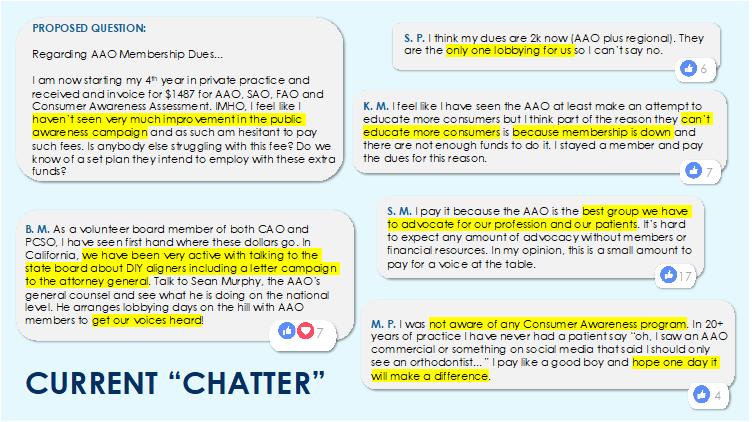
5 minute read
How would You Treat This Patient? – Timothy Shaughnessy


A 12 1/2-year-old boy presented for orthodontic treatment. The medical and dental history was insignificant. The patient’s chief complaint was his overbite and misalignment of the maxillary incisors.
The INITIAL facial photographs (Figure 1) exhibit mild facial convexity and lip protrusion, but with lip competence. The INITIAL intraoral photographs (Figure 2) reveal a Class II dentition, deepbite, and mild incisor misalignment .
The panoramic radiograph (Figure 3) shows a healthy permanent dentition with developing third molars. The cephalometric radiograph and analysis (Figure 4a and 4b) confirm an increased ANB value, along with other Class II skeletal measurements. Figure 1: Initial Facial Photographs
It could be argued that the maxilla is more protrusive than the mandible is retrusive. In fact, when this boy postured his jaw forward to a Class I dental position, his profile became less attractive. Although the patient has a dental deepbite, the skeletal vertical dimension is reasonably normal. Both the maxillary and mandibular incisors are over erupted, relative to the posterior occlusal plane.
How would YOU treat this patient?
The Treatment Plan
When devising the treatment plan for this patient, the clinician must decide how the occlusion is to be corrected. Will growth modification be incorporated, or tooth movement only? Will it be necessary to extract any permanent teeth? If so, which teeth, and how will this affect the anchorage requirements? The best treatment option should not only produce an esthetic and functional outcome, but it should be predictable and time efficient. If patient cooperation can be minimized, this would be even better.
The Treatment Plan Options
Figure 2: Initial Intraoral Photographs
Attempt to treat the patient via non-extraction therapy. This would require maxillary arch distalization and/or differential growth of the jaws following incisor decompensation. Mesial movement of the mandibular dentition would be an undesirable outcome. Although the mandibular incisors are measurably proclined, they are minimally misaligned and arguably in a reasonably stable initial position. Headgear for growth modification would be more ideal than a functional appliance, at least from the standpoint of mandibular incisor position. Any need for Class II elastics would also be a negative for the same reason. This plan is highly dependent on patient cooperation and favorable differential growth of the jaws.
Figure 3: Pretreatment Panoramic Radiograph
Figure 4a: Pretreatment cephalometric radiograph and analysis


Figure 4b: Pretreatment cephalometric radiograph and analysis
Extract maxillary premolars and treat the patient to an Angle’s Class II molar relationship with a Class I canine relationship. This plan makes it a moderate anchorage situation because the molar relationship is not a full cusp Class II. In addition, a headgear could be used for both anchorage control and growth modification. Any amount of differential growth of the jaws would decrease the maxillary posterior anchorage demand.
Extract maxillary first premolars and mandibular second premolars and like Option 1, treat this patient to a Class I molar and canine occlusion. This plan will likely result in more retraction of the anterior teeth than the other two options. On a positive note, differential movement of the molars can contribute to Class I molar achievement. Class II elastics could be used in this effort without the same risk to mandibular incisor position as a mandibular non-extraction treatment plan.
The Treatment Plan Chosen
Maxillary right and left first premolars were removed. The lower arch was treated non-extraction. A small amount of mandibular incisor proclination was expected with leveling. This plan required the least amount of patient cooperation, whether headgear or Class II elastics. It also seemed to have the least chance of failure, i.e., not achieve Class I canine occlusion. Fixed appliances were initially placed on the maxillary teeth only to initiate bite opening. At +6 months, fixed appliances were placed in the mandibular arch. Following bite opening in both arches, the patient was referred to an oral surgeon for extraction of maxillary first premolars. The oral surgeon declined
of the mandible for Class II correction instead. The surgeon cautioned the family that the proposed extraction approach would cause sleep apnea, as a result of retracting the maxillary teeth and “compromising the airway”.
A 6-month delay occurred as a result of the parents’ confliction regarding the proposed treatment plan and the disagreement between the specialists. This patient’s mom ultimately chose the extraction option because her older son was similarly and successfully treated in Texas prior to the family’s relocation to Georgia. The patient’s pediatric dentist agreed to extract the maxillary first premolars. Cervical-pull headgear was worn during en-masse space closure with a .016 x .022 closing loop arch wire in .018 brackets. The patient never wore Class II elastics.
The FINAL facial photographs (Figure 5) obtained at age 15
illustrate an esthetically pleasing outcome. to extract the requested teeth, and urged the patient and his mother to consider surgical advancement
Figure 5: Final Facial Photographs
Figure 6: Final Intraoral Photographs
The FINAL intraoral photographs (Figure 6) show good alignment of the teeth and a nice functional result. The chosen treatment plan was predictable and efficient, and lowest on the need for patient compliance to obtain an excellent outcome. The patient continues to sleep well at night.
The total time in orthodontic treatment was 2 1/2 years, extended by six months when the extraction/sleep controversy ensued. The patient was referred to a different oral surgeon for extraction of the mandibular third molars. The maxillary third molars were not extracted because it appeared that they would erupt and function with the opposing second molars. The post-treatment panoramic radiograph (Figure 7) also demonstrates good root parallelism, including the maxillary extraction sites.
The posttreatment cephalogram, its tracing and the cephalometric values (Figure 8a and 8b) confirm a reduction in the skeletal Class II discrepancy and no evidence of maxillary incisor retroclination as a result of space closure.

Figure 7: Posttreatment Panoramic Radiograph
Figure 8a: Posttreatment Cephalogram


Figure 8b: Posttreatment Cephalometric Values