1 minute read
Advancing Life: Biomedical Technology

from 2023 Biology Edition
by scienceholic

two-dimensional techniques to scan a specific body part before representing the information pixel by pixel
Although CT scans use x-rays to form images, the creation of the images are not computerized Instead, they are turned into slices Slices are cross-sectional images that allow the doctor to piece each slice together to create a 3D image of the body. The x-rays used are motorized x-rays rather than a fixed x-ray tube (standard xray method), which rotates the patient around to gather information from every section of their body
Advertisement
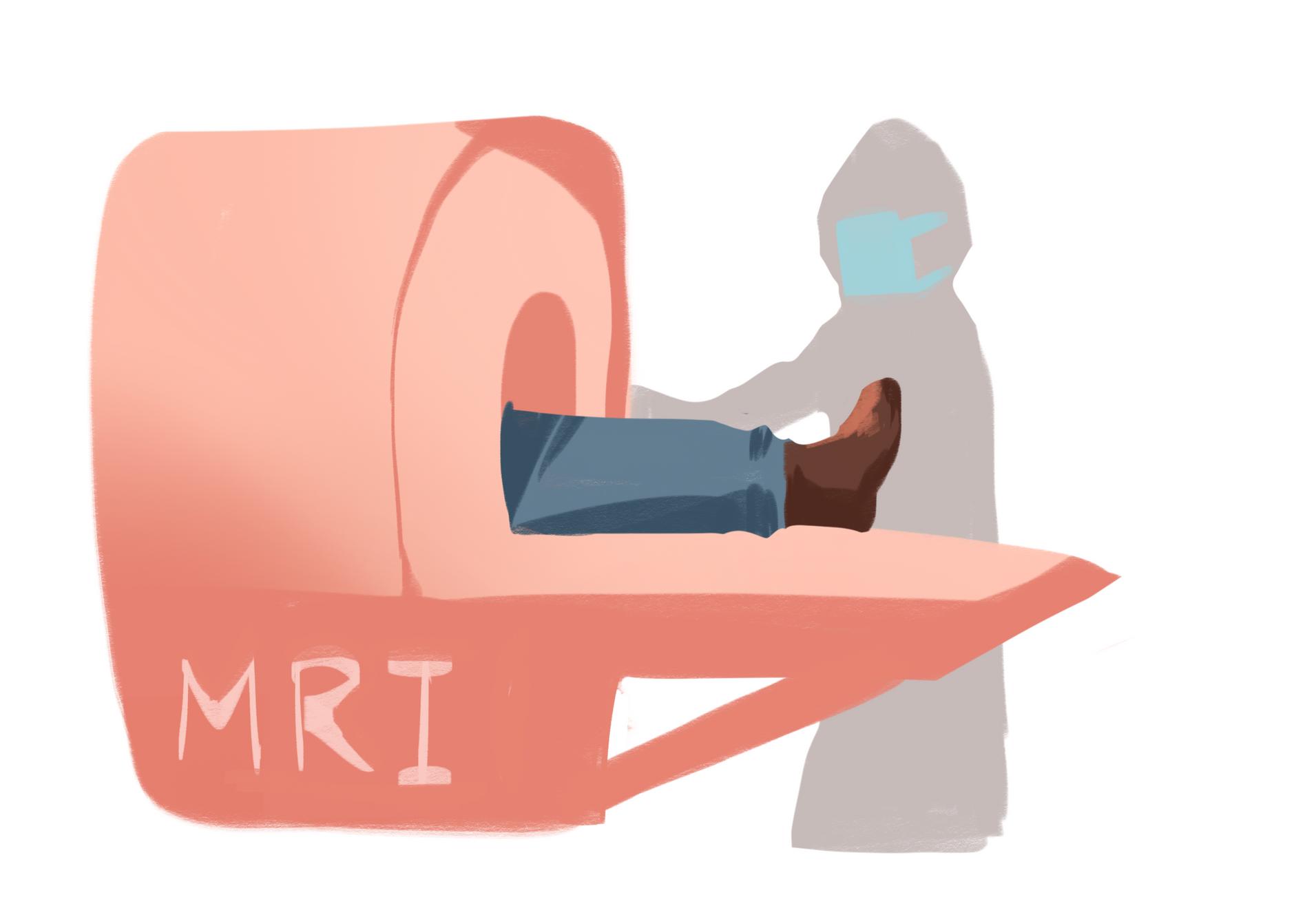
The Science behind MRI
While CT scans use x-rays and multiple detectors to form a 3D model of the human body, magnetic resonance imaging (MRI) uses non-invasive technology to create a 3D model of the body. Rather than using ionized radiation, which can harm the body after large/multiple doses, the MRI uses strong magnets to create images of the body
Because all protons spin to create a small magnetic charge, the strong magnets from MRI machines can rotate the protons to align with the magnetic field A radiofrequency pulse is then used to force the proton into a 90 or 180 degree realignment with the static magnetic field. After the radiofrequency pulse is removed, the protons will be back to their initial state of being pulled by the magnetic field and releasing electromagnetic energy while rotating Because different areas of the body release different frequencies of this energy, the waves bouncing off of those parts are collected by the machine which allows the MRI to differentiate which parts these come fro and generate an image based off of that.


