1 minute read
A Small Look Into MRI and CT scans
from 2023 Biology Edition
by scienceholic
Author: Jefferson Lin
Editors: Hwi-On Lee and Peggy Yang
Advertisement
Artist: Shihan Gao
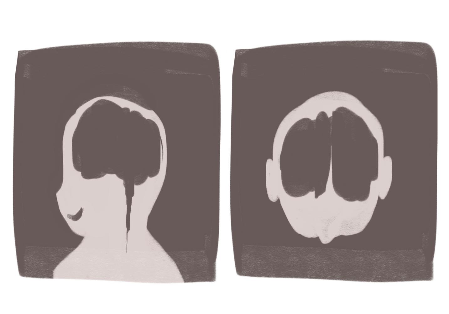
If you have ever suffered the pain of an infection or broken your bone, your doctor has probably suggested you get scanned. Whether they conduct an MRI or CT scan, these scans allow the doctor to determine where the issue is and the possible treatments to fix the problem. But how exactly do these scans work? How is a machine able to see through skin and map the entire body’s skeletal system?
The Science behind CT scans
The creation of computed tomography (CT) was initially theorized during a vacation trip when the scientist, Godfrey Hounsfieldand, thought of creating images of a closed box. Decades later, the CT is now a vital part of examining the human body to discover diseases which standard tools and machinery cannot find What was initially a plan to reconstruct the materials within a closed box became the foundation for CT scans. But the glaring question returns how do they work?
Before we understand CT scans, we need to understand the creation of x-rays, the core methodology behind CT scans. Xrays, which were created nearly over half a century before CT scans, are a type of radiation that can pass through the body
Because the body contains a variety of organs (like your liver and lungs), the energy admitted from x-rays, which is ionizing radiation energy, is absorbed differently depending on the organ. This information is collected by multiple detectors which work in harmony by using


