
Learning
in 2D/3D Medical Image Processing 1st Edition Rohit Raja (Editor)
Visit to download the full and correct content document: https://ebookmeta.com/product/artificial-intelligence-and-machine-learning-in-2d-3d-m edical-image-processing-1st-edition-rohit-raja-editor/

More products digital (pdf, epub, mobi) instant download maybe you interests ...
Image Processing and Machine Learning, Volume 2: Advanced Topics in Image Analysis and Machine Learning 1st Edition Erik Cuevas
https://ebookmeta.com/product/image-processing-and-machinelearning-volume-2-advanced-topics-in-image-analysis-and-machinelearning-1st-edition-erik-cuevas/
Image Processing and Machine Learning, Volume 1: Foundations of Image Processing 1st Edition Cuevas
https://ebookmeta.com/product/image-processing-and-machinelearning-volume-1-foundations-of-image-processing-1st-editioncuevas/

Artificial Intelligence and Machine Learning for EDGE Computing 1st Edition Rajiv Pandey
https://ebookmeta.com/product/artificial-intelligence-andmachine-learning-for-edge-computing-1st-edition-rajiv-pandey/
Artificial Intelligence and Machine Learning in Public Healthcare: Opportunities and Societal Impact Kc Santosh
https://ebookmeta.com/product/artificial-intelligence-andmachine-learning-in-public-healthcare-opportunities-and-societalimpact-kc-santosh/
Machine Learning in 2D Materials Science 1st Edition
Parvathi Chundi
https://ebookmeta.com/product/machine-learning-in-2d-materialsscience-1st-edition-parvathi-chundi/

Artificial Intelligence, Machine Learning and Blockchain in Quantum Satellite, Drone and Network 1st Edition Thiruselvan Subramanian
https://ebookmeta.com/product/artificial-intelligence-machinelearning-and-blockchain-in-quantum-satellite-drone-andnetwork-1st-edition-thiruselvan-subramanian/

Blockchain Tethered AI: Trackable, Traceable Artificial Intelligence and Machine Learning Karen Kilroy
https://ebookmeta.com/product/blockchain-tethered-ai-trackabletraceable-artificial-intelligence-and-machine-learning-karenkilroy/

Coherence: In Signal Processing and Machine Learning
Ramírez
https://ebookmeta.com/product/coherence-in-signal-processing-andmachine-learning-ramirez/
Medical Image Reconstruction From Analytical and Iterative Methods to Machine Learning 2nd Edition Gengsheng Lawrence Zeng
https://ebookmeta.com/product/medical-image-reconstruction-fromanalytical-and-iterative-methods-to-machine-learning-2nd-editiongengsheng-lawrence-zeng/
Artificial Intelligence and Machine Learning in 2D/3D
Medical Image Processing

Artificial Intelligence and Machine Learning in 2D/3D
Medical Image Processing
Edited by Rohit Raja, Sandeep Kumar, Shilpa Rani, and K. Ramya Laxmi
First edition published 2021 by CRC Press
6000 Broken Sound Parkway NW, Suite 300, Boca Raton, FL 33487-2742
and by CRC Press
2 Park Square, Milton Park, Abingdon, Oxon, OX14 4RN
© 2021 Taylor & Francis Group, LLC
CRC Press is an imprint of Taylor & Francis Group, LLC
Reasonable efforts have been made to publish reliable data and information, but the author and publisher cannot assume responsibility for the validity of all materials or the consequences of their use. The authors and publishers have attempted to trace the copyright holders of all material reproduced in this publication and apologize to copyright holders if permission to publish in this form has not been obtained. If any copyright material has not been acknowledged please write and let us know so we may rectify in any future reprint.
Except as permitted under U.S. Copyright Law, no part of this book may be reprinted, reproduced, transmitted, or utilized in any form by any electronic, mechanical, or other means, now known or hereafter invented, including photocopying, microfilming, and recording, or in any information storage or retrieval system, without written permission from the publishers.
For permission to photocopy or use material electronically from this work, access www.copyright.com or contact the Copyright Clearance Center, Inc. (CCC), 222 Rosewood Drive, Danvers, MA 01923, 978-750-8400. For works that are not available on CCC please contact mpkbookspermissions@tandf.co.uk
Trademark notice: Product or corporate names may be trademarks or registered trademarks, and are used only for identification and explanation without intent to infringe.
Library of Congress Cataloging in Publication Data
Names: Raja, Rohit, editor.
Title: Artificial intelligence and machine learning in 2D/3D medical image processing/ edited by Rohit Raja, Sandeep Kumar, Shilpa Rani, K. Ramya Laxmi.
Description: First edition. | Boca Raton: CRC Press, 2021. | Includes bibliographical references and index. | Summary: “Medical image fusion is a process which merges information from multiple images of the same scene. The fused image provides appended information that can be utilized for more precise localization of abnormalities. The use of medical image processing databases will help to create and develop more accurate and diagnostic tools’‐‐ Provided by publisher.
Identifiers: LCCN 2020039590 (print) | LCCN 2020039591 (ebook) | ISBN 9780367374358 (hardback) | ISBN 9780429354526 (ebook)
Subjects: LCSH: Diagnostic imaging Data processing. | Imaging systems in medicine.
Classification: LCC RC78.7.D53 .A78 2021 (print) | LCC RC78.7.D53 (ebook) | DDC 616.07/540285 dc23
LC record available at https://lccn.loc.gov/2020039590
LC ebook record available at https://lccn.loc.gov/2020039591
ISBN: 978-0-367-37435-8 (hbk)
ISBN: 978-0-429-35452-6 (ebk)
Typeset in Times New Roman by MPS Limited, Dehradun
Preface......................................................................................................................
Introduction ...............................................................................................................
Editors ......................................................................................................................
Contributors ...........................................................................................................
Chapter 1 An Introduction to Medical Image Analysis in 3D ............................ 1
Upasana Sinha, Kamal Mehta, and Prakash C. Sharma
Chapter 2 Automated Epilepsy Seizure Detection from EEG Signals Using Deep CNN Model 15
Saroj Kumar Pandey, Rekh Ram Janghel, Archana Verma, Kshitiz Varma, and Pankaj Kumar Mishra
Chapter 3 Medical Image De-Noising Using Combined Bayes Shrink and Total Variation Techniques ......................................................................... 31
Devanand Bhonsle, G. R. Sinha, and Vivek Kumar Chandra
Chapter 4 Detection of Nodule and Lung Segmentation Using Local Gabor XOR Pattern in CT Images ......................................... 53
Laxmikant Tiwari, Rohit Raja, Vineet Awasthi, and Rohit Miri
Chapter 5 Medical Image Fusion Using Adaptive Neuro Fuzzy Inference System ..................................................................... 73
Kamal Mehta, Prakash C. Sharma, and Upasana Sinha
Chapter 6 Medical Imaging in Healthcare Applications 97
K. Rawal, G. Sethi, and D. Ghai
Chapter 7 Classification of Diabetic Retinopathy by Applying an Ensemble of Architectures..........................................................
Rahul Hooda and Vaishali Devi
Chapter 8 Compression of Clinical Images Using Different Wavelet Function ............................................................................. 119
Munish Kumar and Sandeep Kumar
Chapter 9 PSO-Based Optimized Machine Learning Algorithms for the Prediction of Alzheimer’s Disease ...................................... 133
Saroj Kumar Pandey, Rekh Ram Janghel, Pankaj Kumar Mishra, Kshitiz Varma, Prashant Kumar, and Saurabh Dewangan
Chapter 10 Parkinson’s Disease Detection Using Voice Measurements ........ 143
Raj Kumar Patra, Akanksha Gupta, Maguluri Sudeep Joel, and Swati Jain
Chapter 11 Speech Impairment Using Hybrid Model of Machine Learning .......................................................................... 159
Renuka Arora, Sunny Arora, and Rishu Bhatia
Chapter 12 Advanced Ensemble Machine Learning Model for Balanced BioAssays 171
Lokesh Pawar, Anuj Kumar Sharma, Dinesh Kumar, and Rohit Bajaj
Chapter 13 Lung Segmentation and Nodule Detection in 3D Medical Images Using Convolution Neural Network .................. 179
Rohit Raja, Sandeep Kumar, Shilpa Rani, and K. Ramya Laxmi
Preface
In this volume of Medical Image Processing, the study is concerned with interactions of all forms of radiation and tissue. The development of technology is used to extract clinically useful information from medical images. Medical Image fusion is a process which merges information from multiple images of the same setting and the resulting image retains the most valuable information and features of input images. Medical Image fusion can extend the range of operations, reduce uncertainties, and expand reliability. In the Medical Imaging field, different images exist of the same component of the same patient with dissimilar imaging devices, and the information provided by a variety of imaging modes is much adulatory each other’s. The fused image provides appended information that can be utilized for more precise localization of abnormalities. Image Fusion is a process of combining the relevant information from a set of images into a single image, such that the resulting fused image will be more informative and consummate than any of the input images. Image fusion techniques can improve the quality and increase the application of these data. The most important applications of the fusion of images include medical imaging, tiny imaging, remote sensing, computer optics, and robotics. Feature of this book are
• The book highlights the framework of robust and novel methods for medical image processing techniques.
• Implementation strategies and future research directions meeting the design and application requirements of several modern and real time applications for long time.
• The book meets the current needs of the field. Advancement in Artificial Intelligence and Machine Learning in Medical Image processing are seldom reviewed in older books.
• Real Time Applications
We express our appreciation to all of the contributing authors who helped us tremendously with their contributions, time, critical thoughts, and suggestions to put together this peer-reviewed edited volume. The editors are also thankful to Apple Academic Press and their team members for the opportunity to publish this volume. Lastly, we thank our family members for their love, support, encouragement, and patience during the entire period of this work.
Rohit Raja Sandeep Kumar
Shilpa Rani
K. Ramya Laxmi
Introduction
The main scope of this volume is to bring together concepts, methods and applications of medical image processing. The concept of the study is concerned with the interaction of all forms of radiation and tissue. The development of technology is used to extract clinically useful information from medical images. Medical Image Fusion is a process which merges information from multiple images of the same setting, and the resulting image retains the most valuable information and features of input images. Medical Image Fusion can extend the range of operations, reduce uncertainties and expand reliability. In the Medical Imaging field, different images exist of the same component of the same patient with dissimilar imaging devices, and the information provided by a variety of imaging modes is much adulatory each other’s. The fused image provides appended information that can be utilized for more precise localization of abnormalities. Image Fusion is a process of combining the relevant information from a set of images into a single image such that the resulting fused image will be more informative and consummate than any of the input images alone. Image fusion techniques can improve the quality and increase the application of these data. The most important applications of the fusion of images include medical imaging, tiny imaging, remote sensing, computer optics, and robotics.
This book will target undergraduate graduate and postgraduate students, researchers, academicians, policy-makers, various government officials, academicians, technocrats, and industry research professionals who are currently working in the fields of academia research and the research industry to improve the quality of healthcare and life expectancy of the general public.
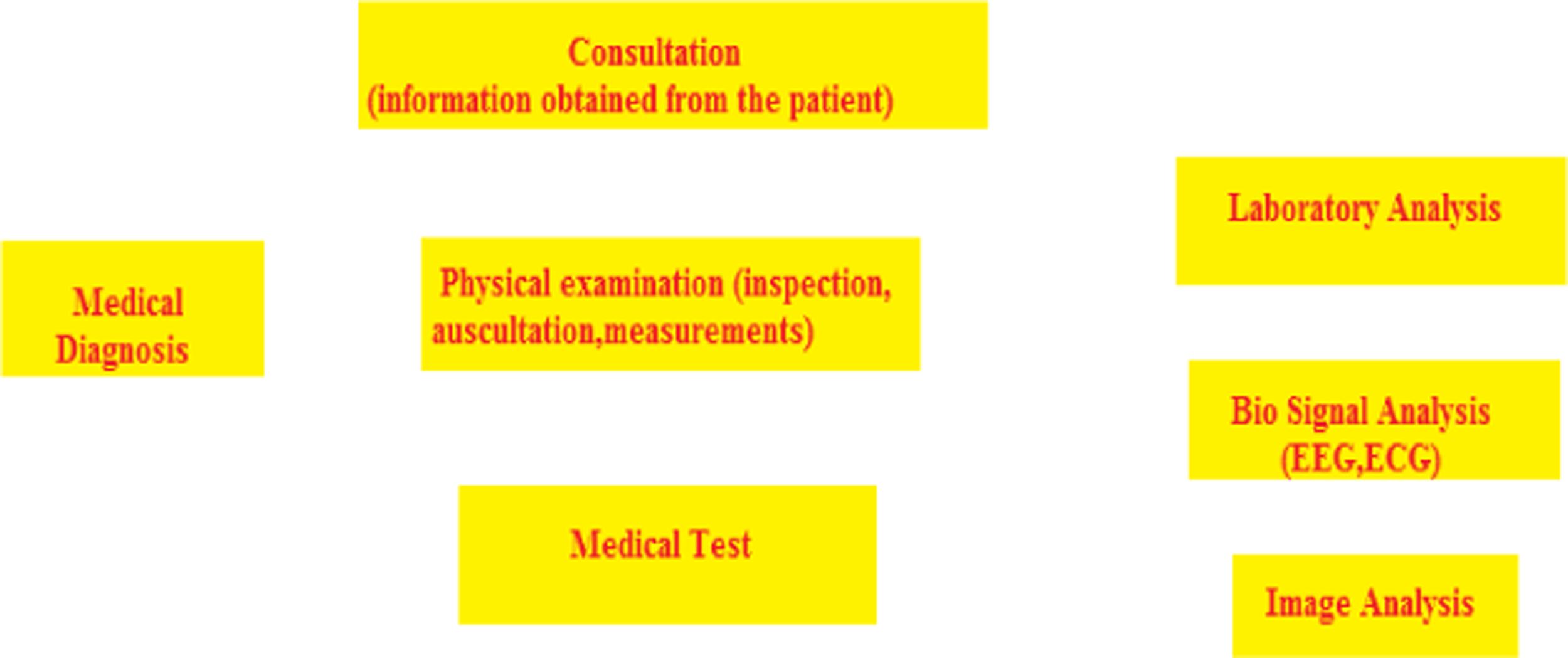
Chapter 1: This chapter introduces Biomedical Image processing, which has experienced dramatic growth and has been an fascinating area of interdisciplinary exploration, incorporating knowledge from mathematics, computer sciences, engineering, statistics, physics, biology, and medicine. Computer-aided analytical processing has already come to be a vital part of the scientific process. 3D imaging in medical study is the method used to acquire images of the body for scientific purpose in order to discover or study diseases. Worldwide, there are countless imaging strategies performed every week. 3D medical imaging is efficaciously growing because of developments in image processing strategies, including image recognition, investigation, and development. Image processing increases the proportion and extent of detected tissues. Currently, misperception has been produced among 2D and 3D machinery in health. This section presents the dissimilarity between these technologies and the software of both simple and complex image evaluation methods within the medical imaging discipline. This section also reviews how to demonstrate image interpretation challenges with the use of unique image processing systems, including division, arrangement, and registering strategies. Furthermore, it also discusses special kinds of medical imaging and modalities which contain CT test (pc tomography), MRI (scientific Resonance Imaging), Ultrasound, X-Ray, and so on. The important goals of this
investigation are to provide a foundation for the creation of fundamental concepts and strategies for medical image processing and to encourage the pursuit of additional study and research in medical imaging processing. We will introduce the 3D Clinical Image Graph Processing and summarize related research depictions in this area and describe recent ultra-modern techniques. The software of 3D scientific imaging and 3D has been a success in offering answers to many complex scientific issues. As technology spreads, its applications continue to grow inside the industry.
Chapter 2: Epilepsy is a neurological afflictions which has affected around 1% of humanity. Epilepsy is generally recognized by the tendency of the cerebrum to generate abnormal electrical activity and other actions creating disturbances in the conventional working behavior of the mind. To gather information about the electrical activity of the human brain, EEG is the preferred option. In this research paper, we have proposed a novel deep Convolutional Neural Network model with back-propagation algorithms, wherein the model analyzes the signal to classify and segregate it into three unique classes namely pre-ictal, normal and seizure. The database employed for this study is the popular, publicly available benchmark Bonn University database. For analyzing the potential and how well the model has performed, we used sensitivity, specificity, and accuracy as the main performance metrics. Here, a 10-fold cross-validation technique was applied. The study resulted in an accuracy of 97.33%, sensitivity of 96.00% and specificity of 98%. The results were then analyzed with other existing work in this field.
Chapter 3: Digital image processing brought about a tremendous revolution in many fields, the medical field being one of them. Medical images are used to detect abnormalities and diseases in the human body; simply by analyzing them. During the acquisition process; noise signals may be introduced in these images, which will negatively impact the diagnostic process. Noise signals degrade the image quality by suppressing useful information present in the form of edges, fine structures, textures and so on. Hence, it is essential to suppress noise signals because noisy images may lead to false interpretations by radiologists. Suppression of noise signals from medical images is called “Medical Image De-noising”. Some examples of medical images are Magnetic Resonance Images (MRI), Computed Tomography (CT) images, Ultra-Sound (US) images and so on. These images are corrupted by various noise signals. For example, MRI images are affected severely by noise, known as Rician noise; CT images are corrupted by Gaussian noise and US images are affected by multiplicative noise called speckle noise. To remove these unwanted noise signals, various filters have been proposed but none of these methods can be used as a global de-noising technique because any filter which can remove one noise effectively fails to remove others. Hence, it is necessary to develop a filter that can remove many noise signals because any image may be corrupted by more than one noise. This requirement motivates the researchers to achieve such a goal. In this chapter, a framework has been proposed to de-noise medical images, which reduces the effect of additive white Gaussian noise. It consists of various spatial domain filters, particularly the median filter and median modified Wiener filter. It also uses adaptive wavelet thresholding and total variation technique in parallel whose results are fused together using wavelet based fusion technique. This process is known as
shrinkage combined enhanced total variation technique as it enhances the quality of de-noised images.
Chapter 4: In recent times, for the detection of nodules and lung segmentation, many Computer Aided Diagnosis systems have been designed to assist the radiologist. The two main factors which affect the accuracy and effectiveness of the detection of lung cancer are the nodule who has similar intensity and they are connected to a vessel and the nodule with typical weak edges, hence it is difficult to define the boundaries. In the present work the main objective is to handle the two above-mentioned problems with the use of a CADe system for segmentation and detection of nodule forms with CT Scan Images using LGXP method and Morphological Image processing. The advantage of using Local Gabor XOR Pattern (LGXP) and modified region growing algorithm for extensive feature set like texture contrast, correlation and shape are extracted. The present work has been analyzed using the data of different subjects of varying ages to reduce the number of errors and to decrease the time needed to examine the scan by a radiologist. There are five problems that can be associated with CADe for lung cancer detection: The first problem is the detection of noises and loss-less information in the CT image. Due to the presence of noise, the performance of the system may degrade. We have taken 10 CT scans or subjects from LIDC-IDRI, which include 5 different cases. The results found are highly satisfactory.
Chapter 5: Image fusion is a procedure which consolidates data from numerous images of a similar setting and the resulting images contain valuable highlights of information Images. Fusion of Images can broaden the scope of activity, diminish vulnerabilities and improve consistent quality. Currently in Medical Imaging fields, various image of a similar segment some portion of a similar patient with divergent symbolism gadgets, and the data gave by an assortment of imaging modes is a lot of adulatory one another. The melded Image gives added data that can be used for increasingly exact restriction of variations from the norm. This part concentrates on computerized Image Fusion and proposes a model for blending images, utilizing ANFIS, executes and look at execution. The proposed model is isolated into four sections: Read Image, Separate Image into shading channel (for RGB Images), Applying to ANFIS, and Combining shading channels.
Chapter 6: The advent of medical imaging has had a tremendous impact on the detection of various types of diseases. In this regard, medical image processing has made a large contribution in identifying numerous diseases, as well as reducing the human effort required in various healthcare applications. Currently, digital sensors are also used, along with standard image modalities such as Magnetic Resonance imaging (MRI), Compute Tomography (CT), ultrasound, X Rays. In the past, these diseases were examined by doctors or radiologists and thus were more prone to human error. In recent years, researchers have successfully used various image modalities for detecting and diagnosing the diseases. The various image modalities that are used to automate the process of detecting these diseases are described in this chapter. This chapter also emphasizes ecent advancements in the field of Medical Imaging, as well as describing future trends in the state-of-art image processing algorithms in providing efficient and affordable healthcare services.
Chapter 7: Diabetic Retinopathy is a major cause of peventable permanent blindness worldwide. To reduce the number of individuals suffering from this disease, annual screening must be performed on all diabetic patients. However, manual screening of all patients is not possible, as it requires greater numbers of medical personnel around the world. On the other hand, if adequate facilities are not made available, then there will be a steep increase in undiagnosed and untreated cases of Diabetic Retinopathy. Thus, a system is needed for automatic diagnosis of Diabetic Retinopathy. The system would refer cases with a high probablility of having the disease to the expert ophthalmologist. This system would be helpful in reducing the rate of eyesight loss and enable a proper and exact diagnosis. Here, an ensemble is proposed that classifies the images into normal and abnormal images. Ensemble consists of three different deep learning architectures. The ensemble’s work and performance are computed using contrasting parameters and is found to be better than all the individual architectures.
Chapter 8: The examination and compression of clinical imagery is a significant area in Biomedical Engineering. The examintion of clinical images and data compression is a quickly developing field with emerging applications in health care services. Currently, digital techniques and applications in healthcare services are utilized for the diagnosis of patients. These techniques providing information about patients in the form of medical images require huge amounts of disk space to store the clinical information. Moreover, if any diagnostic center wanted to send this information to an expert for diagnostic purposes through a network, there is a requirement of larger bandwidth for the purpose of transmission. Therapeutic knowledge grows very rapidly and henceforth hospital requirements to accumulation vast amounts of patient information and data. Clinical images remain the most vital statistics about patients. Accordingly, diagnostic laboratories and hospitals have a massive quantity of patient images and data that require a corresponding massive storage space. More often, transmission bandwidth is inadequate to transmit all the pictures and data over an information channel. Thus, an image compression technique has been used to overcome these types of problems in the clinical field. In this paper, compression is performed with various kinds of wavelet functions to form a clinical picture and we propose the utmost fitting wavelet role which can achieve perfect reduction to a specified sort of clinical picture. For examine the routine of the wavelet role by means of the clinical pictures the loss of data amount is fixed. So that there is no information loss in the examination picture and determined their compression percentage in rate. The wavelet which provides utmost reduction in size of clinical picture with less loss of information has chosen for that image category.
Chapter 9: Alzheimer’s Disease (AD) is one of the most common types of diseases amongst older adults and constitutes one the leading cause of death in senior citizens. To prevent Alzheimer’s and provide early treatment, we have to accurately diagnosis Alzheimer’s Disease and its prophase, which is called Mild Cognitive Impairment (MCI) in the healthcare sector. To recognize the type or stage of disease, it is essential to classify medical data and potentially develop a prediction model or system. The framework that we have developed consists of Machine Learning methods with PSO optimization and has been successfully
applied to the classification of AD and Dementia. For the prediction of Alzheimer’s Disease, we have used seven Machine Learning Algorithms such as Support Vector Machine Classification, Random Forest Classification, XgBoost Classifier, Decision Tree Classification, Adaboost Classifier, K-Neighbour Classifier, and Logistic Regression. Our best-proposed method is the Random Forest Classifier, which achieves the best accuracy of 85.71%.
Chapter 10: This chapter introduces Parkinson’s syndrome, a dynamic condition of the central nervous system (CNS), affecting development & initiating shocks and solidness. It has five phases and influences more than millions of people each year throughout India. It is neuro-degenerative condition that influences the hormone known as dopamine creating neurons in the cerebrum. XGBoost is an additional Machine Learning calculation strategic considering velocity & implementation. XGBoost signifies extreme Gradient Boosting and depends upon a decision tree. In listed task,s we import XGB Classifier from xgboost library, which is a usage of scikit learn APIs for XGBoost arrangement. To assemble the model to precisely distinguish the onset of Parkinson’s syndrome in individuals. In this Python AI venture, utilizing the Python libraries scikit learn, xgboost, pandas and NumPy, we assemble models utilizing a XGB Classifier. We’ll stack the information, obtain the highlights and names, scale the highlights, at that point split the dataset, construct a XGB Classifier, and afterward ascertain the exactness of our model.
Chapter 11: Speech impairment is a technique in which speech sound signals are produced that are effective to communicate with others. Speech impairments can be any type; from mild impairment such as in occasionally struggling over a few words, to severe impairment, such as not being capable of producing speech sounds signals at all. The basic outcome is to study the hybrid model of machine learning for speech impairment. For effective machine learning results, it uses a specific model, effective techniques, knowledge parameters and advanced tree algorithms for displaying the valuable results. We rely on speech as one of the primary methods of communicating with others. Speech impairments in childhood can have a negative influence on social development. Speech impairments at all stages of life can lead to embarrassment and shame. The result of learning patterns on various human affliction diagnoses supports established medical specialists on the effects of starting treatment early, even though some results exhibit the same factors. In this paper, Parkinson’s dataset from the UCI library is used with the top four speechrelated parameters. It obtains a higher accuracy level with a hybrid model compared with the other classifiers.
Chapter 12: This chapter introduces the use of Machines and Computers in the medical field. Currently, machine learning, artificial intelligence, plays an important role in the medical field, as well in recent medical measures, the handling of patient knowledge, and medical background. This proposed article aims to provide a capable method to accurately predict the biopsy result. First, the authors applied different classifiers available in WEKA and prepared graphs for 10 different algorithms; namely Random Subspace, J48, SMO, Bagging, Simple Logistics, LWL, Multiclass Classifier Updateable, lbk, Naive Bayes, Naive Bayes Updateable. On the basis of the group, the Machine Learning mold of Simple Logistics was advanced. Practical outputs indicate that the advanced Simple Logistics machine
learning model has improved results in comparison to ensemble-based machine learning methods. The proposed work describes a good method of performing bioassays on high-dimensional balanced data.
Chapter 13: This chapter looks at the Lung, the most important organ in our cellular respiration system and which is situated in the chest cavity. Lungs are a set of spongy organs which allow us to breathe properly. Lungs are responsible for providing oxygen to the human body and sending carbon dioxide out of the body. The exchange of these gases is called respiration. In contemporary life lung cancer is a common disease and the cause of a greater number of deaths around the world. Lung cancer is the most deadly cancer other than breast cancer, bone cancer and so on. The primary cause of lung cancer is smoking. Although nonsmokers can also get lung cancer, the rate is ten times less than it is for the person who smokes. Diagnosing the lung tumor in the early stages is a very difficult task but if detected in the last stage, the only treatment option is to remove the cancerous lung. Therefore, early detection is vital.
Editors
Rohit Raja is working as associate professor in the IT Department at the Guru Ghasidas, Vishwavidyalaya, Bilaspur (CG). He has done PhD in Computer Science and Engineering in 2016 from C. V. Raman University, India. His primary research interests include face recognition, signal processing and networking and data mining. He has successfully filed 10 patents. He has 80 research publications to his credit in various international/national journals (including IEEE, Springer etc.) and in proceedings of reputed international/national conferences (including Springer and IEEE). He was invited thrice as a guest in Scopus indexed IEEE/Springer conferences. He has been invited four times, being an expert, in various colleges and universities in India. He has the following awards to his credit: “Excellence in Research” Award under the category of HEI Professor by Auropath Global Awards, 2019; the “Distinguished Research Award”, I2OR Awards 2019, during the 4th International Conclave on Interdisciplinary Research for Sustainable Development 2020 (IRSD 2020); and the second runner up award in grand finale of Smart India Hackathon, 2017, Ministry of Skill Development and Entrepreneurship, Government of India.
He is a member of professional international society including IEEE, ISTE, CSI, ACM etc. He is editor/reviewer of many peer-reviewed and refereed journals (including IEEE, Springer, enderscience).
Sandeep Kumar is presently working as a professor in the Department of Electronics & Communication Engineering, Sreyas Institute of Engineering & Technology, Hyderabad, India. He has good academic and research experience in various areas of electronics and communication. His areas of research include embedded system, image processing, biometrics and machine learning. He has successfully filed eight patents – seven national and one international. He was invited thrice as a guest in Scopus indexed IEEE/Springer conferences. He has been invited four times, being an expert, in various colleges and universities in India. He has published 70 research papers in various international/national journals (including IEEE, Springer etc.) and in the proceedings of the reputed international/national conferences (including Springer and IEEE). He has been awarded the “Best Paper Presentation” award in Nepal and in India, respectively, 2017 and 2018. He has been awarded the “Best Performer Award” in Hyderabad, India, 2018. He has also been awarded the “Young Researcher Award” in Thailand, 2018, and the “Best Excellence Award” in Delhi, 2019. He is an active member of 17 professional international societies. He has been nominated in the board of editors/reviewers of 25 peer-reviewed and refereed journals (including IEEE and Springer). He has conducted three international conferences and six workshops. He is also attended 24 seminars, workshops and short-term courses in IITs etc. He is a research guide for a number of Ph.D and M.Tech students.
Shilpa Rani is presently working as an assistant professor in the Department of Computer Science and Engineering, Neil Gogte Institute of Technology, Hyderabad, India. She has good academic and research experience in various areas of computer science. Her area of research includes image processing, IOT,
and big data. She has successfully filed three patents. She has published a number of research papers in various international/national journals (including IEEE, Springer etc.). She has been awarded the gold medal in 2012 during M.Tech. She is an active member of 10 professional international societies. She has been nominated in the board of editors/reviewers of four peer-reviewed and refereed journals. She has also attended 15 seminars, workshops and short-term courses in JNTUH and others. She has published two text books Logical & Functional Programming, Ashirwad Publication, 2009–2010 and Software Engineering, VAYU Education of India, 2011. Her third book has been accepted at an international level by Taylor & Francis, USA
K. Ramya Laxmi has worked as team lead and senior software developer at InfoTech Pvt Ltd for four years. Presently she is working as associate professor in the CSE Department at the Sreyas Institute of Engineering and Technology, Hyderabad. Her research interests cover the fields of data mining, machine learning and image processing. She has good academics and research experience in various areas of Computer Science. She has good hands-on-experience in PHP and Python and has knowledge of tools such as Pentaho and Weka, and is apt at analysing statistics using R-Programming.
Contributors
Renuka Arora
Guru Kashi University
Bathinda, India
Vineet Awasthi
Department of Information Technology
Dr. C. V. Raman University Bilaspur, India
Rohit Bajaj
Chandigarh University India
Devanand Bhonsle Sstc Bhilai, India
Vivek Kumar Chandra Csit Drug India
Vaishali Devi Govt. College Alewa, India
Saurabh Dewangan Nit Raipur, India
D. Ghai
Lovely Professional University Phagwara, India
Akanksha Gupta It Department
Ggv Central University Bilaspur, India
Rahul Hooda Govt. College Alewa, India
Swati Jain Govt. J. Y. Chhattisgarh College Raipur, India
Rekh Ram Janghel Nit Raipur, India
Maguluri Sudeep Joel Sreyas Institute of Engineering and Technology Hyderabad, India
Dinesh Kumar Guru Kashi University India
Munish Kumar Eiepl Delhi, India
Prashant Kumar Nit Raipur, India
Sandeep Kumar ECE Department Sreyas Institute of Engineering and Technology Hyderabad, India
K. Ramya Laxmi CSE Department SIET Hyderabad, India
Kamal Mehta
Computer Science and Information Technology
Mukesh Patel School of Technology and Management
Nmims Shirpur, India
Rohit Miri
Department of Information Technology
Dr. C. V. Raman University
Bilaspur, India
Pankaj Kumar Mishra
Rcet
Bhilai Durg, India
Saroj Kumar Pandey
Nit Raipur, India
Raj Kumar Patra
Cse Department
Cmr Technical Campus Hyderabad, India
Lokesh Pawar Chandigarh University India
Rohit Raja Department of IT
Ggv Central University
Bilaspur, India
Shilpa Rani
CSE Department
NGIT
Hyderabad, India
K. Rawal
Lovely Professional University
Phagwara, India
G. Sethi
Lovely Professional University Phagwara, India
Anuj Kumar Sharma
BRCMCET
MD University
India
Prakash C. Sharma Department of Information Technology Manipal University Jaipur, India
G. R. Sinha Mit Mandley, Myanmar
Upasana Sinha
J. K. Institute of Engineering Bilaspur, India
Laxmikant Tiwari Department of Csit
Dr. C. V. Raman University Bilaspur, India
Kshitiz Varma Csvtu
Bhilai Durg, India
Archana Verma Nit Raipur, India
1 An Introduction to Medical Image Analysis in 3D
Upasana Sinha, Kamal Mehta, and Prakash C. Sharma
1.1 INTRODUCTION
3D Image Analysis is the visualization, processing and evaluation of 3D photo statistics through geometric transformation, filtering, picture segmentation and other morphological operations. 3D conception forms the basis of contemporary radiology. 3D experimental imaging is a modern visual imaging scientific expertise that affords an enriched image of the interior body for scientific assessment making use of 3D imaging modalities. 3D scientific imaging provides more effective pictures of blood vessels and better images of bones. It is undisputable that 3 Dimensional (3D) imaging is continuously improving with the continued enhancement of instrumentation.
1.2 COMPARISON BETWEEN 2D AND 3D TECHNIQUES IN MEDICAL IMAGING
2D and 3D refer to the genuine dimensions in a computer workspace. 2D is “flat”; using horizontal and vertical (X and Y) dimensions; the image graph has solely two dimensions and turns into a line. 3D provides the depth (Z) dimension. This 0.33-
dimension permits rotation and visualization from a couple of perspectives. It is in effect the distinction between an image and a sculpture.
For example, taking the pattern image graphs of echocardiography, there is the volumetric method to statistics acquisition in 2D and 3D (Figure 1.1).
Medical imaging has developed extensively since the early days of CT scanners and mammography equipment. With 3D scientific imaging, healthcare professionals were able to obtain access to fresh angles, resolutions, and small detail that provided an outstanding portrait of the physical section in query, at the same time as reducing the amount of radioactivity in patients [1, 2, 3]. In recent decades, the quantity of 3D scientific imaging has doubled in number every month to about one hundred thirty instances per day by 2018. The science of scanning has become a superior technology in creating statistical units that can make 3D images clearer with greater decision precision and much less noise and artifacts. Medical imaging has superior technological know-how in particular when it comes to these slice counts; it permits us to enlarge the precision of the pictures that we are shooting and, additionally signify the 3d mannequin of the anatomy, which used to be a substitute no longer feasible in the early days of the process (Figure 1.2).
1.3 IMPORTANCE OF 3D MEDICAL IMAGE
As we are all aware, medical imaging encompasses distinctive imaging modalities (a kind of technology used to gather structural or purposeful pictures of the body) such as radiography, ultrasound, nuclear prescription, computed tomography (CT), magnetic resonance and seen light. This requires techniques to image graph the body for diagnostictic and therapeutic purpose and performs an essential function in enhancing medical treatment. This proves that clinical imaging is regularly justified in the follow up of an ailment already recognized or treated [4, 5].
Medical imaging, in particular X-ray, primarily built investigations plus ultrasonography, stays necessary for a range of scientific putting and by altogether predominant stages of fitness precaution. In communal fitness and protective remedy by way of suitable as in each healing and relaxing care, good choices rely on the right analyses. However, medicinal/scientific decisions might also
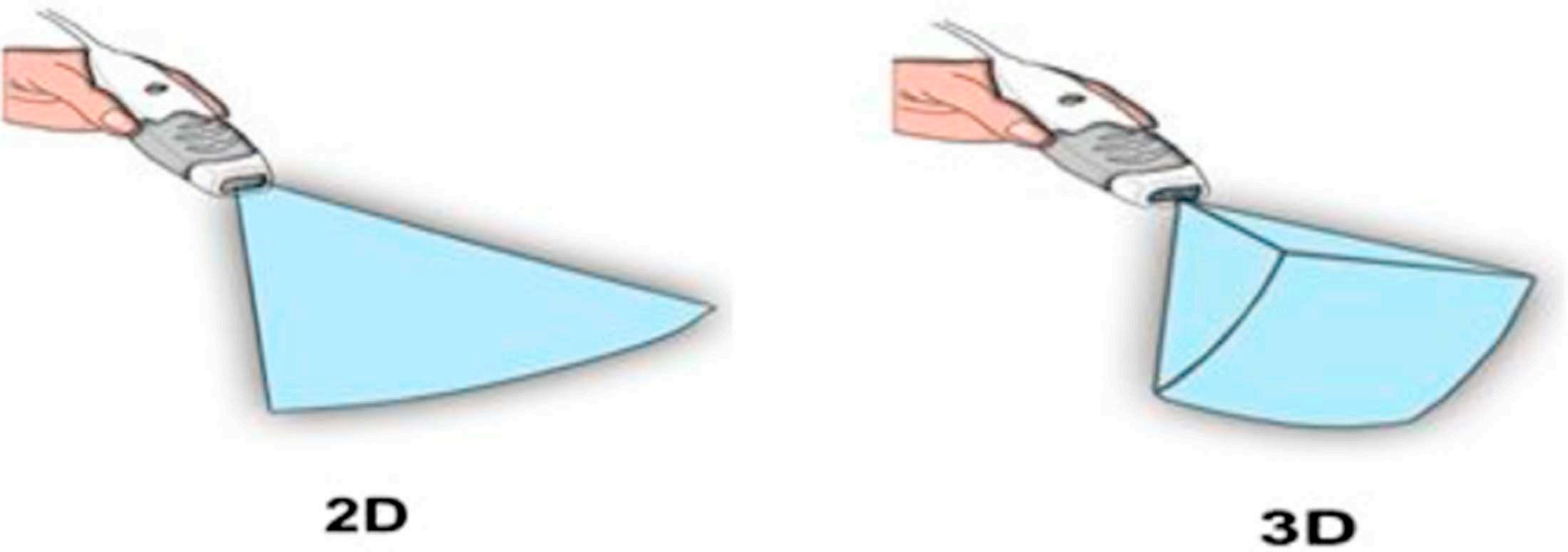
1.1 2D (left panel) and 3D (right panel) Echocardiography Visualization Image.
FIGURE
remain adequate former to therapy of numerous circumstances, the practise of diagnostic imaging offerings is dominant in confirming, efficiently measuring and authenticating publications of several ailments as nicely as in an evaluating reaction to treatment. Through accelerated health care coverage plus the growing accessibility of clinical apparatus, the range of world imaging-based strategies continues to grow significantly. Accurate and safe forms of imaging remain necessary in clinical practice and can reduce the use of pointless procedures. For instance, some medical interventions can be prevented if easy diagnostic imaging such as ultrasound is available.
It is well known that 3D picture processing is a tremendous tool for extent calculation, measurement, and quantitative analysis. It starts off evolved from 3D fashions of the patient, routinely recognized and extracted from anatomical structures, analysis, and surgical simulations can be supported. Moreover, with the usage of augmented actuality capabilities, it is feasible to merge preoperative or intraoperative information with reality, which is a precious device in the discipline of image guided surgery. 3D technological know-how has changed scientific imaging developing the opportunity for talent mapping with excessive decision microscopy. It has the capacity to discover character neurons, hint connections between them, and visualize organelles’ internal neurons.
The fundamental and first step in 3D image processing is the division of a picture which organizes pixels into substances or collections. 3D image division makes it practical to make 3D versions for more than one object and function with quantitative evaluation aimed at the extent, mass, and different factors of identified substances.
New images are taken, whether by CT, MRI, or microscopy image diagram as a 3D range of voxels/pixels. Individual voxel takes a greyscale vary from 0 to 65535 in the sixteen-bit pixel instance or 0 to 255 in the eight-bit pixel case. A segmented image, on the different hand, provides a less complicated explanation of substances that allows an introduction of 3D level methods or shows point data. When the fresh image graph is conveniently displayed as 3D evaluation, then imagining requires clearly described objective limits after growing models. Taking as an instance, to generate a 3D version of humanoid intelligence from an MRI image, the
FIGURE 1.2 Medical Imaging.
intelligence wishes to be recognized first inside the image graph and before its periphery manifest and used for 3D translation. The pixel recognition method remains known as image division, which recognises the qualities of pixels and describes the limitations for pixels that go to an identical group. Moreover, dimensions and numerical evaluation for restrictions such as region, boundary, quantity, and extent can be acquired effortlessly once objective limits are distinct.
1.4 MEDICAL IMAGING TYPES AND MODALITIES
Different kinds of medicinal imaging contain:
i. CT (Computed Tomography)
CT or CAT (pc axial tomography) images are the shape of X-ray that generates 3D images for analysis. It makes use of X-rays towards frame supply section images. A scanner with CT takes an outsized round establishing aimed at the affected person lying on a motorized desk. The X-ray delivers in addition a sensor then it rotates around the affected idividual, generating a slight ‘fan-shaped’ ray of X-rays that permits via part of the patient’s physique to make a picture. Those pictures are then assembled into single, or multiple images of interior organs and tissues. The CT scans supply higher transparency in comparison to traditional X-rays through greater unique picture graphs of the internal organs, bones, mild tissues, and blood vessels within the frame. The advantages of the use of CT scans outweigh the risks, which similar to X-rays, include cancer, damage to an unborn child, or allergic reaction to the chemicals in contrast material. In many instances, using a CT scan removes the need for experimental surgical operations. It is essential that when scanning children, the radiation dosage is lower than that used for adults. In many hospitals, a paediatrics CT scanner is available for that purpose.
ii MRI (Magnetic Resonance Imaging)
MRI scans generate diagnostic image graphs without emission of dangerous radiation. Magnetic resonance Imaging (MRI) makes use of strong magnetic placed besides radio waves to produce pictures of the body which cannot be detected by X-rays or CT scans, i.e., it enables joints, ligaments and soft tissue to be visible [6, 7, 8]. The MRI is frequently used to observe interior detail to detect strokes, tumours, spinal cord accidents, aneurysms, and intelligence function. We realize the majority of the human body consists of water, and every water molecule consists of a hydrogen nucleus (proton) which become allied in a magnetic field. An MRI scanner provides a secure magnetic field to support the proton ‘spins’. A radio frequency is then applied which propels the protons to ‘flip’ their spins in advance than return to their proper arrangement. Protons in particular body organs revert to their regular spins at dissimilar rates so the MRI can differentiate among numerous types of tissue and detect any deformities. In what way the molecules ‘flip’ then arrive back at
iii
their ordinary spin arrangements is noted and processed into a picture. MRI doesn’t use ionizing radiation and is gradually being cast-off at some stage in pregnancy and not using a thing results at the unborn infant mentioned. But there are dangers related to using MRI scanning, and it isn’t endorsed as a primary analysis.Due to the strong magnets used, it is not suitable for individuals with any kind of steel implant, synthetic joints, and so on because of the chance they might be dislodged or heated up in the magnetic field.
ULTRASOUND
Ultrasound remains the most secure method of scientific imaging and takes a large variety of packages. There aren’t any risk to the use of ultrasound, and it remains one of the best low-cost types of medical imaging available to us. Ultrasound makes use of sound waves instead of ionizing emission. High-frequency sound waves travel through the body with the aid of a transducer. Those waves then bounce back once they hit denser surfaces in the body and that is used to generate an image for prognosis. Another type of ultrasound often used is the ‘Doppler’ – an extraordinary method of the use of sound waves that allows the bloodflow via arteries and veins to be visible. Due to the absence of risk in ultrasound, it is the first choice of imaging in pregnancy. However, because its uses are considerable – emergency prognosis, cardiac, spine, and internal organs – it often is the first imaging option for patients.
iv. X-ray
X-ray imaging – X-ray consitutes the oldest and most used form of imaging; indeed, most people have had at least one X-ray in their life. In 1895, it was discovered that X-rays are a form of electromagnetic radiation. X-rays work on a wavelength and frequency that we are not able to view with the bare human eye, but it can penetrate through the pores and skin to create an image of what’s underneath. Generally used for detecting skeletal problems, X-rays can be used to detect cancers through mammography and digestive troubles through barium swallows and enemas. X-rays can be used extensively use as they are low cost, rapid, and effortless for the affected person to bear. But there are risks related to the use of radioactivity for X-ray imaging. In all instances, the affected individual who has an X-ray receives a dose of radiation. The exposure is brief, but there is a slight risk of radiationprecipitated cancers or cataracts later in life or harm to an embryo or foetus in a pregnant woman. Most of the dangers are moderated through using X-rays where strictly necessary and properly safeguarding the rest of the body with a protective shield.
1.5 COMPUTER VISION SYSTEM WORKS IN 3D IMAGE ANALYSIS
The corporation of the laptop vision tool is noticeably application based. There are capabilities that are discovered in many computer imaginative and prescient structures (Figure 1.3).
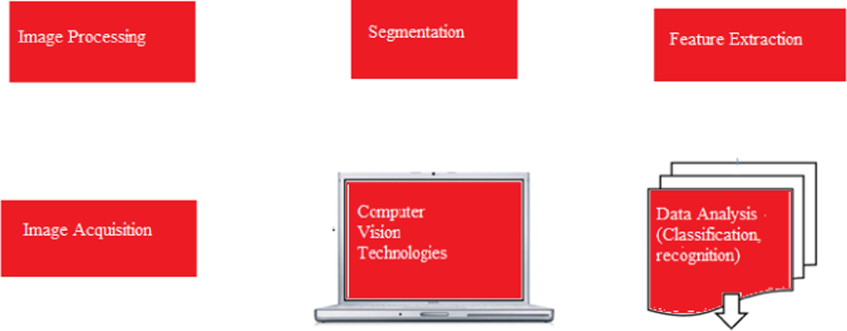
Image Acquisition
Pre-processing
Feature Extraction
Detection/Segmentation
High level processing
Decision Making
Image Processing
Image Acquisition: It is a system of transforming the analogy global round us into double records collected of 0’S and 1’S, inferred as virtual images; e.g., client 3D cameras & laser range detectors.
Image Processing: Image acquisition is considered to be first step of image processing and second step in computer imaginative and perceptive. Procedures are utilized for binary statistics obtained in the primary stage to deduce low-level records on additives of the image graph. For example, the sort of statistic remains categorised by using picture ends, aspect sides, or sections. They are the primary geometric element that assembles substances in snapshots. They constitute the best geometric factors that assemble substances in images.
This 2nd step generally includes superior carried out arithmetic algorithms and strategies.
Low level image processing algorithm includes:
i. Edge detection. This is a hard and fast mathematical technique with the goal of identifying factors in a virtual photograph in which the photo brightness changes sharply or, more officially, has discontinuities. The factors at which image brightness changes are usually prepared into a set of curved line segments termed edges [9]. It is one of the vital steps in image processing, picture evaluation, and a computer imaginative and prescient approach.
Segmentation: Our primary effort is to extend the correctness of segmentation. The early stage consists of utilizing a number of filters (imply, Gaussian blur, region detection)
FIGURE 1.3 Computer Vision System.
and bit operations similar to histogram equalization and standardisation [10, 11]. These actions also need to be utilized in each photograph one at a time, or there are versions of these systems in 3D. An act in 3D is characterized by means of the use of a chain of 2d pictures (slices) organized in a row. Three coordinates each have a voxel. The initial two coordinates, x, and y characterize one pixel on a slice and the 0.33 one, z, represents the order of slice. At primary, the 3D image graph is geared up for segmentation. The aim of this method is to break up the image graph into continuous factors. Those components can be overlapping and collectively can cover the entire picture. Capabilities are calculated for each such segment.
Medical image graph: Division is the technique of automatic or semicomputerized recognition of interior boundaries a second before the 3D image. The crucial state of clinical picture division is the excessive inconsistency in the medical image. The analysis of the situation shows the maximum modes of the variant. Moreover, numerous special modalities (X-ray, CT, MRI, microscopy, pet, SPECT, Endoscopy, OCT, and plenty of greater) stay castoff towards generating medical images. An anticipated final outcome of the segmentation can now be used to build up similarly to diagnostic insights. Feasible purposes remain the computerized dimensions of organs, mobile telephone counting, or simulations established totally on the removed edge records.
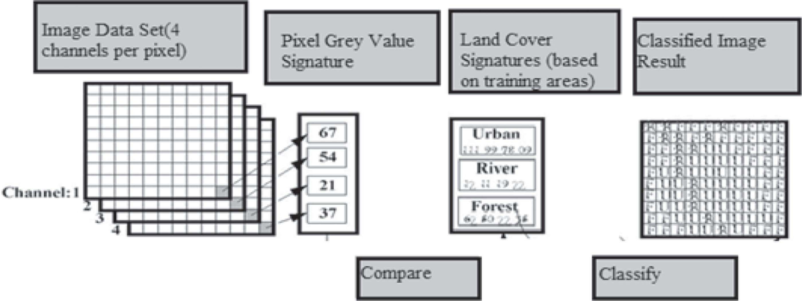
Classification: Probably image graph remains the most essential segment in digital picture evaluation [10]. It is the primary class to have a “pretty image” or a picture, showing the importance of shades illustrating a number of factors of the basic terrain. However, this is ineffective until it is understood what the descriptions suggest. (PCI, 1997). Primary type strategies are supervised classification and unsupervised type.
By supervised type, we recognise instances of the material modules (i.e., land cowl type) of the hobby within the image and are referred to as “training websites” (Figure 1.4). The image graph handling software application tool is then used to strengthen a statistical description of the reflectance for each reality magnificence. This level is often termed as “signature evaluation” and can additionally include creating a description as clean as they mean or the vogue of reflectance on each band, or as complex as special analyses of the imply modifications and covariance over all bands. As soon as a statistical representation has been finished for every report class, the picture is then categorized with the means of analysing the reflectance for each pixel and creating a preference around which of the initials it most resembles. (Eastman, 1995)
Unsupervised type is a technique that inspects a big range of unidentified pixels and splits into a wide sort of class primarily founded mostly on herbal groupings current day in the picture values. In the evaluation of supervised classification, the unsupervised class no longer requires analyst-targeted coaching facts. The easy statement remains that value inner a assumed cover kind ought to be shut collected inside the dimension vicinity (i.e., Have comparable grey levels), whereas information in one in all a kind commands need to be relatively properly separated (i.e., Have very precise grey ranges) (PCI, 1997; Lillesand and Kiefer, 1994; Eastman, 1995). The programmes that cease end outcome from the unsupervised category are spectral ranked which primarily built on herbal alliances of the image
graph values, the uniqueness of the spectral category will not be known in the beginning and one will have to compare categorised statistics to some shape of reference information (including large scale imagery, maps, or internet site online visits) to decide the identification and informational values of the spectral training. Consequently, within the supervised approach, to define useful statistics instructions and then take a look at their spectral reparability; inside the unsupervised method, the computer defines a spectrally separable class, and then describe their statistics price. (PCI, 1997; Lillesand and Kiefer, 1994).
Unsupervised type is becoming more well-known in groups involved in prolonged-time period GIS database upkeep. The motive is that there are actually structures that use grouping methods which are surprisingly short and minute within the nature of operational parameters [10]. Therefore, it is possible to train GIS assessment with only a familiarity with far-flung detecting to undertake classifications that meet regular map accuracy standards. With appropriate ground reality accuracy evaluation tactics, this device can grant a remarkably rapid capability of producing quality land cover facts on a continuing foundation.
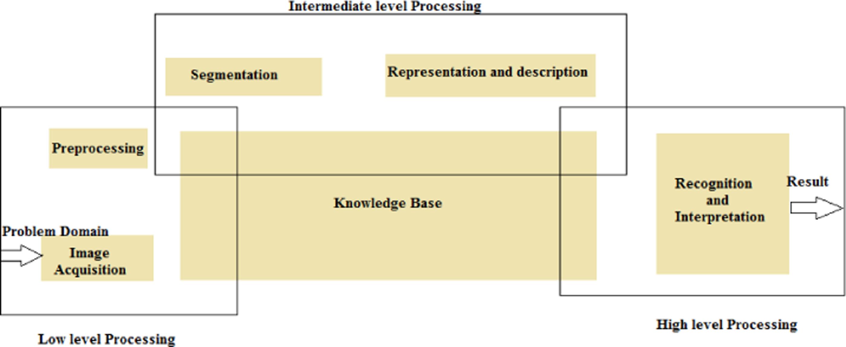
High Level Pre-processing: The final phase of the computer image process is the investigation of the records, which will permit the building of results. High-level algorithms are functional, by means of exchanging the image data and the low-level data computed in earlier steps (Figure 1.5).
Examples of high-level image analysis are:
1. 3D scene mapping
2. Object recognition
3. Object tracking
1.6 VARIOUS TECHNIQUES IN 3D IMAGE PROCESSING IN MEDICAL IMAGING
There are many techniques one could use whilst processing 3D image records. Those strategies range based on the tasks to be accomplished– together with importing, visualizing, processing, and analysing the statistics.
FIGURE 1.4 Steps in Supervised Classification.
1.5 Levels of Pre-processing.
3D Image Imp or t Read in files and metadata Pro cessing
G eometric transformation
Image Registration Filtering and Enhancement

3D IMAGE PROCESSING
Rendered Volumes 2D slices with 3D coordinate system
Image Analysi s Vi sualiz ation
Image Segmentation Region Analysis
1.6 Key Components of a 3D Image Processing Workflow.
3D picture processing is generally utilized in medical imaging to read DICOM or NIfTI pictures from radiographic resources like MRI or CT scans. One may also use 3D image graph processing strategies in microscopy to detect and examine tissue samples or hint neurons (Figure 1.6).
Image Import and Visualization: 3D image information can originate from a range of tools and document layouts. To successfully import and visualize 3D images, it is vital to obtain access to the underlying data and metadata for the images. One could imagine 3D images using a range of strategies depending on the facts needed to be examined. In a few programs, one could imagine the 3D statistics as a reduced quantity.
Image Filtering and Enhancement: 3D images generally include undesirable noise that confuses or deemphasizes the purposes of the sizes that one is involved in. Making use of image filters, normalizing image evaluation, or performing morphological operations are not unusual methods for doing away with noise after 3D images.
FIGURE
FIGURE
Image Registration: While functioning with datasets of 3D pictures, the images are generally occupied from one kind of tool, or as a device is moving, that could present misalignment via rotation, or skew and scale variations. We are able to put off or lessen this misalignment by the use of 3D geometric variations and image graph registration techniques. Picture registration is the procedure of aligning greater images of the same scene. This technique includes designating one image graph because of the reference image, also called the constant image graph, and making use of geometric modifications or nearby displacements to the other pictures in order that they align with the reference. Medical image fusion refers to the fusion of medical pictures obtained from different modalities. Scientific picture fusion enables medical analysis through a manner of improving the first class of the pictures.
Filtering and Enhancement: We will lessen noise or beautify pictures by the use of image graph filtering methods like Gaussian filtering, box filtering, or picture morphology.
Image Analysis: Picture evaluation is the extraction of significant records from pictures; particularly from digital pictures via virtual image processing systems. Image research tasks may be as easy as reading bar coded tags or as state-of-the-art as recognising a person from their face.
1.7 TYPES OF MEDICAL IMAGING COMPRESSED BY 3D MEDICAL VISUALIZATION
Cinematic Rendering Offers a Clearer Picture of Complex Structures: For instance, when specialists are searching for methods to learn about complex areas of the body, including the heart, new technological know-how identified as cinematic rendering can help. Advanced with the aid of Eliot Fishman, Director of diagnostic imaging and physique CT and professor of radiology and radiology science at John Hopkins medicines, the technological information yields realistic pictures from the unification of 3D CT or 3D MRI scans by volumetric conception by way of distinct computer-generated image knowledge. This technique helps physicians whilst diagnosing illness, supervising surgical treatment, and planning a course of action. Cinematic rendering allows healthcare specialists to understand masses extra of the texture of the analysis (Figure 1.7).
Related to how ray locating makes someone’s pores and skin appear larger and permeable within the films, cinematic rendering offers a detailed appearance of the texture of tumours, which allows the delivery of extra data for medical doctors to determine whether or not or not or no longer is a tumour cancerous. “With these textures, the greater precisely we can render and visualize them as people—the texture of the anatomy or the tumor—I assume the richer the statistics for medical doctors to interpret,” Powell says.
Tomosynthesis Recovers Breast Cancer Recognition: Breast imaging has evolved from 2D mammography to 3D chemosynthesis (from time to time known as 3D mammography), which allows radiologists to capture images at numerous perspectives and show tissues on numerous depths at a greater level than would be
possible with a set of pictures only. This technique could permit radiologists to view images in 3D in a much more realistic manner, as noted by Harris.
“Tomosynthesis has been proven to enhance the care for breast most cancers detection and is extra sensitive, especially in sufferers at excessive danger or with dense breasts,” Harris explains. “It helps to differentiate matters that may be misinterpreted that are probably different artifacts.
Artificial Intelligence Takes Medical Imaging to the Next Level: The last five years have brought about massive advancements in imaging, due to the powerful mixture of talent and 3D clinical imaging. At the GPU technology conference in March 2018, Nvidia introduced mission Clara, a “digital scientific AI supercomputer” that uses enhanced calculating competence than may be done with 3D volumetric rendering, in keeping with the work of Powell.
“AI should inject efficiency into clinical imaging, in particular when it comes to detecting organs or anomalies. For example, via combining photograph visualization and AI, cardiologists can measure ejection fraction—the share of blood pumped thru the coronary heart every time it contracts—in a lots shorter length of time barring having to kind via big statistics units and observe the anatomy via sight.”
Usually, cardiologists and radiologists have the practice so that they really theoretically capture what's happening, but AI is in a position to deliver a correct, tough-number dimension to truly extending the opportunities that the analysis is as proper as it is able to be, Powell says [1, 12, 13].
3D Computing Tomography Angiography Maps Vascular Anomalies: At Massachusetts General Hospital, Harris researches 3D computed tomography angiography (CTA), in which medical experts can imagine arterial and venous vessels by way of a CT method. Professionals like Harris and his team practise CTA to record stenosis, aneurysms, dissections, and extraordinary vascular anomalies. On the side of 3D imaging, scientific experts can get an improved experience of what they’re observing in analysis and pathology, as well as any potential artifacts.
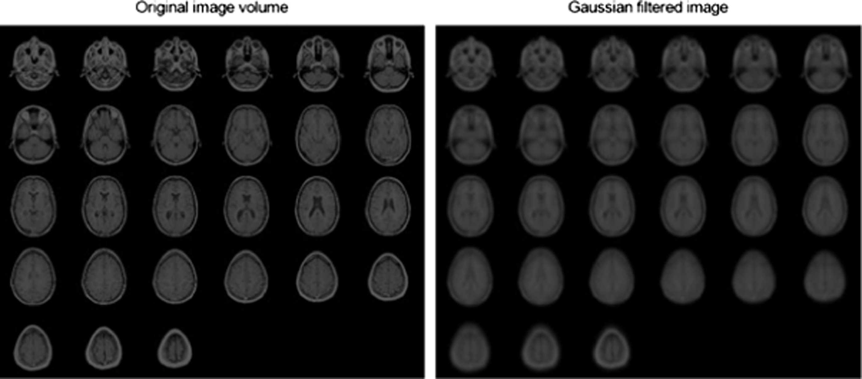
FIGURE 1.7 MRI Images of a Human Brain Using 3D Gaussian Filtering.