ISSUE 76 DECEMBER 2024
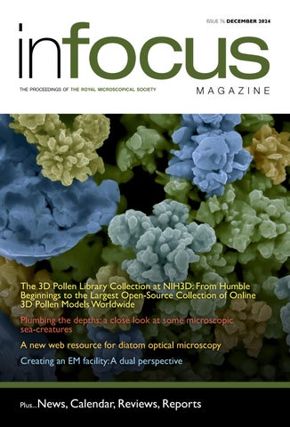
The 3D Pollen Library Collection at NIH3D: From Humble Beginnings to the Largest Open-Source Collection of Online 3D Pollen Models Worldwide Plumbing the depths: a close look at some microscopic sea-creatures A new web resource for diatom optical microscopy Creating an EM facility: A dual perspective
Plus...News, Calendar, Reviews, Reports 1
ISSUE 76 DECEMBER 2024