
Histochemical Stains: Enhancing Understanding of Tissue Biology
Histochemical stains play a pivotal role in the field of histology, providing researchers and diagnosticians with valuable insights into the structure and composition of tissues. From identifying specific cell types to diagnosing diseases, histochemical stains are indispensable tools in both research laboratories and clinical settings. In this article, we delve into various aspects of histochemical stains, including their applications, methods, and significance.

1. Introduction to Histochemical Stains
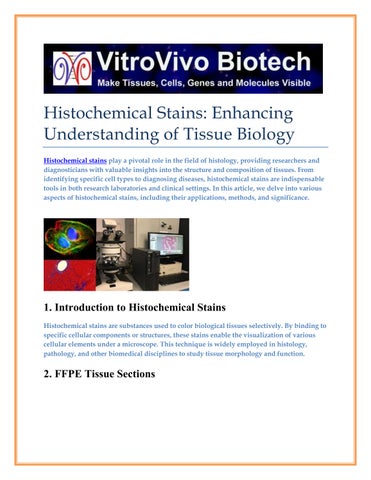
Histochemical stains are substances used to color biological tissues selectively. By binding to specific cellular components or structures, these stains enable the visualization of various cellular elements under a microscope. This technique is widely employed in histology, pathology, and other biomedical disciplines to study tissue morphology and function.
2. FFPE Tissue Sections
Formalin-fixed paraffin-embedded (FFPE) tissue sections are commonly used in histology for preserving and examining tissue samples. Tissues are fixed with formaldehyde to prevent decay and then embedded in paraffin wax for support during slicing. This method allows for long-term storage of tissue samples while retaining cellular structures for staining and analysis.
3. Stains for Mast Cells
Mast cells are a type of immune cell found in connective tissues. Stains specific to mast cells, such as Toluidine Blue and Giemsa stain, are used to identify and quantify these cells in tissue samples. Mast cell stains are crucial in allergy research, as mast cells play a significant role in allergic reactions and inflammation.
4. IHC Kits (Immunohistochemistry Kits)
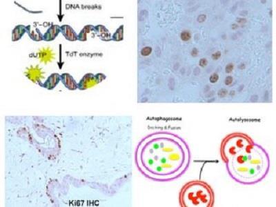
Immunohistochemistry (IHC) kits are essential tools for detecting specific proteins or antigens in tissue sections. These kits utilize labeled antibodies to visualize target molecules within tissues under a microscope. IHC is widely used in cancer diagnosis, where specific protein markers can indicate the presence or progression of tumors.
5. Histology Services
Histology services encompass a range of techniques for studying tissue structure and function. From tissue processing to slide staining, histology services are vital in medical research, diagnostics, and education. These services facilitate the examination of tissues at the cellular level, aiding in the diagnosis of various diseases.

6. Laser Capture Microdissection
Laser capture microdissection (LCM) is a technique used to isolate specific cells or tissues from a heterogeneous sample under direct microscopic visualization. LCM enables the precise isolation of cells for downstream molecular analysis, such as DNA or RNA extraction. This technology is invaluable in studying cell heterogeneity and tissue-specific gene expression.
7. Silver Staining
Silver staining is a technique used to visualize proteins, nucleic acids, and other biomolecules in tissue sections. This method relies on the reduction of silver ions to metallic silver, resulting in dark staining of target molecules. Silver staining is commonly used in neurobiology and microbiology research for highlighting specific structures or pathogens.
8. Histopathology Services
Histopathology services involve the examination of tissue samples to diagnose diseases and evaluate disease progression. Histopathologists analyze tissue morphology and cellular abnormalities to make accurate diagnoses and guide patient treatment. These services are critical in identifying cancerous lesions, infectious agents, and other pathological conditions.
9. Oil Red O Staining
Oil Red O staining is a histological technique used to visualize lipid droplets within cells and tissues. This stain selectively binds to neutral lipids, such as triglycerides and cholesterol esters, producing a distinctive red coloration. Oil Red O staining is commonly used in metabolic research to assess lipid accumulation and distribution in various tissues.
In conclusion, histochemical stains are indispensable tools in biomedical research and diagnostics, enabling the visualization and analysis of tissue structures and cellular components. From identifying specific cell types to diagnosing diseases, the applications of histochemical stains are vast and diverse. By understanding the principles and techniques of histochemical staining, researchers and healthcare professionals can gain valuable insights into tissue biology and pathology.
FAQs (Frequently Asked Questions)
1. What are histochemical stains used for?
o Histochemical stains are used to visualize cellular structures and components in biological tissues for research and diagnostic purposes.
2. How are FFPE tissue sections prepared?
o FFPE tissue sections are prepared by fixing tissues with formaldehyde and embedding them in paraffin wax for slicing and staining.
3. What is the significance of IHC kits in cancer diagnosis?
o IHC kits enable the detection of specific protein markers in tissue sections, aiding in the diagnosis and characterization of cancerous lesions.
4. Why is laser capture microdissection important in molecular biology?
o Laser capture microdissection allows for the isolation of specific cells or tissues for molecular analysis, enabling researchers to study gene expression and cell heterogeneity.
5. Which stains are commonly used for visualizing lipid droplets?
o Oil Red O staining is commonly used to visualize lipid droplets within cells and tissues in metabolic research.