

ArticulAte
a journal exploring the integration of art & science in healthcare
TABLE OF CONTENTS
Rachell Chon
Osteopathic? Sounds like a Made-up Word
CASE REPORTS
Nicholas Afshari; Stephen Walker
Unraveling the Complexity of Chronic Appendicitis: A Case Study and Surgical Insights
Madison Garlock
Paroxysmal Spasmodic Dysphonia in a 51-Year-Old Male
Hinal Rathi; Carson Bridgman; Taimoor Hassan, MD; Dharmista Chaudhary, MD
Initial Stroke-like Presentation Turned Out to be Bacterial Meningitis with No Growth on Cerebrospinal Cultures
John Abdel Sayed, MD; Hinal Rathi; Carson Bridgman; Derar Albashaireh, MD
New Onset Rheumatic Heart Disease in a Morbidly Obese Patient Without Traditional Risk Factors: A Challenging Diagnosis and Management Dilemma
Ryan P Barney; Sean J Henderson, DO; Sean R Schofield
Leiomyosarcoma of the Female Urethra: A Review and Novel Case Report
Kendrick Rubino; Corrine Ricci; Ellice Goldberg, DO; Amanda Brooks, PhD
A Unique Presentation of Actinic Keratosis: The Benefit of Regular Skin Exams
Alex Ignatenko
“A Case of the COVID Toes” COVID-19 Induced Granulomatous Poly-angiitis: A CASE REPORT
Brock K Bakewell; Mark Wardle, DO; Christopher Gordon, MD
90 Degree Patella Malrotation Upon Lateral Dislocation in an 11-Year-Old Male
Kristen Valente, PA-S3; Carrie Chanos, PA-C
The Benefits of Genetic Testing for Prognosis of Disease in a Rare Variant of Grade III Anaplastic Astrocytoma in a Young Adult Male
Julie Steinbeck
Recommendations for Evaluation of Atypical Seizure Presentation: A Case Study of Abnormal Neurogenic-like Presentation of Sick Sinus Syndrome in a 29-Year-Old Female
Riley Stearns; Jenna Buckleitner; Amanda Brooks, PhD
Pain Management in 41-Year-Old Male Patient with Klippel-Trenaunay Syndrome
Brytani White, PA-S III; Sarah Neguse, PA-C
De novo Familial Adenomatous Polyposis with Undiagnosed von Willebrand’s Disease: A Case Report
Alyssa Funk
Investigation into Management Deficits in a Patient with Life-threatening GPA, and Future Management Course................................................................................................................................................
ETHICS & PERSPECTIVES
Gabby Costain
The Unintended Consequence of the BMI Calculator
Steven Gawrys, DO
Critical Lessons from Conducting Sports Medicine Research
Calli Cahil, MA
Overlooked Identities: The Case for Gender Minority Inclusion in the NIH Revitalization Act……...........…..49
Ashley Rousseau
Miss Diagnosis: ADHD in Female Patients
Mark Wardle, DO
Reducing Risk for Helping Hands: Preparedness and Prevention Surrounding Global Health Outreach Experiences……………………………………...……………………….......………………………………127
Bradley Stephen O. Thornock, PhD
On Suckers: A Virtue Epistemological Approach to Anti-Vaccination
Arpit Danewalia, Maison Evensen-Martinez
Tackling the Epidemic of Medical Misinformation: Nurturing Trustworthy Voices in the Social Media Era
RESEARCH
Gubler K; Evensen-Martinez M; Muller ME; Roberts TAM; Santiago M; Arias DC; Gawrys SP; White AB; Steele JL; Wardle M.
Hispanic Health Needs Assessment of Southern Utah
Dallin Trout, PA-C; Darcy Solanyk, PA-C
Analysis of Guidelines Regarding Mental Health Hold Requirements and Duration of Adults from Mountain States
ARTS & HUMANITIES
Megan Elizabeth Dekok, Human Nature (No.1)
Jennifer L Hellier, PhD, Knitted Dissected Rat
Kenton Felmlee, 6L, 2 lbs, and a whole lot of tears
Isabella Contolini, Lungs
Anna Jacobs, She’s Every Woman
Richard Stevens, The Curse of Aion
Gianna Tarka, Denver Paper Fashion Show
Phillip Kong, A Personalized 3D Printed Model to Aid in Patient Education of the ALIF Procedure
Christy Wornom, My Heart Folds
Synneva Collett, Crocheted Blanket
Lon Van Winkle, PhD, Goodbye (to a group of
Rachell Chon, The Train *To our earth that suffers
Brandon Wilkinson, Beyond the Wall
Corey Thorsheim, HUMAN
Peter Wooley, Fixer
Corey Thorsheim, Growing Pains
Joshua Hansen, The Patient and The Doctor
Corey Thorsheim, Pixelated Flower
Cheyenne Bair, The River of Life
Jenna Buckleitner, Year One—Transformation, Year Two—Melting, Year Three—Excuse
Rachell Chon, Clicked for Me
Hannah Vedova, TO MY HUSBAND
Corey Thorsheim, Kea Bird
Laura Sullivan, Ceramic Hearts
Anna Jacobs, Delivery
Gianna DeCosmo, New Beginnings
Anna L Megenhardt, Ephemeral Embrace: A Symphony of Lillies
Ben Graf, Psychology, The Memory, Vagabond, Masquerade

Dear Readers,
Welcome to Articulate, a peer-reviewed journal dedicated to a multifaceted exploration of medicine. Our mission is to showcase the scientific, artistic, and humanistic endeavors of the healthcare community and to foster a unique blend of interdisciplinarity, creativity, and collaboration. While we remain committed to traditional research, Articulate also recognizes the profound impact of art and poetry on understanding the human experience of medicine. By bringing together scientific inquiry and the expressive power of art, we aim to enrich medical discourse and inspire a holistic understanding of health and healing.
The concept of Articulate arose from the question of what defines an academic healthcare journal. Topics such as research, healthcare policy, ethics, and education immediately came to mind, traditionally represented as a collection of research manuscripts, case reports, and editorial articles. At the same time, we asked what might be missing and how we could best honor osteopathic philosophy. Just as our opening reflection, “Osteopathic? Sounds like a made-up word” by Rachell Chon, explores how learners define the word osteopathic, we explored different definitions of scholarly activity. In the end, connecting art and humanities with scientific inquiry shows our multifaceted approach to professional development, understanding that the humanistic aspects of medicine are as important as the scientific in the care of both patients and practitioners, and never forgetting the person behind the practitioner.
We are proud to present the first volume of Articulate, which represents both professional and personal identities in the study and practice of medicine. We invite readers to explore the journal in chronologic order, where case reports, research articles, and perspectives are interspersed with art and poetry. We welcome discussion, dialogue, and investigation into the interconnectedness of art and medicine. By approaching similar topics in diverse formats, we hope the published articles and features provoke thought, spark creativity, and inspire practice. Thank you to the Articulate editorial board, all section and student editors, the RVU Marketing Department, and the RVU administration for supporting this project. Finally, we would like to acknowledge the talent and diligent work of all contributors. Please enjoy Volume I of Articulate
Thank you,

Nicole Michels, PhD Editor in Chief Chair of Medical Humanities
Rocky Vista University
Dear RVU Community,
This first issue of Articulate, Rocky Vista University’s peer-reviewed journal of the science and art of medicine, is the culmination of a vision, and multi-year effort to create an opportunity for publication of scientific manuscripts of merit, perspective pieces, and arts – paintings, sculptures, poems, essays, and other creative forms of expression from members of our community.
I am impressed by the quality of the submissions selected for publication. The artwork, in many forms, the perspective pieces, the case reports, and the research papers are outstanding, and warrant a home for publication and dissemination.
Creating a journal is no small endeavor. Conceiving of the concept, calling for submissions, deciding what to include, what to refer back to the authors for revisions, what to reject, edit, layout, a million formatting decisions, etc. takes experience, effort, skills, and diplomacy. I want to thank editor-in-chief, Nicole Michels, PhD; co-managing editors Hope Ruskaup, MFA and Alexis Marosi Horst, MA; section and student editors; and the editorial board for all their hard work in launching Articulate.
Lao-tzu said, “The journey of a thousand miles begins with one step.” What you have before you is a look into the early part of a journey. The work of the authors, creators, peer-reviewers, editors, and staff may seem like “one step” to some. I assure you, it is the result of many miles travelled. Future issues of Articulate will be built on the work of this inaugural issue.
Please enjoy this first issue of Articulate!
Sincerely,

David A. Forstein, DO FACOOG (dist.) President, CEO, and Interim Provost Professor of
Obstetrics and Gynecology
Rocky Vista University
Osteopathic? Sounds like a made-up word.
By Rachell Chon
“What was the word again? Osteopathic?” the patient asked, as I was drawing her blood for a volunteer event. She had never heard of “DOs,” before and was skeptical of how we were different than “regular doctors.” She had been a St. George resident for decades and was surprised to hear there was a medical school right across the street from the clinic. I was half-way through my first semester as a medical student and was just starting to understand what an osteopathic physician was myself.
I nervously straightened my badge on my scrubs and started to jumble words into an elevator pitch for osteopathic medicine. I paraphrased Still’s osteopathic principles, threw in snippets of “fascia,” “holistic medicine,” and sprinkled in terms like “somatic dysfunction,” and “tissue texture,” into my underdeveloped monologue.
I was not confident in my answer, and frankly, my patient wasn’t either.
To my disappointment, our conversation dwindled into silence as she responded, “Osteopathic…well, to me, it still sounds like a made-up word.
to medicine. We osteopaths take in the big picture. It’s this meshing of traditional, evidence-based medicine with a fully comprehensive twist. There’s an interconnectedness to every individual, and that’s our focus when we diagnose and treat our patients.”
Vitor Da Costa, OMS-II:
“An osteopathic physician is a trained physician who can diagnose and prevent illness or injury using a combination of conventional medical techniques and osteopathic manipulative treatment. This is a handson approach to diagnoses and treatment for patients who have improper body mechanics, so we can help alleviate and get the body to be able to heal itself.”
Catherine Arnold, OMS-II:
Osteopathy, with its emphasis on the interconnectedness of the body’s systems and a holistic patient care strategy, contrasts with the reductionist approach associated with allopathic medicine. As osteopathic students, we will all have an opportunity to share what distinguishes osteopathy from other healthcare philosophies and may have the chance to further enhance the medical journey of our future patients.
I reached out to a few students and two fellows who were willing to do a short interview before the summer of 2023 at Rocky Vista University on the Utah campus.
In a few sentences, I asked them how they would have responded to this same patient.
Jessica Vergara, OMS-II:
“Well, you caught us!
It’s just one of those hard-to-pronounce medical words we like to throw around. We hardly call our MD counterparts allopathic physicians, but that’s what they’re called.
Osteopathic is just a word describing an approach
“We learn the same things as our allopathic counterparts, but learn an additional component called osteopathic principles. In practice, we try to focus on the holistic nature of all aspects of health, not just solely on symptoms people may be presenting with. For example, we account for mental and physical health to view each patient as a whole person. We try to touch on various aspects of your lifestyle, so we can treat specifically to your needs whenever we can. We do a lot of the same things that allopathic doctors do, but also have hands-on training so we can use osteopathic techniques to physically manipulate parts of the body to encourage the healing process and help the body heal itself.”
Ciro Valdez Garcia, OMS-II:
“Well, osteopathy is an approach to medicine where we focus on the body’s structure and its function. From our practice, we emphasize on the body’s natural ability to heal. Our goal as osteopathic physicians is to enhance this healing ability so that our patients can heal faster and have better healthcare outcomes overall. In doing so, we really want to emphasize the whole body, rather than just the one illness, as so many factors can contribute to what a patient may be presenting with. We take the time to ask more individualized questions and also utilize our skillset in osteopathic manipulative medicine we can really improve the care in our patients and their healthcare outcomes.”
Kirra Rivera, OMS-II:
“Like all words, osteopathic is a made-up word--but it still has meaning, right? To break it up into parts,
“osteo,” means bone and “path,” means disease. An osteopathic physician does all the things an allopathic or medical doctor does, but we have additional training in musculoskeletal medicine, where we do an extra set of treatments with our hands. This specific skillset allows us to address common issues such as back pain or headaches. Instead of treating solely with medication--although we may include it in our treatment plan--we try to solve your pain from the bottom-up. In other words, we try to figure out why your back is in pain in the first place, and we have more tools to do just that.”
Brandon Ciak, OMS-II:
“Just like an MD, I’m a certified doc who can diagnosis, prescribe, and act as a healthcare provider for patients. The main differences are that DOs tend to hold more of a holistic approach to patient care as well as receiving extra hands-on training in the field of osteopathic manipulative treatments, or OMT. Just think of it as an extra “tool” in my tool bag that I get to carry around when assessing and treating patients!”
Arsany Fahim, OMS-II:
“If a patient said to me, “osteopathic sounds like a made-up word,” I would first think of why they might have said that. They may just be unfamiliar and uncomfortable with the new term, or they could be skeptical of my abilities in treating them. I would probably crack a bad joke and say, “All words are made-up,” to start the conversation. But I think it’s important to not give the patient a sermon on the definition of osteopathy—they may not be interested in that. I think patients are more interested in making sure I am a good doctor who they can trust with their health. That is the message I hope to get across, the association with “osteopathic,” and a “good doctor.” To do that, you can’t just say it. You have to show them.”
Rabail Abbas, OMS-II:
“Osteopathic medicine focuses on the biomechanics of the body. We aim to heal with a perspective of the mind, body, and spirit when we view the body as a whole, functional unit. We try to incorporate everything when we are creating a treatment plan for each patient, including their lifestyle, and prioritizing their quality of life. I like to call us the “spotlight operators,” because we shine the light on the body of where it needs healing and help encourage the body to bring healing to itself. Osteopathic principles
are really intertwined with emphasizing the human condition, instead of just focusing on prescriptions and treatments.”
Madison Lee, OMS-II:
“An osteopathic physician is just like any other physician completing medical school; however, we get specialized training in osteopathic manipulation therapy or OMT. With this added skillset, it acts as another toolbox into our repertoire so we can use on patients for different somatic dysfunctions. This includes disease with the musculoskeletal or nervous system. With our experience, the principles of osteopathy give us another tool that we can use to effectively treat and help our patients.”
Alex Seegrist (OPP Student Fellow, UT):
“You’re probably more familiar with the traditional allopathic route to become a physician. The allopathic route will grant you a MD degree while the osteopathic route will grant you a DO degree, doctor of osteopathic medicine. The main difference is that DOs complete additional training in osteopathic manipulative medicine which utilizes the body’s structure and function to regulate its ability to selfheal. Both MD and DO medical school graduates have to go through the same residency training to become clinical physicians and surgeons.”
Adam Berry (Senior Anatomy Student Fellow, UT):
“That’s a great question. You may be more familiar with an MD, who are doctors of allopathic medicine, which sounds like doctor of osteopathic medicine. Like our allopathic colleagues, we get the same medical training and can pursue all the same professional tracks as surgeons, pathologists, family doctors, etc. Although our scope of practice is the same as MDs, osteopathic references our additional training in using our hands to manipulate the musculoskeletal system.”
*****
The responses all included nuanced differences between osteopathic and allopathic medical approaches, but each student included small details of their own personal philosophies and approaches to patient care. Overall, there was an emphasis in connection to human touch, showcasing a
readiness to connect with patients on a more personal level, which is reflective of the osteopathic approach itself—viewing patients as complete beings, rather than a collection of symptoms.
In modern medicine, it is not only our responsibility to help patients understand their healthcare options, but also relay what we understand about health and the human body. My hopes are to encourage myself and osteopathic students to explain osteopathic medicine in a way that can resonate with patients, reassuring them of their competence while highlighting the unique capabilities DOs can bring to the table.
If I had the chance to meet the same patient again, my response would be the following:
“Studying to be an osteopathic doctor is like being a double-major in two separate, but similar, fields at a university or college. We are trained to be medical doctors who can clinically and ethically treat patients with the same knowledge about physiology, anatomy, and pharmacologically to treat disease. But we are also trained to be experts in applying these concepts to the dynamics of our body structure and movement. By viewing disease from multiple perspectives, we are trained to see each disease alongside each individual patient.”


Human Nature (No. 1)
By Megan Elizabeth Dekok
As an amateur horticulturalist, I feel inspired to utilize preserved plant specimens from the garden to add dimension and texture to classic depictions of human anatomy. My goal is to emphasize medicinal and native plants; in this piece I included foxglove flowers featured most prominently in the auricles of the heart. Foxgloves (Digitalis lantana) are used in the pharmaceutical production of digoxin, a cardiac glycoside drug indicated for treatment of congestive heart failure. The medicinal use of this plant was first documented in herbal medicine of the mid-eighteenth century. The dynamic interplay between botany and medicine as scientific disciplines has always been fascinating to me, and through art I hope to continue exploring this relationship. A more subtle trope within this piece highlights medical waste and the impact of the healthcare industry on climate change. I included expired medical tape as a textural background element and utilized skin marking pens to enhance the contrast within the anatomy. Both of these efforts serve to extend the lifespan of single-use medical supplies. This piece was sold at auction with all proceeds benefiting Safehouse Denver
Medium: Mixed Media on Illustration Board
Dimensions: 30’’ x 20’’
Case Report
Abstract
Unraveling the Complexity of Chronic Appendicitis: A Case Study and Surgical Insights
Nicholas Afshari OMS-III, Stephen Walker OMS-III
Rocky Vista University College of Osteopathic Medicine
Chronic appendicitis, a less prevalent variant of acute appendicitis, remains a subject of ongoing debate within the medical community. Unlike the rapid onset characteristic of acute appendicitis, chronic appendicitis may manifest with insidious symptoms persisting over an extended period, potentially spanning months, before becoming clinically significant. While the recognition of chronic appendicitis may not fundamentally alter treatment paradigms, early diagnosis holds promise in averting avoidable secondary complications.
Here, we present the case of a 29-year-old female who experienced minor symptoms over the course of several years, with a recent two-month exacerbation preceding presentation. Despite the protracted nature of her symptoms, she ultimately underwent appendectomy, revealing evidence suggestive of chronic inflammation. This delayed diagnosis resulted in secondary complications that could have been mitigated with timelier recognition of chronic appendicitis.
Chronic appendicitis remains a topic of evolving understanding within the medical community. This chronic inflammatory condition, distinct from the acute form, poses unique diagnostic challenges and prompts a reevaluation of traditional diagnostic criteria. A better understanding of this condition can lead us to a faster diagnostic time, leading to a better outcome and management.
Background
Chronic appendicitis, characterized by persistent inflammation or fibrosis of the appendix, presents a clinical challenge due to its comparatively less understood nature in contrast to acute appendicitis, a well-documented surgical emergency. While acute appendicitis has been extensively studied, with a clear lifetime risk documented at 8.6% in males and 6.9% in females, the epidemiological profile of chronic appendicitis remains inadequately defined. Fibrosis, a common component of chronic appendicitis, refers to the formation of excess fibrous connective tissue in the appendix. This fibrous tissue can lead to narrowing or obstruction of the appendiceal lumen, contributing to chronic inflammation and recurrent symptoms. Unlike acute appendicitis, where risk factors such as recurrent stool obstructions, chronic or recurrent infection, and malignant processes are well established, the risk factors for chronic appendicitis are less delineated. Recent studies suggest that chronic appendicitis may be diagnosed when symptoms resembling acute appendicitis persist for longer than 7 days in the right lower quadrant. Bridging the knowledge gap surrounding chronic appendicitis is essential for achieving accurate diagnosis and optimal management of this enigmatic condition. Understanding its prevalence, associated risk factors, and distinct clinical features is crucial for clinicians to provide timely and appropriate interventions.
Patient Presentation
We present the medical journey of a 29-year-old White woman who sought emergency medical care due to a two-month history of persistent right lower
quadrant pain. The patient reported uncertain duration, possibly extending over years, with localized pain that fluctuated but intensified suddenly over 24 hours. The pain was sharp and exacerbated upon palpation at McBurney’s point. Accompanying symptoms included nausea without vomiting, stable bowel movements, and a normal white blood cell count (WBC) of 8.5. Notably, a CT Abdomen and pelvis with contrast revealed a fluid-filled appendix, extensive periappendiceal fluid, and a 2 cm appendiceal abscess at the base. During a laparoscopic appendectomy, distinct characteristics emerged: the appendix exhibited a porcelain-like appearance indicative of chronic inflammation. The organ was severely indurated and fibrosed, presenting a challenging rock-hard texture, making extraction complicated. Moreover, the chronic inflammation had led to a friable cecum, hindering permanent closure of the appendiceal base with staples and sutures. Consequently, a right colectomy with ileocolic anastomosis was performed to ensure complete closure and facilitate full recovery. Upon pathology analysis, the appendix revealed a diffuse fibrotic surface, underscoring the chronic nature of the inflammation.
Discussion
The enigmatic nature of chronic appendicitis presents significant challenges in clinical practice, impacting diagnostic approaches and treatment decisions. Physicians must carefully consider how to utilize the insights gained from cases like ours to optimize patient care. In addressing the first prompt regarding the use of this information by physicians, it is imperative to recognize the importance of maintaining a higher index of suspicion for chronic appendicitis, particularly in cases where patients present with recurrent or protracted abdominal symptoms consistent with appendiceal inflammation. While acute appendicitis typically manifests with sudden and severe symptoms, chronic appendicitis may exhibit a more insidious onset, often mimicking other abdominal conditions. Therefore, clinicians should maintain vigilance and consider chronic appendicitis in the differential diagnosis, especially when patients present with recurrent right lower quadrant pain or exhibit atypical symptoms that persist over an extended period. However, it is crucial to note that the decision to intervene surgically for chronic appendicitis should not solely
rely on the presence of chronic symptoms. Instead, treatment should be guided by the patient’s clinical presentation, severity of symptoms, and the presence of complications. While some patients may benefit from early surgical intervention to prevent further complications, others may be managed conservatively until symptoms become more acute or complications arise.
Conclusion
In conclusion, our case report sheds light on the intricate nature of chronic appendicitis, a condition demanding a nuanced approach in both diagnosis and surgical intervention. The observed impact on peritoneal tissue and the heightened risk of surgical complications, as evidenced by the presence of friable and fibrosed tissue, accentuate the clinical challenges posed by this condition. This case emphasizes the imperative for heightened clinical awareness, timely diagnosis, and tailored surgical strategies in managing chronic appendicitis effectively. Further research is crucial to unravel the complexities of this condition, paving the way for enhanced patient outcomes and refined medical practices.
References
Douglas Smink, MD, MP. Appendectomy. In: UpToDate, Connor RF (Ed), Wolters Kluwer. Accessed 10/01/2023.
Mussack T, Schmidbauer S, Nerlich A, Schmidt W, Hallfeldt KK. Die chronische Appendizitis als eigenständige klinische Entität [Chronic appendicitis as an independent clinical entity]. Chirurg. 2002;73(7):710715. doi:10.1007/s00104-002-0437-1
Holm N, Rømer MU, Markova E, Buskov LK, Hansen AE, Rose MV. Chronic appendicitis: two case reports. J Med Case Rep. 2022;16(1):51. Published 2022 Feb 9. doi:10.1186/s13256-022-03273-2
Knitted Dissected Rat
By Dr. Jennifer L Hellier, PhD
Media: Synthetic fiber, paper, ink, pins, aluminum, and vinyl pad Size: 13-1/8” x 9-3/8” x 2-1/4” D
As a PhD student and postdoctoral fellow, my research focus was elucidating the anatomical and physiological changes in temporal lobe epilepsy. This meant I needed to create a reliable animal model that consistently developed seizures following an initial insult and retained these seizures throughout its life. These rats taught me the skills of problem solving, humility, and patience. To pay homage to these amazing animals, I chose to knit a dissected rat to show others the beautiful anatomy that lies within a Sprague-Dawley outbred rat.

t h i c s & P
The
By Gabby Costain
Reflecting on the beginnings of my aversion to doctor’s visits takes me back to a poignant moment at 15 years old. Anticipation hung heavy in the air as I knew what awaited me during those visits – the inevitable weigh-in, the classification of my BMI as overweight, and the subsequent receipt of a pamphlet prescribing healthier eating habits and increased exercise. At that delicate age, wrestling with orthorexia and eating disorders, these seemingly well-intended pamphlets became formidable obstacles in my journey towards recovery.
Orthorexia nervosa is an eating disorder characterized by an obsession with eating foods that one considers healthy. This obsession can lead to restrictive eating patterns and extreme concern about purity and quality of food. I began developing such eating habits in high school, when I was continuing my pursuit to play Division 1 soccer in college. I felt pressure to perform at the highest level. This type of pressure can exacerbate orthorexic tendencies as athletes strive to maintain peak physical condition, often leading to rigid dietary restrictions and an unhealthy fixation on “clean” eating. Upon appearance, I was very healthy and athletic. However, the BMI calculator does not take into account muscle mass, so every time I went to the doctor’s office, I was labeled as overweight. To the average person, being labeled as overweight may not have a large effect, but to the 9% of the US population that battles with an eating disorder, this seemingly harmless labeling can cause catastrophic effects [1].
Within the realm of healthcare, BMI has emerged as a ubiquitous metric, seeking to screen patients for obesity and assess their risk of developing diseases. This quick and standard screening, calculated by dividing one’s weight by the square of their height, assigns the label of overweight at a BMI over 25 and classifies a BMI over 30 as obese [2]. However, the stark limitation lies in BMI’s failure to account for nuances such as muscle mass and the diverse array of body compositions present in the population. The consequences of this overreliance on BMI reverberate disproportionately, adversely affecting individuals
with higher BMI, especially those in recovery from eating disorders or possessing athletic builds. This is something that I, along with the 28.8 million people in the US that battle eating disorders, could adversely be affected by.
I vividly recall the fragility of what I once believed was recovery at 18. Stepping into a new physician’s office, I had a sense of confidence in my progress, only to have it unravel within moments. As the unfamiliar physician nonchalantly revealed my weight, oblivious to my history with eating disorders, it felt as though I was laid bare, exposed to a vulnerability I believed I had conquered. The label of “overweight” stung with a familiar sharpness, sending shockwaves through years of hard-fought progress. The Electronic Medical Record (EMR) screening of overweight patients, meant to be helpful, felt like a slap in the face, erasing the strides I had made and reducing my complex journey to a simplistic prescription of exercise and healthy eating. In those fleeting moments, the essence of my recovery seemed to dissolve, leaving behind a raw and wounded sense of self. For those navigating the delicate terrain of eating disorder recovery or possessing athletic builds, these unexpected messages can serve as landmines, detonating the stability they’ve painstakingly cultivated. It’s a stark reminder of the intricacies of eating disorders, where seemingly innocuous encounters can trigger a regression in progress and inflict deep wounds on mental and emotional well-being. It underscores the critical need for healthcare providers to approach such delicate matters with sensitivity and awareness, recognizing that recovery is a nuanced journey that requires support, understanding, and a commitment to empowering individuals rather than reinforcing harmful stereotypes.
As physicians, our commitment to the oath of “doing no harm” compels us to uphold a standard of care that continually evolves with advancements in medical knowledge. In this regard, emerging tools such as calculators integrating hip and weight circumference offer a more nuanced and accurate evaluation of a patient’s health status. Notable examples include the Conicity Index [3] and the A Body Shape Index [4], both of which surpass the limitations of the traditional BMI measurement. These alternative calculators incorporate additional measurements that provide a more comprehensive assessment of body composi-
tion and overall physical health. For instance, the Conicity Index, which incorporates waist circumference, has demonstrated superior accuracy in predicting 10-year cardiovascular risk [5]. Furthermore, research comparing the predictive abilities of the Body Shape Index to the conventional BMI has revealed the former’s significantly higher efficacy in identifying increased cardiovascular risk [6]. By embracing these alternative screening methods, we not only promote healthier lifestyles but also avoid the unintended detrimental effects often associated with the use of BMI. It is imperative that we conscientiously explore and adopt such alternatives to BMI, ensuring our practices align with our commitment to patient well-being and the principles of medical ethics.
Alternatively, EMRs could incorporate features enabling patients to opt out of receiving potentially triggering messages, such as those targeted towards individuals labeled as “overweight.” Moreover, if recommendations are slated for inclusion in the patient summary, engaging in a discussion during the visit becomes essential, mitigating the potential for unexpected messages that may evoke feelings of shame or inadequacy. As physicians, we must remain mindful of the 28.8 million Americans currently grappling with eating disorders, recognizing that these messages have the potential to deter them from seeking further healthcare, echoing the impact it had on my own experience. It’s imperative that we cultivate an environment of sensitivity and support, ensuring that our interactions uplift and empower patients on their journey towards holistic well-being.
As I get closer to my medical school graduation, memories of the apprehensive 15-year-old girl who dreaded visits to the doctor’s office flood my mind. This girl is emblematic of the 28.8 million Americans grappling with eating disorders, many of whom share her aversion to medical settings. It is incumbent upon us as physicians to elevate our standards of care, ensuring that our actions are deliberate and devoid of unintended harm. The use of the BMI calculator serves as a prime example of this imperative. In medicine, alternatives abound, and it is our duty to steadfastly pursue practices that promote the well-being of our patients rather than perpetuate potential harm.
1. “Eating Disorder Statistics: ANAD - National Association of Anorexia Nervosa and Associated Disorders.” ANAD National Association of Anorexia Nervosa and Associated Disorders, 18 Dec. 2023, anad.org/eating-disorder-statistic/.
2. Centers for Disease Control and Prevention. (2022, June 3). Defining adult overweight & obesity. Centers for Disease Control and Prevention. https://www.cdc.gov/obesity/basics/adult-defining.html
3. Shenoy, U., & Jagadamba. (2017b, April). Influence of central obesity assessed by Conicity Index on Lung age in Young Adults. Journal of clinical and diagnostic research : JCDR. https://www. ncbi.nlm.nih.gov/pmc/articles/PMC5449779/
4. Bertoli, S., Leone, A., Krakauer, N. Y., Bedogni, G., Vanzulli, A., Redaelli, V. I., De Amicis, R., Vignati, L., Krakauer, J. C., & Battezzati, A. (2017, September 25). Association of Body Shape Index (ABSI) with cardio-metabolic risk factors: A cross-sectional study of 6081 Caucasian adults. PloS one. https://www.ncbi.nlm.nih.gov/pmc/articles/PMC5612697/
5. Motamed N, Perumal D, Zamani F, Ashrafi H, Haghjoo M, Saeedian FS, Maadi M, Akhavan-Niaki H, Rabiee B, Asouri M. Conicity Index and Waist-to-Hip Ratio Are Superior Obesity Indices in Predicting 10-Year Cardiovascular Risk Among Men and Women. Clin Cardiol. 2015 Sep;38(9):527-34. doi: 10.1002/clc.22437. Epub 2015 Sep 7. PMID: 26418518; PMCID: PMC6490781.
6. Aoki KC, Mayrovitz HN. Utility of a Body Shape Index Parameter in Predicting Cardiovascular Disease Risks. Cureus. 2022 Apr 6;14(4):e23886. doi: 10.7759/cureus.23886. PMID: 35541302; PMCID: PMC9083219.
By Kenton Felmlee
I knew med school would be hard, but I didn’t realize how much it’d take. People said make sure you’re ready, it’s more than you think. But that didn’t tell me exactly if I had what I needed. So I looked up the recipe to become a doctor.
I was surprised when I saw it, I had never seen a bake time so long. 4 years to preheat, 4 more to bake, a final 4 to make sure it’s done. Then I saw the ingredients, and the recipe must be wrong. It said everything you’ve got, and a little more too.
“Just know the whole pathway”, “make sure you take breaks”. But the human body never stops and it feels like I can’t either. I signed up to be a doctor, but I didn’t realize how much it’d take.
Lectures about aging, while my parents are doing it.
Miles away from the ones I love, where was that in the recipe?
Where were the long hours of feeling not enough?
Where in the directions did it say to become someone you’re not? To put away all your feelings and who you are, to become a doctor?
I feel like a stranger among family and friends I haven’t talked to in months. Like an outcast or an alien, Student Doctor, I think they call it. But it doesn’t feel real. Disorders and diseases we learn to cure, But where is the cure for Imposter Syndrome?
Or is this how everyone feels, empty and alone, together only in misery? But I’ve already preheated, I’m just starting to bake. Only three and some years and a doctor I’ll make. I have the ingredients the recipe said, and I’m willing to give them.
Sometimes they add themselves, without me even trying. On the long nights, when my eyes scream from staring at the same slides, My tear ducts start to leak, like a dam about to break. I let them go, slowly down my face.
They’re refreshing, rolling into nothingness, leaving only a trail. I wonder how many times I’ll have to feel this way, hopeless but trying. Working towards something, yet it feels like I’m fighting. And then I wipe them away and start again.
The long nights in the library, among my peers and my lectures Every day, I add just a little to the recipe hoping one day it’ll be enough. It’s an expensive recipe to become a doctor: 6 liters, 2 lbs, and a whole lot of tears
Case Report
Paroxysmal Spasmodic Dysphonia in a 51-Year-Old Male
Madison Garlock, OMS-III Rocky Vista University
Introduction
Abstract
Spasmodic Dysphonia (laryngeal dystonia) is a lifelong condition that causes the muscles of the voice to spasm. This creates breaks and pauses within sentences or between words, making it difficult to speak and be understood. The pauses or breaks can be as bad as every word, often increasing in intensity when frustration or anxiety occurs. This case study is focused on a 51-year-old male who had been experiencing changes in voice after specific triggers such as a poor night’s rest, an emotional event, or a respiratory illness. The events last for 1-3 days, whereafter he regains full function and ability to speak. The patient’s inciting event occurred years earlier just prior to a bloodletting procedure in treatment of polycythemia vera.
Spasmodic Dysphonia (SD) is a rare speech disorder that is hypothesized to be neurologic in origin, specifically the basal ganglia (Cleveland Clinic). The exact cause of SD is unknown; however, most cases result from a trigger in the brain and nervous system, sometimes from psychological stress (Penn Medicine). It is characterized by task-specific voice dysfluency resulting from selective intrinsic laryngeal musculature hyperfunction and is typically a sporadic phenomenon (Lin and Sadoughi, 2020). Usually, the voice disruptions gradually increase over several months then become consistent and remain chronic without further progression (Brin et al., 1998). The vast majority of those affected are female, with some estimates as high as 80% (Adler et al., 1997). SD is rare; it affects roughly 50,000 people in North America and usually starts during middle age (30-50) (Mount Sinai). It is task specific, meaning it only occurs during speaking and does not affect emotional expression such as laughter, crying, and shouting (Bloch et al., 1985).
Currently there is no cure for SD, but treatment can be used to reduce symptoms. The current gold standard treatment includes a small dose Botulinum toxin injection into the muscles of the larynx (Ludlow, 2010). Oral medications have not been proven to provide consistent relief, but a number of products are aimed to settle muscles or nerves that present excess activity such as carbidopa/levodopa, lorazepam, clonazepam, gabapentin, and diazepam (Dysphonia International, 2019). Surgical approaches have fo-
cused on partial or total denervation including myectomies (Genack et al., 1993; Goding et al., 2000; Woo et al., 1990; Shaw et al., 2003), unilateral recurrent laryngeal nerve avulsion (Netterville, Stone and Rainey, 1991; Weed et al., 1996), and bilateral denervation with reinnervation of the thyroarytenoid (Berke et al., 1999; Allegretto et al., 2003) (Ludlow, 2019).
Case Study
The patient is a 51-year-old male who presented with broken and strained speech. The breaks in speech occurred between every word, increasing in intensity when the patient became more anxious or frustrated. He had been experiencing these episodes a few times per year and speculated they were caused by a lack of sleep or an inciting emotional event. The patient had an extensive medical history including over 20 surgeries involving his neck, spine, and knees. He had chronically high levels of white blood cells (WBCs), RDW, creatinine, immature granulocytes, neutrophil abs, monocyte abs, neutrophils, lymphocytes, and C-Reactive Protein (CRP) (fig.1). The patient had a history of Polycythemia vera, diagnosed 3 years earlier and Factor V Leiden, diagnosed around the same time.
The primary event occurred years early when the patient was entering the hospital for a bloodletting procedure in treatment of Polycythemia Vera (PV). PV is a rare blood cancer that causes the bone marrow to produce an excess of red blood cells (RBCs), resulting in a thickening of the blood (Mayo Clinic). The patient has a known genetic JAK2 mutation, assumed to be the cause of the PV. He had been bloodletting for the previous year and was becoming low on iron as documented by his hematologist. Before beginning the treatment, he began having trouble speaking and felt weak.
On exam, the patient was in some distress as he was unable to complete full sentences but was alert and oriented to person/place/time. He showed no loss of deep tendon reflexes (DTR) in all extremities and no signs of neurological deficits. The patient was able to speak in sentences when he used a higher voice or if he tried to sing. His normal voice resulted in the inability to say more than one word, often having to repeat the word. The patient would frequently get frustrated, and the dysphonia would increase in inten-
sity, inhibiting the patient from starting words—often looking out of breath with a spastic appearance to his diaphragm. When the patient was not attempting to speak, he involuntarily made small grunts or loud short breaths.
Differential diagnoses focused on a neurological origin which was proven to not be the root cause as all imaging was found to be normal including a head CT and ECG. Chronic Promyelocytic Leukemia was ruled out given the chronically high nature of the patient’s past labs and his known PV diagnosis from years prior. It was thought that the patient could be experiencing a panic attack, but that was quickly ruled out as the patient’s heart rate and blood pressure remained around his normal values.
The patient was then admitted following a list of neurological tests which all returned normal.
A benzodiazepine was given to the patient to relieve distress regarding his loss of fluency. This quickly reduced symptoms and allowed the patient to speak in longer sentences, still showing some signs of a spastic diaphragm. Following the original event, the patient experienced a few days of slightly disfluent speech. He complained of small stuttering events when flustered but a much milder presentation. After two days, the patient completely returned to his regular fluency.
The next few months, the patient experienced these attacks at a higher rate, some being brought on by slight triggers. This was to be expected, as a rise in symptoms after an inciting event has been documented in the literature. The patient was instructed to continue treatment with benzodiazepines during these episodes and had regular neurological testing. These attacks then tapered off and he experienced them less often and less severely. He attempted to avoid his known triggers which include: respiratory illness, poor sleep, fatigue, stress, anxiety, and other strong emotions. During an episode, when he would take long breaks from talking, such as a day, he would recover faster (the next day).
Table 1. Patient labs were chronically high in inflammatory markers
Discussion
Patients with SD can often go undiagnosed for years with symptoms. A study conducted by Creighton et al. in 2013 reported that in a cohort of 107 patients, it took 4.43 years to be diagnosed after going to a physician with vocal symptoms. Recent history of major stress/depression and upper respiratory tract infection prior to onset of symptoms was observed in 58% and 21% of patients respectively (Ozgursoy et al., 2020). This may explain why the patient’s blood count was high and may have triggered the attack. Usually, the voice disruptions gradually increase over several months then become consistent and remain chronic without further progression (Brin et al., 1998) as was found in this patient.
For muscle tension dysphonia, there is usually an inciting event that causes it to develop. These events may include: surgery, virus, inflammatory illness, lesions, and neurological conditions such as multiple sclerosis and Parkinson’s disease (Penn Medicine). Although it has been described in the literature, the symptoms have not been well defined and may appear similar to those of vocal tremor or muscle tension dysphonia (MTD). Thus, patients with SD might not be easily identified by local clinicians for treatment (Barkmeier and Ludlow, 2001).
Spasmodic dysphonia is rare; some estimates are as low as 1 per 100,000 cases (Nutt et al., 1998), but an accurate diagnosis is difficult which causes a significant roadblock to research. Although there has not been a genetic basis found for this diagnosis, Schweinfurth et al., 2002 found that in a case series, 20% of patients experiencing SD were also found to have other focal dystonia, such as writer’s cramp, which this patient also complained of.
A major problem with treatment of SD includes the balance between adequately reducing vocal fold hyperadduction while not producing aspiration during swallowing or aphonic speech (Salassa et al., 1982).
Currently, Botulinum toxin is the gold standard treatment for SD. A study of 10 patients with SD treated with unilateral thyroarytenoid muscle injections using electromyography on both sides of the larynx before and after treatment showed a significant decrease in speech symptoms (Bielamowicz and Ludlow, 2000).
Speech-related changes in regional cerebral flow as measured by Ali et al., 2006 before and after Botulinum toxin injection in 10 age and gender matched volunteers found that Botulinum toxin treatment results in more efficient cortical processing of sensory information, making this information more available
to motor areas that use it more effectively in regulating laryngeal movements.
Surgical treatments are also an option for patients experiencing SD, but individuals are warned of an initial increase of side effects such as breathiness and swallowing difficulties (Ludlow, 2010). Surgery is the next step after continuous Botulinum injections as, “A large portion of patients have limited relief for a relatively short period of time due to early breathiness and loss-of-benefit before reinjection” (Ludlow, 2010). In a procedure explained by Berke et al., 1999, the adductor branch of the recurrent laryngeal nerve is denervated bilaterally, and its distal stumps are reinnervated with branches of the ansa cervicalis nerve. It was found that in 21 patients receiving this procedure, 19 of them reported the overall severity to be “absent to mild.”
Christy Ludlow, 2019, states the level of knowledge of the pathological mechanisms and the pathways involved in this and other focal dystonias is limited compared to progressive neurodegenerative disorders. As the disorder is not progressive, yet results in a chronic disability, a different type of molecular mechanism is likely involved and needs to be determined.
While there is currently no cure for SD, voice therapy and chemodenervation with Botulinum toxin injections remain the mainstay of treatment (Khan 2023). No medications have been proven to provide constant relief from SD, but a number of products are used to settle muscles or nerves that are spasmodic such as lorazepam, clonazepam, gabapentin, diazepam, and other benzodiazepines (Dysphonia International).
diazepines, which do not cure the speech impairment but alleviate a significant burden of it. He takes long breaks from talking, such as a day, which hastens his recovery.
If patients are unwilling to undergo procedures such as Botulinum toxin injections or surgery, benzodiazepines may be a temporary fix for patients with mild episodes causing substantial anxiety. Patients may also be instructed to take a “vocal rest” during the height of their episodes to lessen distress and give the larynx an opportunity to conclude its spasms. For patients wishing for a permanent or longer fix, Botulinum toxin is the current gold standard treatment with surgical options being more of a permanent option.
Physician awareness for SD should be increased as it has been found within the literature that lack of awareness among practitioners and a lack of well-defined diagnostic criteria can make it difficult for patients with SD to receive a diagnosis and subsequent treatment (Creighton et al., 2015). With current roadblocks, such as a small patient population and poor criteria, providers should be aware of the existence of this diagnosis and know its symptoms at the minimum. Although this is not a life-threatening illness, it can be significantly debilitating for patients and has relatively non-invasive treatments that notably relieve distress and disfluency.
Conclusion
We believe that the patient was in a low iron state with high inflammation markers (a known cause for SD), which caused a neurological change, leading to an alteration in voice and the events he now experiences. These neurological changes are more profound and reemerge when the patient is in a weakened state such as sleep deprivation, high emotions, or a physical illness. The patient has been treated with benzo-
Adler CH, Edwards BW, Bansberg SF. Female predominance in spasmodic dysphonia. J Neurol Neurosurg Psychiatry. 1997;63:688
Ali SO, Thomassen M, Schulz GM, Hosey LA, Varga M, Ludlow CL, Braun AR. Alterations in CNS activity induced by botulinum toxin treatment in spasmodic dysphonia: an H215O PET study. J Speech Lang Hear Res. 2006 Oct;49(5):112746. doi: 10.1044/1092-4388(2006/081). PMID: 17077220.
Allegretto M, Morrison M, Rammage L, et al. Selective denervation: reinnervation for the control of adductor spasmodic dysphonia. J Otolaryngol. 2003;32:185–189.
Barkmeier JM, Case JL, Ludlow CL. Identification of symptoms for spasmodic dysphonia and vocal tremor: a comparison of expert and nonexpert judges. J Commun Disord. 2001 Jan-Apr;34(1-2):21-37. doi: 10.1016/s00219924(00)00039-3. PMID: 11322567.
Bielamowicz S, Ludlow CL. Effects of botulinum toxin on pathophysiology in spasmodic dysphonia. Ann Otol Rhinol Laryngol. 2000 Feb;109(2):194-203. doi: 10.1177/000348940010900215. PMID: 10685573.
Berke GS, Blackwell KE, Gerratt RR, et al. Selective laryngeal adductor denervation-reinnervation: a new surgical treatment for adductor spasmodic dysphonia. Ann Otol Rhinol Laryngol. 1999;108:227–231.
Bloch CS, Hirano M, Gould WJ. Symptom improvement of spastic dysphonia in response to phonatory tasks. Ann Otol Rhinol Laryngol. 1985;94:51–54
Brin MF, Blitzer A, Stewart C. Laryngeal dystonia (spasmodic dysphonia): observations of 901 patients and treatment with botulinum toxin. Adv Neurol. 1998;78:237–252.
Carroll, Thomas M. (2023, November 28). Laryngeal tremor workup. Approach Considerations. https://emedicine.medscape.com/article/867463-workup
Creighton FX, Hapner E, Klein A, et al. Diagnostic delays in spasmodic dysphonia: a call for clinician education. J Voice 2015;29:592-4. https://doi. org/10.1016/j.jvoice.2013.10.022 10.1016/j.jvoice.2013.10.022
Genack SH, Woo P, Colton RH, et al. Partial thyroarytenoid myectomy: an animal study investigating a proposed new treatment for adductor spasmodic dysphonia. Otolaryngol Head Neck Surg. 1993;108:256–264
Goding GSJ, Pernell KJ. Doxorubicin chemomyectomy: effects on evoked vocal fold tension and mucosal wave. Ann Otol Rhinol Laryngol. 2000;109:294–300.
Karatayli Ozgursoy S, Vargas ER, Heckman MG, Rutt AL. Demographics and coexisting tremor, cervical dystonia and vocal fold disorders in a group of patients with spasmodic dysphonia. Acta Otorhinolaryngol Ital. 2020 Jun;40(3):198-203. doi: 10.14639/0392-100X-N0284. PMID: 32773781; PMCID: PMC7416374.
Professional, C. C. medical. (n.d.). Spasmodic Dysphonia. Cleveland Clinic. https://my.clevelandclinic.org/health/diseases/21838-spasmodic-dysphonia
Pennmedicine.org. (n.d.). https://www.pennmedicine. org/for-patients-and-visitors/patient-information/conditions-treated-a-to-z/spasmodic-dysphonia#:~:text=What%20Is%20the%20 Cause%20of,is%20caused%20by%20psychological%20stress.
Khan HA. Use of Botulinum Toxin in Spasmodic Dysphonia: A Review of Recent Studies. Cureus. 2023 Jan 7;15(1):e33486. doi: 10.7759/ cureus.33486. PMID: 36628391; PMCID: PMC9825114.
Lin J, Sadoughi B. Spasmodic Dysphonia. Adv Otorhinolaryngol. 2020;85:133-143. doi: 10.1159/000456693. Epub 2020 Nov 9. PMID: 33166970.
Ludlow CL. Spasmodic dysphonia: a laryngeal control disorder specific to speech. J Neurosci. 2011 Jan 19;31(3):793-7. doi: 10.1523/ JNEUROSCI.2758-10.2011. PMID: 21248101; PMCID: PMC4940852.
Netterville JL, Stone RE, Rainey C, et al. Recurrent laryngeal nerve avulsion for treatment of spastic dysphonia. Ann Otol Rhinol Laryngol. 1991;100:10–14.
Nutt JG, Muenter MD, Aronson A, Kurland LT, Melton LJ., 3rd Epidemiology of focal and generalized dystonia in Rochester, Minnesota. Mov Disord. 1988;3:188–194.
Mayo Foundation for Medical Education and Research. (n.d.). Polycythemia Vera. Mayo Clinic. https://www.mayoclinic.org/diseases-conditions/polycythemia-vera/symptoms-causes/syc20355850
Medications. Dysphonia International. (2019, December 8). https://dysphonia.org/about-sd/treatment-for-sd/medications/
Salassa JR, DeSanto LW, Aronson AE. Respiratory distress after recurrent laryngeal nerve section for spastic dysphonia. Laryngoscope. 1982;92:240–245.
Schweinfurth JM, Billante M, Courey MS. Risk factors and demographics in patients with spasmodic dysphonia. Laryngoscope. 2002;112:220–223.
Shaw GY, Sechtem PR, Rideout B. Posterior cricoarytenoid myoplasty with medialization thyroplasty in the management of refractory abductor spasmodic dysphonia. Ann Otol Rhinol Laryngol. 2003;112:303–306.
Spasmodic dysphonia treatment NYC. Mount Sinai Health System. (n.d.). https://www.mountsinai. org/locations/grabscheid-voice-swallowing-center/conditions/spasmodic-dysphonia
Weed DT, Jewett BS, Rainey C, et al. Long-term follow-up of recurrent laryngeal nerve avulsion for the treatment of spastic dysphonia. Ann Otol Rhinol Laryngol. 1996;105:592–601
Woo P. Carbon dioxide laser-assisted thyroarytenoid myomectomy. Lasers Surg Med. 1990;10:438–443.
t h i c s &
By Steven Gawrys, DO
Residency program directors are emphasizing research more than ever given the pass/fail nature of Step 1 and Level 1.1,2 This trend is true in the field of sports medicine as well.3 However, many medical students start medical school with little understanding of how to embark on seeing research projects through to completion.4 Many essential resources are available both online and through medical school faculty to help teach medical students how to create and carry out a research project.4,5 To complement the information available in these resources, this perspective piece aims to highlight lessons learned firsthand from conducting research with the author’s research group not otherwise outlined in other resources. The three most important concepts this perspective piece will focus on are working as a research team, learning to set realistic goals, and implementing patience and persistence in research efforts.
Numerous studies demonstrate that teamwork is a central concept for all aspects of medicine, including research.6,7 For example, in the sports medicine publication by Gawrys, 2023, the study required numerous people to accomplish the goal of publication.8 One team member brought experience working with research approval and IRB navigation, another brought experience in writing manuscripts, another brought knowledge of statistical analysis, and another brought contacts and knowledge of the organization of the club athletics in the subject population. Working as a team, research members gained new insights into each other’s skill sets and delegated to manage the busy workload and enable the efficient completion of tasks.
Goals that are specific, measurable, attainable, relevant, and time-bound (SMART) have been implemented and have had extensive benefits in both healthcare and research settings, including encouraging behaviors that lead to successful outcomes.9-12 To begin learning the research process, the group focused on setting small, attainable goals, starting with submitting posters and letters to the editor.13 Starting our original research with posters for research conferences, both non-specialty specific conferences, such
as the Utah Osteopathic Medical Association, and sports-specific conferences, such as the American Osteopathic Academy of Sports Medicine (AOASM), enabled our research to start with smaller goals. Letters to the editor enabled the group to learn the publication process and facilitated the transition of the original research from posters to publications.
For more robust research projects, setting smaller and more attainable goals encourages meaningful, consistent, and longitudinal engagement from the team as a whole throughout the busy demands of medical school. As seen in the Gawrys 2023 project, smaller goals can include breaking down the project into phases such as 1) Research Idea Brainstorming; 2) obtaining research approval documentation; 3) background research collection; 4) timeline planning; 5) IRB drafting, submission, and approval; 6) methods preparation; 7) team recruitment; 8) methods execution; 9) data collection and analysis; 10) manuscript drafting; and 11) journal and conference research and submission, especially concerning the sports medicine specific journals.8 Without breaking down research into small steps, the project initially seemed overwhelming and promoted procrastination and discouragement.
In conducting research, persistence and patience are essential because of the inherently extensive process that research must undergo to be published.14,15 While letters to the editor conducted by the group have taken several months, some of the original research has taken over a year.8,13 The research team often felt pressured by deadlines to apply for leadership positions, presenting at research conferences, club membership responsibilities, sub-internships, the everyday demands of the medical school curriculum, and residency applications. Being mindful of each member’s schedule and looking at sports medicine-specific deadlines were essential for the planning. For example, the AOASM conference deadline for research submissions is typically in February of each year.16 Therefore, the group needed to be mindful of completing quality research well beforehand to submit it on time. If the research team did not show patience, low-quality or incomplete research could have diminished the contribution to the literature and not adequately demonstrated the group’s qualifications on a CV. Understanding a realistic project timeline and giving adequate time, often months to years, for
quality research to develop is a crucial trait for medical students to understand.
Conclusions
There are many lessons to understand in carrying out research. Three critical concepts for medical student researchers to implement include working as a team and delegating, setting attainable goals, and implementing patience to produce research successfully. This perspective piece offers one glimpse of the understanding needed. This opinion calls for others to contribute to the literature available to enable medical students to conduct research and contribute to the body of literature available to the medical community.
1. Wolfson RK, Fairchild PC, Bahner I, et al. Residency Program Directors’ Views on Research Conducted During Medical School: A National Survey. Acad Med. 2023;98(10):1185-1195. doi:10.1097/ ACM.0000000000005256
2. Cotter EJ, Polce EM, Williams KL, Spiker AM, Grogan BF, Lang GJ. Current State of Research Gap-Years in Orthopedic Surgery Residency Applicants: Program Directors’ Perspectives. Iowa Orthop J. 2022;42(1):19-30.
3. Fellowship Data & Reports. NRMP. Accessed January 8, 2024. https://www.nrmp. org/match-data-analytics/fellowship-data-reports/
4. Ho A, Auerbach A, Faulkner JJ, Guru SK, Lee A, Manna D. Barriers to research opportunities among osteopathic medical students. J Osteopath Med. 2023;123(4):187194. Published 2023 Feb 1. doi:10.1515/ jom-2022-0116
5. How to Conduct Research as a Medical Student. The DO. Published February 1, 2022. Accessed December 2, 2023. https:// thedo.osteopathic.org/columns/how-toconduct-research-as-a-medical-student/
6. Sangaleti C, Schveitzer MC, Peduzzi M, Zoboli ELCP, Soares CB. Experiences and shared meaning of teamwork and interprofessional collaboration among health care professionals in primary health care settings: a systematic review. JBI Database System Rev Implement Rep. 2017;15(11):2723-2788. doi:10.11124/ JBISRIR-2016-003016
7. Kunaviktikul W. Optimizing healthcare quality: teamwork in education, research, and practice. Int J Evid Based Healthc. 2016;14 Suppl 1: Optimizing healthcare quality: teamwork in education, research, and practice:S1. doi:10.1097/ XEB.0000000000000098
8. Gawrys SP, Wong WJ, Parker LM, Bradshaw JT, Starr EG, Wilde B. Educational intervention promotes injury prevention adherence in club collegiate men’s lacrosse athletes. J Osteopath Med.
2023;123(11):537-541. Published 2023 Jul 28. doi:10.1515/jom-2022-0200
9. Bovend’Eerdt TJ, Botell RE, Wade DT. Writing SMART rehabilitation goals and achieving goal attainment scaling: a practical guide [published correction appears in Clin Rehabil. 2010 Apr;24(4):382]. Clin Rehabil. 2009;23(4):352-361. doi:10.1177/0269215508101741
10. White ND, Bautista V, Lenz T, Cosimano A. Using the SMART-EST Goals in Lifestyle Medicine Prescription. Am J Lifestyle Med. 2020;14(3):271-273. Published 2020 Feb 17. doi:10.1177/1559827620905775
11. Epton T, Currie S, Armitage CJ. Unique effects of setting goals on behavior change: Systematic review and meta-analysis. J Consult Clin Psychol. 2017;85(12):1182-1198. doi:10.1037/ ccp0000260
12. Lenzen SA, Daniëls R, van Bokhoven MA, van der Weijden T, Beurskens A. Disentangling self-management goal setting and action planning: A scoping review. PLoS One. 2017;12(11):e0188822. Published 2017 Nov 27. doi:10.1371/journal. pone.0188822
13. Gawrys SP, Bradshaw JT, Parker LM. Standardization of osteopathic manipulative treatment in telehealth settings to maximize patient outcomes and minimize adverse effects. J Osteopath Med 2022;122(7):377-378. Published 2022 Mar 15. doi:10.1515/jom-2021-0266
14. Powell K. Does it take too long to publish research?. Nature. 2016;530(7589):148151. doi:10.1038/530148a
15. Singh S. From the Desk of the Editor … Why does it take so long to publish your research?. J Conserv Dent. 2021;24(6):529. doi:10.4103/jcd. jcd_92_22
16. 2024 Clinical Conference | AOASM. Accessed January 9, 2024. https://aoasm. org/2024-clinical-conference/

Lungs
By Isabella Contolini
This piece arose out of my mutual love for both crafting and anatomy. Knitting and crochet were one of the things that kept me sane during first year. I was inspired to knit organs after getting to see the real ones in anatomy lab. The human body is incredible, and this is a way I can remind myself of that since keeping real organs in the house is not very practical. I plan to make other organs as well, but so far these are the only ones I have completed that are life-size. I followed a pattern for the lungs, but the trachea & bronchi were made free-form. They are knitted with acrylic yarn and stuffed with polyfill.
She’s Every Woman
By Anna Jacobs, OMS IV
Black maternal mortality rates are on the rise in the United States, and I wanted to create a piece that inspires conversation. In the painting, a woman is kneeling, using an ultrasound to image her own baby. This represents the fact that black women often have to advocate for themselves in the healthcare sphere, as healthcare workers sometimes fail to advocate for their black patients. The dark background represents the darkness of the ultrasound machine, and the colors used in her hair and in the umbilical cord represent the color Doppler used to measure systolic and end diastolic flow.
The title comes from the day I showed the finished piece to my preceptor, an advocate for black women and an MFM specialist. I had told her that the woman was not modeled after anyone in particular, but she needed a name. “She doesn’t need a name, she’s every woman.” was her response. That stuck with me, because it is true. Her image should remind you of someone: a patient, a friend, or a spouse. Someone who has faced hardship, experienced motherhood, or been a self advocate. She IS every woman.
Acrylic on gallery wrap canvas. 24” x 32”

Creative Writing
The Curse of Aion
By Richard Stevens
I remember in my youth the feeling of life. It was a sweet taste, tinged with bitter, reflecting the duality of all things. Though it was light as fire and marked by the shadows it cast, I thought that it was eternal. It felt eternal. It roared with the might of a lion in its prime. One that would never succumb to disease. Death was the shadow that surrounded the fire. Though it watched and waited, it would never dare to tread where the light splashed merrily, and so it was subject to the eternal flame. It was only when I had seen the flame start to flicker that I wondered if it could go out.
The first time I had seen it falter was when I was in high school, when my grandfather had passed. On chemo for stage IIIC diffuse-type gastric adenoma, my grandda found himself in the hospital for a completely different reason the day he died. He had been feeling unwell since the day before. Nauseous, tremulous, and constantly having to hobble to the restroom to keep his bile off of the living-room floor; it was little surprise that his ordinarily tan skin was practically shock white. Still, he tried to say he was
fine. At least, he did until fever spiked a few hours later. At that point, he didn’t have much of a grasp of what was going on around him. I had helped to carry him to the car to drive him to the hospital and remember being caught completely off guard by how rigid he felt, even as we hauled him off.
In the hours that followed, he continued to deteriorate, his mental state deteriorating to the point he could hardly communicate sensibly with concerned loved ones. All the while, his breath became more and more labored. It was around this time that we knew he would pass. The doctors had said as much after their tests had come back. They had tried antibiotics, but it wasn’t slowing my grandda’s sickness as much as they had hoped. Ultimately, he passed away, or at least that’s what my mother had said as she had held his hand, feeling for the thready pulse in his wrist to finally give way. I wasn’t so sure, though. I had caught snippets of what the doctors had said. Things like Bacterial meningitis, E coli, hematogenous spread, increased cerebral pressure, and multi-organ failure. Though I had failed to grasp the whole picture their words painted, I understood enough to see that the doctors didn’t believe that my grandfather had passed peacefully as my mother claimed. The doctors and their jargon wove a narrative around a body breaking beyond recovery. With that description, my mental image of Death took its first step into the firelight. It still stayed near the edges of the light, preferring where shadows hid everything beyond
from those illuminated by life’s light. Nevertheless, it greeted any and all who made the mistake of sitting just shy of where the light ended, fatigue keeping them from moving closer to that enticing light. It was understandable, but it didn’t have to be this way. So long as we stoked life’s light and extended its reach, there would be no need for anyone to wander off near where Death waited.
Seeing this alternative, I knew what I needed to do. I would devote my life to keeping the lives of men and women aflame. I would stave off Death’s cold touch until passing was inevitable. I would study Death in its many forms so that its sibling, Life, could hold claim to the souls that pawed at her evergreen gown.
Years later, that dream took me to a university. There, I met my future wife, and together we left for the west coast where I could research diseases in a California lab. For a few years, it seemed like I had everything I could ever want. My wife, Izzy, and I were both happy together. I had kept the promise and was now learning more about disease to help save lives from Death’s long-reaching unknown. Izzy and I even found out that she would soon be expecting. We never thought that life’s joys would ever leave us. We were young then, too, and didn’t realize how quickly life changed. Of course, there’s no greater teacher than experience, and I experienced it all firsthand. #
“Herb,” Izzy called, her voice little more than a rasp, “Herb, I’m cold. Do you have any blankets in this place?”
Of course, she was cold. We kept the labs cold to prevent the growth of the unsavory pathogens we studied. It was always cold here, but after years of studying in this clinically sterile office space, I had just accepted the cold as part of my life. Izzy had been able to live a warm life under the California sun, working as a dental assistant until her pregnancy had prevented her from continuing. Another month and a half, and we would be able to hold our son in our arms. At least, that’s what we had thought before my research team had to abandon our previous project to study a water and insect-borne parasite that spread like Pestilence atop his stark white horse. I had told Izzy about the abrupt change just two days before the news showed the world how devastating this parasite was to our species.
Getting up from the microscope I had been staring at, watching to see how the parasite reacted in the presence of other microbes, I went to a closet near me and grabbed several lab coats. As I slung each one over my shoulder, a series of names came to mind. Bill. Jeana. Ted. Phill. Laural. They wouldn’t be needing them anymore. Bringing the coats over to where Izzy had propped herself up against a wall, I did my best to cocoon her with the lab coats. Even as I did, I felt how cold and clammy she was. Her fever
was definitely worsening.
“Thank you, my darling,” she said, stroking my face gently with the tips of her fingers, her touch feeling feeble. “Were you able to see how other microscopic species interact with the Aion parasite?”
“I was,” I said, being careful to keep from mentioning how the Aion parasite quickly used enzymes to digest all of the other organisms’ nuclei. It was the same pathologic process the Aion parasite induced in us.
The parasite, which had already spread to almost every body of freshwater, was extremely resilient to our immune system. Much like Trypanosoma brucei, the organism had an external glycoprotein shell that reworked itself with annoying frequency. Far too quickly for our immune system. Before our B cells could produce immunoglobulins against the invader, it had rewritten its antigens so that our antibodies no longer recognized the parasite as a foreign body. We found that we could kill the microscopic reaper by changing the pH of its surroundings, increasing the water temperature it resided in, and creating hypertonic solutions. While we had told the public that this information might help us keep our water safe, we had known we were giving them false hope. It was far too easy for the parasite, Trypanosoma Aion, to pollute a water source. Besides, what good did it do to keep a water source clean when it was all too likely that the food we ate was going to be
infected by the smallest fly bite.
“I know you’re going to find a cure,” Izzy said, her confidence causing me to choke up.
Rubbing my red eyes, I croaked, “I’ll try my best.”
“I know you will,” her voice full of quiet assurance despite the pain she was in, “You always do.”
I nodded back to her, feeling a piece of myself die as I did. I couldn’t tell her the truth. I couldn’t tell her that I was on the verge of giving up entirely. She didn’t need to worry about that. Not now. I couldn’t let my face show how this lab had slowly killed any hope I had that I would be able to help find a cure to this new pathogen.
During those weeks, my colleagues and I had worked with the organism. Our moods had slowly grown grim in the face of our research. From what we could see, we knew we weren’t likely to find a treatment before it ate its way through the seven billion people on this planet. As soon as the creature got into your body, it would quickly infiltrate any nucleotide rich cells. If it came by a water source, a patient would slowly begin to deteriorate as the creature ruptured as many cells as it could, using the wave of nucleotides that cascaded from its victim to help it rapidly reproduce, sending thousands upon thousands of new parasites to continue feeding off the infected host only to take up new hosts when the flies and
crows had come for the corpse they had left behind. If the parasite entered through the blood, the process was much the same. The only difference was that the Aion parasite destroyed the nervous system first, causing progressive mental impairments to manifest. Our moods had only become grimmer when we had seen that happen firsthand with Ted.
“I just hope that I’m able to see you find the answer,” Izzy said, trailing off as she was overtaken by a coughing fit.
“I hope so...” I started, but the words died in my throat, choked out by the tears I was fighting.
Too. Damn it all, say too. I thought forcefully but to no avail. It didn’t seem to matter. I’m not sure Izzy heard me over the sound of her coughs.
“I just want to thank you for everything, Herb,” Izzy said, her eyes glassy with tears in the sterile lab light, “You’ve given me a life worth living.”
“You’ve done the same,” I said, my voice cracking.
“I’m trying my best to hold on,” She said, a tear rolling down her cheek, “I’m doing my best to keep fighting for you. For our son.” She patted her belly with a sad smile on her face. She then turned to me and asked, “If... If it seems like I won’t make it, do you think you can get me to a hospital? They might be able to find some way of keeping our son alive.”
I nodded once more, unable to tell her the awful truth I had been carrying with me ever since Izzy had gotten sick. The parasite spread very rapidly, and it destroyed every nucleotide-rich cell it could. I knew that the pest must have spread throughout her entire body by this point, including her uterus. The creatures writhing around in my wife had undoubtedly already digested the cells that would have been my son. The brainless cells had stolen his life long before he could ever feel its warmth. The fetus would ultimately be aborted in the next couple of days. Or it would if Izzy lived that long.
“Herb,” Izzy said, her voice so faint it would have been inaudible if there had been anyone there besides us, “Herb, how is it that you’ve managed to stay healthy. Your entire team... this facility... me... We’ve all...” She couldn’t bring herself to say what she was thinking, but I knew all the same.
“There’s a chance I have a resistance to it,” I said, vocalizing a hypothesis I had held for the better part of a month now, “Perhaps there’s a specific DNA sequence the enzymes need in order to bind to our chromosomes and dissolve them. Perhaps I lack it.”
“That’s good,” Izzy said, sounding genuinely happy, “Herb, you’ll be able to figure out what that sequence is. You’ll be able to go on living. I’m so glad you’ll survive this.”
I nodded once more, my thoughts turning over
the question I had been battling since developing this hypothesis. Did I want to survive this? If I had an immunity, then others were sure to as well, even if it was only a small fraction of the population. Humanity would likely survive, albeit as an endangered species. But what would be the point of living if all that I loved in life was stolen from me? I’d already lost my parents and most of my friends to this plague. Now it was going to claim the last of my family. Life would have lost all its warmth and left me in the cold and dark, even if Death hadn’t claimed me as its own yet.
Taking a shaky breath that helped to clear the tears from my throat, I said, “Izzy, no matter what happens, I want you to know that I love you.”
Silence followed my words.
“Izzy?” I asked. Stillness sat on my ears.
“Izzy!” I shouted, rushing to her side. Her eyes were closed. Her lips were parted slightly. No breath escaped them. I pressed two fingers to the side of her now cold neck. There wasn’t a pulse. For a moment, I lay still, shocked to my core.
Finally, I stood. I looked over at my station, where the slide and microscope still sat. In a fit of rage, I took my arms and cleared the counter, cursing the damn parasite as I smashed all the lab equipment resting nearby. Shards of glass flew while flasks and test tubes smashed against the ground, their highpitched shattering serving as a counterpoint to the
otherwise silent tears that scorched my cheeks. At last, I picked up a shard of broken flask. I felt it cut my fingers, the jagged edges biting at my clenched fist. I looked down, surprised to see a steady stream of red, making a faint pitter-patter against the lab floor.
There was something satisfying about seeing the red trail I was leaving as if I was letting all my pain drain from my body. At that moment, I felt a strange clarity.
Taking the shard with me, I sat down next to Izzy. I then took the shard and used it to slash a deep cut into my calf. Immediately, pain began to flee my body in pulses. I watched as my pain pooled where I lay, satisfied. I reached for Izzy’s hand, holding it as I waited to rejoin her.
Though I had been a fool to think Death a passive observer, standing just outside the warm of Life’s radiant light, watching my life leak away had opened my eyes. Death was not merely the company of the old, giving them rest from life when the sweet taste had finally started to sour in their aging mouths. Death was as close as our own shadows and would snuff fire without so much as a warning. It was our burden to live with that knowledge, accept and embrace it, or bury it like a bad memory. It was hard to say which was the right way of handling it. It was hard news, and I had spent so much of my life dancing between the two juxtaposed ideologies, appreciating aspects of both, but never holding fast to one. Well, I was holding fast now. Finally, accepting the true nature of mortal frailty had cast what I
wanted out of life in stark relief. I had had everything I wanted, only to have it fall prey to a frayed mortal coil. Death had claimed it. He had wanted to leave me behind, but acknowledging the truth of the paradigm had made me realize he would accept me as readily as he accepted all. He would even accept me on my own terms. Having lived through the past nightmarish month and a half, I knew what more life would hold for me if I continued to cling to it. More death. More loss. Unless I chose to simply…not experience it. There was an odd sense of peace in that thought. Though it might not have struck a chord with everyone, the sentiment held me spellbound. A bittersweet release. On my own terms.
Author Notes
When I first wrote this story in the Summer of 2019, I recall wanting to let the piece stand alone and speak for itself, as I hope that all of my attempts at semi-cohesive narratives do. The same way I know all of my self-effacing jokes do. Much like Picasso, upon being asked to explain the symbolism behind his painting, I too find that explaining the meaning and intents behind a piece can rob a reader of their own interpretation, which I think will be more valuable than anything I can say directly. That said, with what I hope is a more refined perspective born from the experience five years can give a person, there are certain aspects and themes within the story itself that I thought would be appropriate to comment on. First among those aforementioned comments is an acknowledgement of when the piece was written and under what circumstances. This story was one I wrote in response to a writing prompt while on summer break in between semesters while I studied biology. It was not long after I had read the fascinating and insightful book Parasite Rex. As one who regularly takes concepts from the world around him and extrapolates on them in fictitious settings, I eagerly embraced the idea of doing just that when I decided to utilize a writing prompt that challenged me to write an apocalyptic narrative. It seemed like an ideal excuse to write a story about a perfect pathogen that ravages the world. It was a topic that was occasionally discussed in some of my classes as we talked
about how evolution strategies could act almost like arm race, a problem we are continuing to see in antibiotic therapies. To my younger self, this was a way to explore a concept that had been speculated on by many around me while giving me room to explore it artistically as well. At the time, I had enjoyed drawing upon examples of adaptive traits to an organism I had named after a Hellenistic deity associated with time to hint at the themes I was aiming to explore in this piece, all while mixing in my own dreamed up pseudo-science. Looking back on it now, my perspective back then was not too dissimilar to Herb’s own before the story proper begins. The irony is not lost on me.
Several months later, a novel disease would begin to make its way around the world, severely affecting countless lives in diverse ways as it did. For some, the emergence of this contagion interrupted routine, affected livelihood, and stole moments of significance through the health measures that were put into place. For others, life as a whole was interrupted as either their own life or the life of someone they loved was claimed by COVID-19. The hardships brought on by this pandemic are not ones that I take lightly. As I have made edits to this piece over the years, I have attempted to take the idle introspection of my past self and approach them with the weight and care I see they need. Though, for fear of failing to do this adequately, I wish to state my intentions openly with the piece in this respect. The second thought I want to touch on is how
my recent education has affected how I view this piece. Though the nature of this piece lends itself to comparisons of semi-recent notable events, my personal take on this piece is that it is one exploring the inescapable frailty of human life. Forgive me. I know I said that I don’t think authors should interpret their own pieces, but consistency to me seems as much an art as humor or writing, and some days I’m barely capable of being articulate, let alone skilled. Besides, I’m only being half inconsistent here. In writing this, I am attempting to give you my interpretation of the story as a reader, not the author. A slight but significant difference. Though writers jot down the words to a story, it is in a reader that a story truly lives. As such, if my reading of the story is disagreeable to your own, don’t feel you should be swayed on my account. I offer the following insight merely as an explanation of why I think it valuable for physicians to be versed in both science and the humanities. Outside of helping to educate us in countless fields, art allows us to explore aspects of humanity and our individual selves in a way almost nothing else can, particularly because there are very few physical risks in the making and partaking of art. This is particularly true when exploring themes of death.
In the climax of the narrative, Herb appreciates the double-edged sword of mortality and chooses to end his on his own terms and seems to find peace in doing so. As one who is going into a field that seeks to extend the length and improve the quality of
life, this philosophy differs from my own given the context under which the decision was made. Grief can significantly affect a person’s judgement, and any decision made under its influence is liable to a degree of impulsivity. Nevertheless, in the context of health care, many people will have to make the decision of whether or not to explore life extending treatments or to accept more palliative options, whether for themselves or a loved one. As physicians, we often wish to extend life as long as possible and offer whatever help we can provide. After all, it is why we entered this field. However, there are times when such attempts or interventions not only have little chance of working but might not match up with the treatment goals a patient or their family has. As such, I think it is important to recognize that giving a person power to choose in what feels like a powerless situation can be very affirming for the individual. It is why I hope to be able to use my education and clinical experience to convey to patients what treatment options they have open to ensure they are able to adapt a rational treatment plan that matches their goals.
Denver Paper Fashion Show
By Gianna Tarka
I’ve participated in the Denver Paper Fashion Show for five years now. The show is a locally hosted event that invites individuals or teams to participate as designers with the challenge of using ninety percent paper to create a fashion that will walk the runway. The other ten percent can be anything from hot glue to thread to glitter: really whatever the artist tries to use to assemble the paper into something wearable and add creative detail. The show provides me with an outlet to fulfill my fashion design hobby. Through the demands of medical school, I have found that it is exponentially more vital now to stick with the things that make you happy and help you to recharge. Fashion design is one such outlet for me because it gives me freedom to create in a media I enjoy. I have many other traditionally artistic hobbies, but designing wearable articles is uniquely fulfilling. Being a designer for the show is even more than just doing my hobby though. The show goes to help fund DAVA, Downtown Aurora Visual Arts, which is a free youth arts center that promotes creativity and diversity and provides job training in arts and tech. I love this mission, and I love the challenge of making a design for a show that supports it. Working with paper to such a degree poses a challenge because paper isn’t forgiving. It doesn’t like to mold to the human form the way fabric can. Finding ways to hide how the model gets into the design demands stealth, and then there
is the persistent fear that the design will rip from the motion while onstage. Every year tends to use a new solution for these assembly woes. I’ve used glue dots, hot glue, thick paper, thin paper, and even made paper beads that were woven together this year. In the past, I’ve sketched several options with different themes. For the last two shows, however, we have been given a theme to work from. The 2020 show, delayed to 2022, divided teams into light and dark themes. As a team assigned to the dark theme, my mind went to the movie Alien. For 2023, the theme “Masters of Art” was announced. After much debate with my sister, design partner, and model, Georgia O’Keeffe was chosen as our inspiration. The end result was drawn from her watercolor paintings of flowers. As such, the design was titled, “It’s Yonic, Look it Up” and went to place third overall.


Case Report
Initial stroke-like presentation turned out to be Bacterial meningitis with no growth on Cerebrospinal cultures
Categories
Neurology, Internal Medicine, Infectious Disease
Keywords
Adult Bacterial Meningitis, Stroke
Co-Authors
Hinal Rathi OMS IV, Arkansas College of Osteopathic Medicine, Fort Smith, AR
Carson Bridgman OMS IV, Rocky Vista University, Parker, CO
Taimoor Hassan MD, PGY-1, Parkview Medical Center Internal Medicine Residency Program, Pueblo, CO
Dharmista Chaudhary MD, Attending Physician, Parkview Medical Center Internal Medicine Residency Program, Pueblo, CO
Disclosures: Acknowledgements, COI Responsibility, Human Subjects, etc.
None
Abstract
Bacterial meningitis (BM) is a lethal disease that requires immediate treatment. Recent studies have shown a strong association between BM and stroke. However, there are very few case studies in the literature that have examined this relationship. Here, we present a case of a 71-year-old female who was admitted initially for concerns of stroke. However, throughout her admission, the patient developed BM with negative cerebral spinal fluid (CSF) cultures. Empiric antimicrobial therapy was started, resulting in the complete resolution of symptoms. Through this case report, we want to highlight the need for assessing signs of BM in patients with stroke as early detection and appropriate treatment can aid in preventing morbidity and mortality.
Introduction
Bacterial meningitis (BM) is a neurological emergency. In the US annually, there are around 15,000 – 25,000 cases of BM in adults with community-acquired BM being the most common [1]. In 50% of such cases, Streptococcus pneumonia is the major causing bacteria with Neisseria Meningitis being the culprit in 30% of the remaining cases [1,2]. Risk factors include older age, vaccination status, immunosuppression, diabetes mellitus, alcoholism, small housing space, and nosocomial surgeries [2,3]. Some common clinical symptoms of BM are fever, headache, neck stiffness, vomiting, sensitivity to light, and an altered mental status [2,3]. In patients who have bacterial meningitis, there is a high chance of developing stroke. On the physical exam, classic signs such as Brudzinski and Kernig may be present [4]. Lab findings such as leukocytosis with left shift, increased serum procalcitonin, and C-reactive protein can be seen. Definitive diagnosis is made through lumbar puncture with findings of neutrophilic pleocytosis, decreased glucose, and increased protein levels [4,1]. Additionally, a CT head without contrast should also be obtained in high-risk patients to aid in the timely recognition of bacterial meningitis. Once a single set of blood cultures has been collected, it is important to begin empiric antimicrobial and adjunctive therapy [2,4].
Case Report
A 71-year-old female with a past medical history significant for Type II diabetes presented in the emergency department (ED) after waking up with left-sided weakness, dysarthria, and altered mental status. The patient was a poor historian but according to her son and husband, the patient went to bed the night prior with no neurological deficits and awoke in the morning approximately 8 hours later with left-sided weakness and dysarthria. The patient had no known cardiac or stroke history.
In the ED, vital signs showed a blood pressure of 186/104, heart rate of 85, respiration of 18, temperature of 97.4 degrees Fahrenheit, and SpO2 of 95% on room air. Her physical exam revealed left upper extremity weakness ⅖, left lower extremity weakness ¾, dysarthria, an inability to follow commands, and negative Kernig’s and Brudzinski signs. The last known normal time was over 4.5 hours, which deemed her out of range for thrombolysis per the American Stroke Association protocol [13].
Abnormal lab findings are listed on next page (Table 1). Labs, such as white cell blood count, red cell blood count, hemoglobin, hematocrit, lactate, and procalcitonin, were all within the normal range. The CT head without contrast did not show any hemorrhage. The patient’s ABCD2 score was 6 and she was started on aspirin 81mg and atorvastatin 80mg.
The next morning, her condition deteriorated; her neuro exam showed right-sided weakness, right-sided hemineglect, and left-sided improvement. Her repeat CT of the head without contrast showed a possible right MCA infarct (Figure 1). Neurology was consulted and an MRI brain was requested. Neurology also performed an EEG which was concerning for structural lesion and interictal discharges in the left temporal area, and she was started on Keppra for seizure prophylaxis. There was a concern for multiple strokes.
On day 3 of admission, the patient developed a fever and tachypnea. She was started on ceftriaxone 2000mg IV Q12, ampicillin 2000mg IV Q6, and vancomycin 2000mg IV once. MRI brain showed possible herpes encephalitis signs and acyclovir was added. Infectious Disease was consulted, and they recommended continuing the same regimen. Lumbar puncture revealed cloudy fluids, elevated proteins, WBC count with neutrophil predominance, and elevated glucose (Table 2). A gram stain of the CSF was ordered to determine if alternative antibiotic coverage was needed. The gram stain was negative.
After starting the empiric treatment, her condition started to improve. The patient also tested negative for all strains of Herpes virus, thus ruling out Herpes Meningoencephalitis. Acyclovir, ampicillin, and vancomycin were discontinued. Her neurological symptoms resolved, and she became alert and oriented X 4 within 12 hours. Her CSF culture showed no bacterial growth. ANA and ANCA panels were normal. A repeat CT brain without contrast showed a reversal of all the changes, and she was discharged home without any residual deficits (Figure 2).


Discussion
BM was first described in the 1960s and since then, there have been many advances in diagnosis and treatment [3]. However, even today the mortality rate of BM is around 14-20% [2,4]. This is because BM can have severe systemic complications such as septic shock, acute hypoxic respiratory failure, disseminated intravascular coagulation, and neurological complications such as stroke, edema, hemorrhage, or hydrocephalus [5,6]. If the clinical presentation is atypical, it can lead to delayed diagnosis and treatment, worsening the above complications. [6,7].
There have been various mechanisms explored to understand how neurological complications arise in BM. One mechanism that has been identified is the connection between inflammatory markers, like cytokines, and the coagulation cascade [7]. Inflammation of the meninges in BM triggers the release of cytokines (such as IL-1, IL-6, TNF, IL-8, and IL-10) as part of the immune system’s response to the infection. These cytokines then interact with the complement cascade (C5a-C9 membrane attack complex), which activates the coagulation pathway [6,7,8]. This process leads to hypercoagulability, vasospasm, and damage to the brain’s internal structure. As a result, patients with BM become more susceptible to neurological complications such as cerebral ischemia/stroke [7].
Interestingly, neurological complications are the most commonly reported adverse events in BM patients as per numerous clinical trials and case studies [6,7,8]. For example, according to a study conducted on the Danish population, out of 151 cases of BM, 14% displayed indications of stroke on their CT head imaging. Of these cases, 15 had ischemic lesions, 3 had intracranial hemorrhage, and 4 had a combination of both [9]. Another meta-analysis study was conducted on 1,692 patients with BM derived from 15 cohort studies. The findings were that 16% of these patients had cerebral infarcts. Additionally, the presence of these infarcts was strongly linked to higher mortality rates. [10].
Table 1. Abnormal lab findings that were obtained in the ED.
Figure 1. Initial CT brain without contrast showing a possible right MCA infarct
Figure 2. CT brain without contrast done after starting antibiotics with normal findings
Cell Count
Table 2. Cerebrospinal fluid analysis from the Lumbar Puncture
In our case, during the patient’s follow-up CT scan of their head, it was discovered that they had a right MCA infarct after which she developed fever and tachypnea leading to BM. With her initial presentation of stroke, BM was not on the top of the differential diagnosis. However, there have been cases in the literature where BM has presented itself unconventionally, such as with no initial fever symptom [11]. In a different study of 218 cases, 124 patients were diagnosed with bacterial meningitis despite an absence of cerebrospinal fluid pleocytosis and having positive cultures [12]. Regardless of the specific presentation of symptoms, BM should always be considered as a potential diagnosis, particularly in patients with neurological complications or comorbidities.
Conclusion
Atypical presentation of BM may occur in patients presenting with signs of stroke. Therefore, any patient presenting with stroke or stroke-like symptoms should have BM as a differential diagnosis. Initiating empirical antibiotics and supplementary therapy is crucial, regardless of negative culture results from the initial lumbar puncture. Additionally, it is important to take any changes in neurological exam results seriously, as they may be a sign of meningitis. Prompt action is necessary to conduct a lumbar puncture, as negative results for CSF gram stain and cultures can occur in as little as 30 minutes after administering antibiotics.
References
1. Heckenberg SG, Brouwer MC, van de Beek D. Bacterial meningitis. Handb Clin Neurol. 2014, 121:1361-75. 10.1016/B978-0-70204088-7.00093-6.
2. Mount HR, Boyle SD. Aseptic and Bacterial Meningitis: Evaluation, Treatment, and Prevention. Am Fam Physician. 2017, 96(5):314322.
3. Tyler KL. Chapter 28: a history of bacterial meningitis. Handb Clin Neurol. 2010, 95:41733. 10.1016/S0072-9752(08)02128-3.
4. Brouwer MC, Tunkel AR, van de Beek D. Epidemiology, diagnosis, and antimicrobial treatment of acute bacterial meningitis. Clin Microbiol Rev. 2010, (3):467-92. 10.1128/ CMR.00070-09
5. Murala S, Nagarajan E, Bollu PC. Infectious Causes of Stroke. J Stroke Cerebrovasc Dis. 2022, 31(4):106274. 10.1016/j.jstrokecerebrovasdis.
6. Pfister HW, Feiden W, Einhäupl KM. Spectrum of complications during bacterial meningitis in adults. Results of a prospective clinical study. Arch Neurol. 1993, (6):575-81. 10.1001/archneur.1993.00540060015010
7. Siegel JL. Acute bacterial meningitis and stroke. Neurol Neurochir Pol. 2019, 53(4):242-250. 10.5603/PJNNS.a2019.0032.
8. Khardenavis V, Kulkarni S, Deshpande A. Pneumococcal meningitis-associated bihemispherical acute vasculitic infarcts. BMJ Case Rep. 2017, bcr2017221328. 10.1136/bcr2017-221328.
9. Bodilsen J, Dalager-Pedersen M, Schønheyder HC, Nielsen H. Stroke in community-acquired bacterial meningitis: a Danish population-based study. Int J Infect Dis. 2014, 18-22. 10.1016/j.ijid.2013.12.005
10. Beuker C, Werring N, Bonberg N, Strecker JK, Schmidt-Pogoda A, Schwindt W, Stracke P, Schulte-Mecklenbeck A, Gross C, Wiendl H, Minnerup H, Minnerup J. Stroke in Patients with Bacterial Meningitis: A Cohort Study and Meta-Analysis. Ann Neurol. 2023, 93(6):1094-1105. 10.1002/ana.26618
11. Arora U, Ray A, Vyas S, Vikram NK. Rampant spread of infection in an afebrile immune-competent patient presenting with young-onset ischaemic stroke. BMJ Case Rep. 2020, 13(8). e235495.10.1136/bcr-2020235495.
12. Troendle, M., Pettigrew, A. A systematic review of cases of meningitis in the absence of cerebrospinal fluid pleocytosis on lumbar puncture. BMC Infect Dis. 2019, 19 - 692. 10.1186/s12879-019-4204-z
13. 1. Acute ischemic current treatment approaches stroke for acute ischemic ... American Stroke Association. Accessed August 23, 2023. https://www.stroke.org/-/media/StrokeFiles/Ischemic-Stroke-Professional-Materials/ AIS-Toolkit/AIS-Professional-Education-Presentation-ucm_485538
Overlooked Identities: The Case for
Gender Minority Inclusion in the NIH Revitalization Act
By Calli Cahill MA, PhD Candidate Community Member, Rocky Vista IRB Duquesne University
Abstract: The 1993 National Institutes of Health (NIH) Revitalization Act mandated all federally funded research to include women, minority groups, and subpopulations. While their definition of “subpopulation” is generally thought of as referring to race and ethnicity, it ought to also include sex and gender minorities (e.g., transgender and nonbinary individuals). Not only are these communities more socially at-risk, but they also lack adequate access to healthcare. As a result, gender minorities have been, perhaps inadvertently, excluded from clinical research, leading to dangerous medical gaps. There is a dearth of evidence surrounding drug-on-drug interactions for those treated with gender-affirming hormones, and even fewer psychological studies on gender self-identification. Transgender women, who have the highest rates of HIV, are the most underrepresented in PrEP studies (Sevelius et al., 2016). Modifying clinical documentation for gender inclusivity, recruiting diverse participants, and training clinicians in gender competency under an expanded Revitalization Act will ensure safe, accessible, and inclusive research practices.
Introduction
The National Institutes of Health (NIH) Revitalization Act of 1993 represents a major turn toward inclusivity and medical advancement, requiring all federally funded clinical research to include women and minorities (NIH, 2019). In its 30 years of being public law, it has positively impacted the research industry by contributing to sex-related and race and ethnicity differences in treatments, pathologies, and symptoms (Oh et al., 2015). For example, between 1997 and 2000, the Food and Drug Administration (FDA) suspended ten prescription drugs, eight of which caused increased health risks in women (Kyeong, 2018). Race-related studies have found various examples of physiological differences between races; for instance, 75% of Pacific Islanders cannot convert clopidogrel—an antiplatelet drug, into its active form (Oh et al., 2015). The Revitalization Act is also an attempt to repair the damage done to minorities by the healthcare system, like in the Tuskegee Syphilis Experiments (NIH, 2023). Still, there is plenty of work to increase social equity and improve population health with representative research subjects.
Defining “Subpopulation”
a. Traditional focus on race and ethnicity
The NIH has specified that a vital classification in clinical research is “subpopulations,” which they use to define racial, ethnic, geographic, cultur-
al, or national differences within a more significant minority group (NIH, 2019). The NIH differentiates “minorities” by racial, ethnic, or cultural origins. The focus on including racial minorities is at the forefront, as the NIH has recommitted itself with a strategic plan to increase racial minority participation in clinical studies to 40% of all participants for specific disease categories (NIH, 2023). However, categorizing all minority groups within the parameters of race or ethnicity overlooks other kinds of minorities, namely, sexual and gender minorities or SGM (NIH, 2023).
b. Overlooking the inclusion of gender minorities
The NIH has recognized the importance of SGM inclusion in clinical research in general and even commissioned a Sexual & Gender Minority Research Working Group to coordinate SGM health research endeavors. However, sexual and gender minorities have not formally been added to any new iteration of the NIH’s Revitalization Act under the umbrella of “minority” or ”subpopulation.” The NIH’s definition of “minority” determines that culture and self-identification are fundamental to subpopulation representation (NIH, 2019), components also essential to gender identity and lived experience. Most importantly, the NIH highlights that relevant subpopulations be analyzed and indicated in studies (NIH, 2019).
Not only is self-identification essential to the SGM lived experience, but the SGM community is
highly relevant as far as the NIH’s definition regarding health risks, clinical research gaps, and emergent physiological data. As such, the NIH should recognize SGM as a subpopulation in a revised and expanded Revitalization Act that goes beyond binary male-female distinctions. This policy change would effectively decrease the dangerous medical gaps, increase social equity, and lead to medical, technological, and treatment innovations for transgender, genderqueer, and SGM individuals.
Social Vulnerability and Healthcare Disparities
In 1977, the FDA barred most women of childbearing potential from clinical research to prevent birth defects like those caused by thalidomide (U.S. Department on Health & Human Services, 2020). However, this policy ignored how variables like body size and hormones affect drug metabolism in women. Likewise, racial and ethnic subgroups were underrepresented in research despite bearing a disproportionate burden of disease (Burchard et al., 2015; Ma et al., 2021). These unethical policies exacerbate healthcare disparities, like unequal access to treatment or unknown side effects from drug-hormone interactions. While not perfect, efforts to increase representation, like the Revitalization Act, aim to disperse the risks and benefits of any given intervention more equitably in society (Durant, 2014). Diversity in research has improved the health of women and ethnic minorities, and the SGM community can
also benefit from a reformulation of the Revitalization Act.
Compared with the general population, SGM individuals face disproportionate hardships, including poverty, unemployment, and intimate-partner violence (Liszewski et al., 2018). Many have experienced homelessness, sexual assault, and mistreatment in school or by the police (Liszewski et al., 2018). Alarmingly, a large percentage of non-binary adults have histories of suicidal ideation and attempts as well as avoidance of medical care due to discrimination or refusal (Wilson & Meyer, 2021;Chan, 2019).
Transgender women, who are particularly vulnerable to HIV, are underrepresented in PrEP studies and incorrectly grouped with “men who have sex with men,” ignoring their unique sociocultural differences (Sevelius et al., 2016). These gaps underscore a failure to understand non-binary and transgender lived experiences, leading to a degenerative cycle of continued ill-health and abuse.
Gaps in Clinical Research
Two of the most pressing lapses in SGM healthcare deal with reproductive health and drugon-drug (DoD) interactions. Reproductive health typically revolves around the experience of cisgendered women compounded by highly gendered spaces (Moseson et al., 2020). The current research focus on ciswomen is irrelevant for those who are non-binary and transgender with studies on pregnancy only re-
cruiting ciswomen or leading to misclassification bias that misrepresents the data on female and non-binary populations. Research that includes SGM participants has the ability to open up medical advances for all genders.
Drug-on-drug interactions will affect many SGM patients. Seventy percent of transgender adults have used gender-affirming hormones in their lifetime, which may include a doctor prescribing testosterone or estrogen (Cirrincione & Huang, 2021).
What is unclear is how other drugs may interact physiologically with these hormones. As a best guess, clinicians will apply DoD data from the general population to predict what risks may present in an SGM population, completely ignoring the complexity of gender-affirming care (Cirrincione & Huang, 2021).
With the pharmacokinetic and pharmacodynamic differences in absorption, metabolism, drug transport protein, and kidney elimination between cismen and ciswomen, there are concerning shortcomings in how these would be affected by artificial hormones and exacerbate the current health burdens on this community.
Strategies for Inclusion
The role of the NIH in ameliorating inequities is to be the authoritative voice on inclusive procedures and practices. This would include regulations on how these populations are documented and added to clinical data as part of federally-funded research,
much like it does now for women and ethnic minorities. With the NIH’s support, there would be clinically relevant conclusions on non-binary health outcomes, a closing of the research gap, and an overall awareness of this important population. The NIH is not alone in its efforts to increase inclusion in clinical research; patient-facing practitioners and research teams are also responsible.
Recruitment, data collection, and protocol are three primary areas with practical objectives for safe and accessible inclusion efforts. First, all research recruitment efforts should begin with gender-neutral intake forms and spaces for gender self-identification to avoid misgendering, sex/gender codifications, and stigmatizing medical spaces (Vincent, 2019; Moseson et al., 2020). For example, cisgendered people may use pronouns like “she/her,” but a non-binary individual may prefer “they/them” or no pronouns or titles at all (Liszewski et al., 2018).
Second, non-binary individuals who participate in clinical research should be reported as a separate gender group. Studies that have done so have found clinically significant differences between cisgendered and non-binary participants (Chan, 2019). Moreover, not reporting these metrics results in misrepresentation of the population and presents a barrier for data meta analysis to determine sample size calculations for future studies (Cortina, 2022; Clayton & Tannenbaum, 2016). Therefore, it is not
only a necessity in terms of representation, but also in terms of ethical research practices.
Third, all medical personnel, including researchers, can become proficient in gender literacy and cultural competency through resources at the World Professional Association for Transgender Health and the National LGBT Health Education Center (Liszewski et al., 2018). While it is essential to maintain transparency and openness with patients, the expectation to provide education on their healthcare needs should not fall on the non-binary patient. Furthermore, any research using subjects from the SGM community, should be performed with a spirit of collaboration. For example, researchers should seek to consult an SGM community advisory board and when possible, include SGM researchers on projects that affect their community.
Conclusion
Sexual and gender minorities comprise a relevant subpopulation that requires its health needs to be met with clinical research and a federally-backed recognition of social and clinical disparities. Expanding the Revitalization Act would hold researchers and clinicians to a certain standard of research, documentation, data collection, and recruitment while simultaneously meeting the demand for comprehensive healthcare advancement. The transgender and non-binary communities permeate all races, which further exposes them to specific health-related issues. Gen-
eral health outcomes are poor, if known at all, in the SGM community, aggravating the health imbalances that racial, ethnic, and differently-abled minorities already face.
References
American Psychological Association (APA). (2015). Key Terms and Concepts in Understanding Gender Diversity and Sexual Orientation Among Students. https://www.apa.org/pi/lgbt/ programs/safe-supportive/lgbt/key-terms.pdf
Bruin, D. M., Greenblatt, R. M., Bibbins-Domingo, K., Wu, A. H. B., Borrell, L. N., Gunter, C., Powe, N. R., & Burchard, E. G. (2015). Diversity in Clinical and Biomedical Research: A Promise Yet to Be Fulfilled. PLOS Medicine, 12(12), e1001918. https://doi.org/10.1371/ journal.pmed.1001918
Burchard EG, Oh SS, Foreman MG, Celedón JC. Moving toward true inclusion of racial/ethnic minorities in federally funded studies.
A key step for achieving respiratory health equality in the United States. Am J Respir Crit Care Med. 2015 Mar 1;191(5):514-21. doi: 10.1164/rccm.201410-1944PP. PMID: 25584658; PMCID: PMC4384771.
Chan, P. S. (2019). Invisible Gender in Medical Research. Circulation: Cardiovascular Quality and Outcomes, 12(4). https://doi.org/10.1161/ circoutcomes.119.005694
Cirrincione, L. R., & Huang, K. J. (2021). Sex and Gender Differences in Clinical Pharmacology: Implications for Transgender Medicine. Clinical Pharmacology & Therapeutics, 110(4), 897–908. https://doi.org/10.1002/cpt.2234
Clayton JA, Tannenbaum C. Reporting Sex, Gender, or Both in Clinical Research? JAMA. 2016;316(18):1863–1864. doi:10.1001/ jama.2016.16405
Cortina CS. Inclusion and Reporting of Transgender and Nonbinary Persons in Clinical Trials and Tumor Registries—The Time Is Now. JAMA Oncol. 2022;8(8):1097–1098. doi:10.1001/ jamaoncol.2022.1638
Durant, R. W. (2014). Minority-Group Recruitment Goals in Federally Funded Clinical Research: What’s in a Number? AMA Journal of Ethics. Retrieved January 30, 2024, from https:// journalofethics.ama-assn.org/article/minori-
ty-group-recruitment-goals-federally-funded-clinical-research-whats-number/2014-06
Key Issues Facing People With Intersex Traits. (2021, November 5). Center for American Progress. https://www.americanprogress.org/article/ key-issues-facing-people-intersex-traits/#:~:text=It%20is%20estimated%20that%20 up,identifiable%20sexual%20or%20reproductive%20variations.
Labots, G., Jones, A., de Visser, S. J., Rissmann, R., & Burggraaf, J. (2018). Gender differences in clinical registration trials: is there a real problem? British Journal of Clinical Pharmacology, 84(4), 700–707. https://doi.org/10.1111/ bcp.13497
Liszewski, W., Peebles, J. K., Yeung, H., & Arron, S. (2018). Persons of Nonbinary Gender — Awareness, Visibility, and Health Disparities. New England Journal of Medicine, 379(25), 2391–2393. https://doi.org/10.1056/nejmp1812005
Ma, M. A., Gutiérrez, D. E., Frausto, J. M., & Al-Delaimy, W. K. (2021). Minority Representation in Clinical Trials in the United States. Mayo Clinic Proceedings, 96(1), 264-266. https:// doi.org/10.1016/j.mayocp.2020.10.027
Moseson, H., Zazanis, N., Goldberg, E., Fix, L., Durden, M., Stoeffler, A., Hastings, J., Cudlitz, L., Lesser-Lee, B., Letcher, L., Reyes, A., & Obedin-Maliver, J. (2020). The Imperative for Transgender and Gender Nonbinary Inclusion. Obstetrics & Gynecology, 135(5), 1059–1068. https://doi.org/10.1097/ aog.0000000000003816
NIH. (2023). Diversity & Inclusion in Clinical Trials. National Institute on Minority Health and Health Disparities. https://www.nimhd.nih. gov/resources/understanding-health-disparities/diversity-and-inclusion-in-clinical-trials. html
NIH. (2019). Policy and Guidelines on The Inclusion of Women and Minorities as Subjects in Clinical Research. Grants and Funding. https://grants.nih.gov/policy/inclusion/women-and-minorities/guidelines.htm#:~:text=The%20NIH%20Revitalization%20Act%20 of,and%20minorities%20in%20clinical%20research.&text=The%20statute%20includes%20 a%20specific,and%2C%20in%20particular%20clinical%20trials.
Oh, S. S., Galanter, J., Thakur, N., Pino-Yanes, M., Barcelo, N. E., White, M. J., de NIH Policy and Guidelines on The Inclusion of Women and Minorities as Subjects in Clinical Research | grants.nih.gov. (2019). Nih.gov.
Sevelius, J. M., Keatley, J., Calma, N., & Arnold, E. (2016, March 10). ‘I am not a man’: Trans-specific barriers and facilitators to PrEP acceptability among transgender women. PubMed. Retrieved January 30, 2024, from https://pubmed.ncbi.nlm.nih.gov /26963756/#article-details
U.S. Department on Health & Human Services. (2020). Policy of inclusion of women in clinical trials. OASH Office on Women’s Health. https://www.womenshealth.gov/30-achievements/04#:~:text=In%201977%2C%20 the%20FDA%20issued,populations%20at%20 all%20other%20costs
Vincent, B. (2019). Breaking down barriers and binaries in trans healthcare: the validation of non-binary people. International Journal of Transgenderism, 20(2-3), 132–137. https://doi. org/10.1080/15532739.2018.1534075; Moseson, H., et al. (2020)
Wilson, B. D. M., & Meyer, I. H. (2021, June). Nonbinary LGBTQ Adults in the United States. Williams Institute. https://williamsinstitute.law.ucla.edu/publications/nonbinary-lgbtq-adults-us/
A Personalized 3D Printed Model to Aid in Patient Education of the ALIF Procedure
By Phillip Kong
I was inspired to create this 3D model of a spine to assist my preceptor in explaining the Anterior Lumbar Interbody Fusion (ALIF) surgery to his patients. My goal was to bridge the gap between academic knowledge and practicality, offering a tangible, interactive representation of the spine and this unique surgical procedure.
The model aims to explain the complexities of the ALIF surgery, a procedure that involves a collaboration between general/vascular surgeons and orthopedic surgeons. By providing a hands-on learning experience, the model facilitates a deeper understanding of the surgical steps, the anatomy involved, and the variations in patient anatomy, which are crucial for surgical planning and execution.
The “backbone” of this project lies in the integration of various materials to accurately replicate the human spine. The lumbar spine model, sourced from Thingiverse (by dantana, licensed under Creative Commons - Attribution - Share Alike), was imported into Blender, a 3D modeling software tool, and then cleaned and edited for printability. I designed each of the intervertebral discs and the vessel in Blender myself. The printed model uses PLA plastic for the spine, TPU filament for the intervertebral discs, aorta, and vena cava, and Velcro for attaching the vessels to the vertebral bodies. The choice of materials was deliberate – PLA provides rigidity to represent the bony structures, while TPU, being flexible, simulates the elasticity of the blood vessels and discs. The use of Velcro adds an interactive element, allowing for customization to mimic patient-specific anatomical variations.
On an individual level, this model serves as a personalized educational tool, adaptable to demonstrate variations in patient anatomy, thus enhancing the surgeon’s ability to tailor their approach to each patient. Universally, it underscores the importance of innovation in medical education, emphasizing the need for dynamic and interactive teaching tools in complex fields like surgery. It also speaks to the broader theme of the intersection between technology and healthcare, showcasing how advances in one field can significantly enhance practices in another.


Case Report
New Onset Rheumatic Heart Disease in a Morbidly Obese Patient Without Traditional Risk Factors: A Challenging Diagnosis and Management Dilemma
Categories
Cardiology, Internal Medicine
Keywords
New onset heart failure, Mitral Regurgitation, Rheumatic Mitral Stenosis, Elevated BMI, No prior history, Rheumatic Heart Disease
Co-Authors
John Abdel Sayed, MD, Parkview Medical Center, Pueblo, CO
Hinal Rathi, OMS IV, Arkansas College of Osteopathic Medicine, Fort Smith, AR
Carson Bridgman, OMS III, Rocky Vista University, Parker, CO
Derar Albashaireh, MD, FACC, FSCAI, Parkview Medical Center, Pueblo, CO
*All Authors contributed Equally to the Manuscript
Disclosures: Acknowledgements, COI Responsibility, Human Subjects, etc.
None
Abstract
Rheumatic Heart Disease (RHD) is a rare condition in developed countries, with a reported estimated prevalence of 0.001-0.005%. We present the case of a 47-year-old female from the United States with a body mass index (BMI) of 85.99, who was diagnosed with new-onset heart failure with preserved ejection fraction (HFpEF) secondary to RHD, despite having no prior history of Group A Streptococcal infections and never having lived outside the United States. This case report highlights a potential association between severe obesity and the new onset of RHD in patients from developed countries and examines the challenges of treating RHD in the context of severe morbid obesity. The rarity of RHD in developed countries, as well as its presentation in a patient without traditional risk factors, underscores the importance of increased vigilance in the diagnosis of this disease.
Introduction
Acute Rheumatic Fever (ARF) is an autoimmune disease that occurs 2-4 weeks following Group A Streptococcal infection pharyngitis. Common sequelae of ARF is Rheumatic Heart Disease (RHD), which develops 10-20 years after the initial infection. Risk factors for RHD include previous group A streptococcal infection and living in developing countries with overcrowding, poor sanitation, and limited access to healthcare [1]. Worldwide, RHD currently affects approximately 15.6-19.6 million people, with most cases occurring in India and China. However, the occurrence of RHD in developed nations like the United States, especially in patients who are obese, is not well-documented in the scientific literature [1,2,3].
During ARF, the immune system produces autoantibodies to the streptococcal antigens that cross-react with heart tissue, leading to inflammation and damage to the heart valves by fibrin deposition, especially at the mitral and aortic valves. Over time, this damage can progress, causing the valves to become thickened and scarred, which impairs their function and leads to complications such as atrial fibrillation, heart failure, pulmonary edema, and thromboembolism [3]. The pathophysiology of RHD is complex and involves a combination of autoimmune, genetic, and environmental factors that contribute to the development and progression of the disease. Symptoms of RHD can vary depending on the severity of valve damage and can include shortness of breath, chest pain, palpitations, and swelling of the feet or abdomen [3]. Diagnosis of RHD typically involves a combination of clinical evaluation, medical history, and imaging studies. Confirmation of the disease is done by transthoracic echocardiography (TTE) and transesophageal echocardiography (TEE), which evaluate valve morphology and severity of valve dysfunction [1,3].
The choice of treatment will depend on several factors, including the severity of valve damage, the patient’s age and overall health status, and the presence of any underlying medical conditions. Treatment options for RHD depend on the severity of the disease. Surgical intervention is usually necessary in more severe cases of RHD and includes valve repair or replacement. Percutaneous balloon valvuloplasty (PBV) may also be used to treat stenotic valves by using a balloon catheter to widen the narrowed valve [3].
PBV, when successful, can significantly improve the patient’s quality of life by relieving the symptoms of valve stenosis, such as shortness of breath, chest pain, and fatigue. Furthermore, a study done on 518 patients showed that survival rates in patients undergoing balloon valvuloplasty were improved, with a 5-year survival rate of 89 +/- 1%, a 10-year survival rate of, 79 +/2%-, and 15-year survival rate of 43 +/- 9% [5]. PBV offers several advantages over surgical interventions like valve repair or replacement requiring sternotomy. First, it is a minimally invasive procedure that avoids the risks associated with open-heart surgery, such as infection and prolonged recovery time [5]. Second, it often leads to immediate symptom relief and improvement in valve function, facilitating a quick return to daily activities. Lastly, given its minimally invasive nature, it is often preferable for patients who are considered high-risk surgical candidates, such as the elderly or those with severe obesity, multiple comorbidities, or compromised lung function. This procedure, therefore, presents a valuable therapeutic alternative for patients unsuitable for more invasive surgical options [5,6].
Objective
This case report intends to spotlight an unusual incidence of new-onset RHD in a patient from a developed country exhibiting severe morbid obesity. We explore the complexities and challenges involved in diagnosing and managing associated complications such as rheumatic mitral regurgitation and stenosis, which were further complicated by the patient’s extreme obesity. Through this case, we aim to cast light on the potential yet underexplored connection between obesity and RHD. Our objective is to contribute to the existing body of knowledge concerning the management of RHD, emphasizing the need for refined diagnostic and treatment strategies tailored to high-risk populations like the severely obese.
Case Presentation
A 47-year-old female with a past medical history significant for morbid obesity (BMI of 85.99), panhypopituitarism, adrenal insufficiency, hypothyroidism, obstructive sleep apnea (OSA), and history of pulmonary embolism (PE) presented to the ED with shortness of breath for the past 2 weeks, which had worsened over the last 2 days. It was exacerbated with exertion and relieved by rest and caused dizziness and tunnel vision. It was not accompanied by chest pain, palpitations, cough, fevers, chills, hematemesis, or hemoptysis. She
denied a history of recent travel or exposure to sick contacts. She endorsed occasional alcohol consumption but denied tobacco or any recreational drug use. She was able to perform other daily activities of life independently. Her home medications included Coumadin for venous thromboembolism prophylaxis, bumetanide 1 mg BID for obesity-related peripheral edema, desmopressin 20 mcg taken for diabetes insipidus in the setting of panhypopituitarism, hydrocortisone 10 mg and 5 mg for the adrenal insufficiency, gabapentin 300 mg TID for peripheral neuropathy, atorvastatin 10 mg, levothyroxine 75 mcg, and pantoprazole 40 mg.
In the ED, her vital signs were BP 143/85, HR 74, temp 97.4F, and oxygen saturation 88% on RA. She was placed on a 2L nasal cannula to saturate 90-94%. The patient denied any baseline supplemental oxygen use at home; only uses her CPAP nightly for her OSA without O2. Her EKG showed sinus rhythm with right axis deviation, rate 59, QTc 458. BMP showed sodium 136 mmol/L, potassium 3.9 mmol/L, chloride 100 mmol/L, bicarbonate 30 mmol/L, glucose 114 mg/dL, BUN 9 mg/dL, creatinine 1.17 mg/dL, calcium 9.3 mg/ dL, and anion gap 6. BNP was elevated at 283 pg/mL, and troponin I was 8.3 ng/mL. CBC showed a WBC of 4.4 x 10³ microliter, RBC 3.82 x 10³ microliter, hemoglobin 9.5 g/dL, hematocrit 33.3%, MCV 87.2 fL, MCH 24.9 pg, MCHC 28.5 g/l, and platelet 191 x 103 microliter. Her INR was subtherapeutic at 1.8 and PT at 21.1 seconds. She reported being compliant with Coumadin and was last seen in the Coumadin clinic about 5 weeks ago with a last INR of 2.3 (goal 2-3). CT angiography of the chest was negative for PE and showed air trapping with diffuse opacities in the lungs, enlarged pulmonary arteries, bilateral pulmonary edema, and multi-chamber cardiac enlargement. Ultrasound of the bilateral lower extremities was negative for deep venous thrombosis.
On the general medical floor, the patient was diuresed with IV bumetanide. Further evaluation with a TTE and subsequent TEE revealed severe rheumatic mitral regurgitation, moderate mitral stenosis, moderate left atrial enlargement, and elevated right ventricular systolic pressure with mild dilation. On chart review, a TTE done 2 years prior was grossly normal. The patient denied any history of Group A Streptococcus infection and lived in the US her entire life. She also denied any family history of heart disease or sudden death. Cardiology was consulted and recommended goal-directed
therapy as the patient’s BMI secluded her from percutaneous intervention. She was also deemed extremely high risk for sternotomy given her obesity. Previously, the patient was unsuccessful in completion of the bariatric surgery program since she was unable to fulfill the pre-procedure requirements.


Figure 1-4: TTE The left atrium is moderately enlarged. There is rheumatic mitral stenosis. The degree of mitral stenosis is severe. Right ventricular systolic pressure is elevated. Considering 2D visualization and technical calculations, the left ventricular ejection fraction estimate is 60-65%. The right
ventricle is mildly dilated. Consider TEE for further evaluation of the mitral valve. The right ventricle is mildly dilated. The left atrium is moderately enlarged. The right atrium is enlarged. There is no evidence of an interatrial shunt by color Doppler. The mitral valve leaflets appear thickened.


Figure 5–8: TEE Posterior leaflet thickened and tethered. Moderate Mitral stenosis present. Severe MR present. Moderate sub valvular calcification. By visual estimation, the left ventricle is normal in size, thickness, and function. There is moderate tricuspid regurgitation.
Discussion
The prevalence of obesity has surged worldwide, and it is now recognized as a major public health concern. Obesity has been linked to an array of health complications, including heart disease. Indeed, adipose tissue in obesity can trigger systemic inflammation, which, in turn, contributes to the development of cardiovascular diseases [7,8]. However, the correlation between obesity and RHD, an autoimmune disease that usually ensues following a Group A Streptococcal infection, has not been well explored in the scientific literature. The findings from our case study suggest the need for further investigation into the potential relationship between obesity and RHD.
In this case, the patient’s extreme obesity posed significant challenges to the diagnosis and management of her RHD. The primary diagnostic tool for RHD, transesophageal echocardiography (TEE), and the interpretation of its findings can be technically challenging in morbidly obese patients. In our patient, the TEE revealed severe rheumatic mitral regurgitation and moderate mitral stenosis. It is noteworthy that the patient’s TEE performed two years prior was grossly normal, indicating a rapid progression of the disease.
The management of RHD typically involves a combination of surgical and non-surgical treatments, largely dictated by the severity of the valve damage and the overall health of the patient. However, our patient’s extreme obesity rendered her ineligible for common surgical interventions like percutaneous balloon valvuloplasty or sternotomy due to the heightened risk of complications. Further complicating matters, she had previously been unable to complete the bariatric surgery program, a potential pathway to reduce her BMI and enable safer surgical intervention for her RHD [9]. As such, her treatment was confined to goal-directed medical therapy. The management of the patient’s RHD was further complicated by her other comorbidities including panhypopituitarism, adrenal insufficiency, hypothyroidism, obstructive sleep apnea (OSA), and a history of pulmonary embolism (PE).
These conditions, along with her obesity, can exacerbate the symptoms of heart failure and complicate the management of RHD, stressing the importance of a comprehensive and multidisciplinary approach to the care of these patients.
This case underscores the significance of vigilance and consideration of RHD in the differential diagnosis of patients with unexplained heart failure, even in the absence of traditional risk factors and in developed countries. It further highlights the necessity of a more extensive investigation into the relationship between obesity and RHD and raises important questions about the management strategies for patients who cannot undergo standard surgical interventions due to severe obesity. Moreover, it underlines the importance of preventative measures and weight management in mitigating the burden of cardiovascular diseases.
Lastly, this case emphasizes the need for more research and development of novel, minimally invasive techniques that can be employed in the management of RHD and other cardiovascular diseases in patients with severe obesity, as well as strategies to assist these patients in meeting pre-procedure requirements for procedures like bariatric surgery. It is vital that we continue to improve our understanding of RHD’s occurrence in non-traditional populations and evolve our therapeutic strategies to ensure optimal care for these patients.
Conclusion
This rare case of Rheumatic Heart Disease (RHD) in a severely obese patient in a developed country underscores the need for heightened awareness and a potential paradigm shift in our understanding of RHD risk factors and presentation. It appears that obesity may have an underexplored role in the manifestation and progression of RHD, necessitating further research.
Managing RHD in severely obese patients poses significant challenges due to technical difficulties with diagnosis, the associated comorbidities, and the elevated risk of surgical complications. Therefore, comprehensive, and multidisciplinary approaches, as well as advancements in minimally invasive techniques, are crucial. This case also emphasizes the importance of preventative measures, including weight management, in mitigating the burden of cardiovascular diseases. Hence, there is a pressing need for more research and innovative strategies to ensure optimal care for all patients, regardless of their weight status.
References
1. Marijon E, Mirabel M, Celermajer DS, Jouven X. Rheumatic heart disease. Lancet. 2012 Mar 10;379(9819):953-964. doi: 10.1016/S01406736(11)61171-9. PMID: 22405798.
2. Dooley LM, Ahmad TB, Pandey M, Good MF, Kotiw M. Rheumatic heart disease: A review of the current status of global research activity. Autoimmun Rev. 2021 Feb;20(2):102740. doi: 10.1016/j.autrev.2020.102740. Epub 2020 Dec 14. PMID: 33333234.
3. Carapetis JR, Beaton A, Cunningham MW, Guilherme L, Karthikeyan G, Mayosi BM, Sable C, Steer A, Wilson N, Wyber R, Zühlke L. Acute rheumatic fever and rheumatic heart disease. Nat Rev Dis Primers. 2016 Jan 14;2:15084. doi: 10.1038/nrdp.2015.84. PMID: 27188830; PMCID: PMC5810582.
4. Carapetis JR, Steer AC, Mulholland EK, Weber M. The global burden of group A streptococcal diseases. Lancet Infect Dis. 2005 Nov;5(11):685-94. doi: 10.1016/S14733099(05)70267-X. PMID: 16253886.
5. Fawzy ME, Fadel B, Al-Sergani H, Al Amri M, Hassan W, Abdulbaki K, Shoukri M, Canver C. Long-term results (up to 16.5 years) of mitral balloon valvuloplasty in a series of 518 patients and predictors of long-term outcome. J Interv Cardiol. 2007 Feb;20(1):66-72. doi: 10.1111/j.1540-8183.2007.00212.x. PMID: 17300407.
6. Turi ZG. The 40th Anniversary of Percutaneous Balloon Valvuloplasty for Mitral Stenosis: Current Status. Struct Heart. 2022 Sep 21;6(5):100087. doi: 10.1016/j. shj.2022.100087. PMID: 37288059; PMCID: PMC10242581.
7. Alpert MA, Brown DL, Jneid H, Barzilai B, Huang J, Cannon RO III. Obesity-related increase in mitral regurgitation: a call for action. J Am Coll Cardiol. 2014 Dec 2;64(21):218794.
8. World Health Organization. (n.d.). Obesity and overweight. World Health Organization. https://www.who.int/news-room/fact-sheets/ detail/obesity-and-overweight
9. Kurnicka K, Domienik-Karłowicz J, Lichodziejewska B, Bielecki M, Kozłowska M, Goliszek S, Dzikowska-Diduch O, Lisik W, Kosieradzki M, Pruszczyk P. Improvement of left ventricular diastolic function and left heart morphology in young women with morbid obesity six months after bariatric surgery. Cardiol J. 2018;25(1):97-105. doi: 10.5603/ CJ.a2017.0059. Epub 2017 May 25. PMID: 28541599.
My heart folds
By Christina Wornom
my heart folds
before I have a heartbeat, rhythmically sustaining the ebb and flow of that sanguine life water that nourishes the beds of my being before the rooms have walls and the walls have doors between before that first phone call rings, setting in motion the turn of event an electric line dance bringing energy to each room my heart folds it is very early still, the sun has not yet risen to grace the mountain peaks I cannot see
long ago we had our brain tissue wrapped around our heart tissue it surely means something, though I know not what perhaps then they were not as separate as they seem fate never straying from that programming, they obeyed orders and never dreamt of another path a histological change perhaps this is why they contend so fiercely
Crocheted Blanket
By Synneva Collett
I used to consider myself a crafts person rather than an arts person. However, during one of my humanities classes in college we had a discussion focused on when a craft becomes an artwork, when an artwork becomes a craft, and if there is even a difference between the two. Since then, I have felt that anything that is the result of an effort to create can become a work of art. Since art is a hobby for me, my projects tend to be created with either a practical purpose in mind or just for fun. For example, I crocheted this starburst blanket to compliment a papasan chair and used yarn with variegated colors to create a random pattern. I chose blues and grays since they are calming colors and help create a relaxing and comfortable environment. I added the white border to match the cushion color of the chair and a trim of the variegated colors to bring the pattern together.


Case Report
Leiomyosarcoma of the Female Urethra: A Review and Novel Case Report
Ryan P. Barney; Sean J Henderson, DO1; Shaun R. Schofield2
1Granger Medical Clinic, Provo, Utah 2Rocky Vista University
Abstract: Leiomyosarcoma is a type of soft tissue sarcoma, originating in the smooth muscle and often with poor prognosis. Most frequently occurring in the uterus and abdomen, urinary leiomyosarcoma is quite rare.1,2 In order to increase our fund of knowledge regarding this rare disease, reviews and case studies are helpful, allowing for comparison and analysis as the medical literature grows. After a review of past cases of primary urethral leiomyosarcomas, only a few appear in the literature.3 A case involving a patient presenting with urethral leiomyosarcoma is discussed. It is the goal of this paper to present information that will prove useful to the medical community as we seek a better understanding of and more effective treatments for this cancer.
Introduction:
Less than one percent of all primary tumors of the urinary tract originate in the urethra, and less than two percent of urinary tract cancers are of non-uroepithelial and non-carcinoma character.4 Therefore, incidence of leiomyomas and leiomyosarcomas in the urinary tract, and the urethra in particular, are of specific interest to the understanding of cancers of the urinary tract.
Leiomyosarcomas are highly malignant smooth muscle cancers that account for a tiny proportion of primary urinary tract cancers. Five-year survival rate is 50-60%.1 They can present with dysuria, terminal hematuria, polyuria, dyspareunia, and may be detectable as a palpable mass on the anterior aspect of the vagina in female patients.1,5 Management and treatment of leiomyosarcomas include surgical excision, radiation therapy, and chemotherapy, specifically with the agents doxorubicin and ifosfamide.6 While other treatments may exist that could be more effective, disease heterogeneity and a low number of positive clinical trials make a therapeutic approach to leiomyosarcomas difficult, and treatment efficacy tends to be relatively low.6 This incomplete understanding of the etiology and management of leiomyosarcomas makes reviews and case reports especially important, for the advancement of the field.
Review of Literature:
A PubMed search for articles and case reports concerning incidence of leiomyosarcomas of the male and female urethra, as well as leiomyomas and leiomyosarcomas affecting other regions of the urinary tract, was conducted using keywords “leiomyosarcoma,” “urethra,” “leiomyoma,” “primary,” and related derivatives.
A review of relevant PubMed publications shows fewer than 30 cases of urinary leiomyosarcomas, mostly in the urinary bladder, and only two previously reported cases of primary urethral leiomyosarcomas, one female and one male. Both previous cases, reported by Ozaki and Ahallal, respectively, occurred in patients between the ages of 55 and 65, who presented with dysuria.1,5
In the case of the female patient, the tumor was fully resected, and no metastasis was detected, despite proximity to other pelvic structures.5 In the male patient reported by Ahallal, the tumor originated in the bulbomembranous urethra, and caused significant discomfort, dysuria, and hematuria.1 After resection, the patient experienced local recurrence at
4 months, followed by metastasis to the lungs, resulting in death 7 months after initial surgery.1 This case underscores the significance and high risk of urethral leiomyosarcomas.
Case Presentation:
A 56-year-old Latina female initially presented to her gynecologist for a routine pap smear and with complaints of small bumps palpable on the anterior and lateral walls of the vagina, as well as a mass, reported to be about the size of a small marble, on the anterior aspect of the vagina. This mass constituted the principal concern and was identified as a likely urethral diverticulum or polyp. As the patient reported no dysuria, hematuria, or other symptoms related to the mass, a six-month follow-up was scheduled. At the 6-month follow-up visit, the mass was significantly larger and more noticeable to the patient, though still without pain or discomfort with urination. The patient was referred to urology for further evaluation. The patient reported a significant family history of cancer. The patient’s individual medical history is limited to arthritis and menopause, with the last menstrual period occurring between one and two years prior to patient presentation. Surgical history is limited to appendectomy. The patient reported no history of abnormal pap smear or STI, and denied any abnormal bleeding, urination, or vaginal discharge. Upon presentation to urology, the patient complained of mild dyspareunia, but continued to deny any problems with urination. Urinalysis was normal. Upon exam, a large mass-like, pale-color lesion was observed to be protruding from the urethral meatus, though both appearance and symptomatology were inconsistent with ordinary urethral prolapse or diverticulum. This mass was observed to be positioned directly inferior to the urethra. Further investigation via pelvic MRI with and without contrast was ordered, and 4-week OR visit for suspected diverticulectomy and cystoscopy was scheduled.
Pelvic MRI showed a 2 cm polypoid solid mass located at the anterior urethral meatus, with no evidence of urethral diverticulum. The mass was identified by radiology as a possible urethral caruncle or leiomyoma.
The mass was surgically removed with no resulting damage to the urethra, vagina, uterus, or bladder and sent for pathological analysis. Initial pathology differential diagnosis included leiomyoma with bizarre nuclei and smooth muscle tumor of uncertain malignant potential (STUMP). The specimen,
which had a smooth-mucosal surface on about twothirds of its surface, with the other third constituting a margin or resection, was sent for outside consultation. Outside consultation rendered the final diagnosis of a low-grade urethral leiomyosarcoma (grade 1 of 3). The tumor appeared to have been completely excised, and the patient was referred to radiation oncology and medical oncology for follow-up treatment.
Discussion:
Primary urethral leiomyosarcoma is extremely rare, with fewer than 5 cases having been reported in the literature to our knowledge. Many more cases have been reported involving metastasis or invasion of the urethra following leiomyosarcoma and leiomyoma presentation elsewhere in the genitourinary region.4,7
All patients in the recorded cases presented with mild pain upon either urination or intercourse, and tumors of significant size were discovered and resected. Little information is available regarding patient and family histories of cases in the literature, making cohesive comparison with the present case difficult. We provide the information in hope that other physicians will not delay evaluation of newfound lumps in the vaginal wall and rely less on typical presenting symptoms of bladder/urethral carcinomas. We also hope that as more cases of this cancer are recognized, they will provide the data necessary for a fuller understanding of this disease and its risks and prognosis.
Works Cited:
1. Ahallal Y, Tazi MF, Khallouk A, et al. Primary leiomyosarcoma of the male urethra: A case report. Cases J. 2009;2:207. Accessed May 3, 2022. doi: 10.1186/1757-1626-2-207.
2. Mikuz G. [Non-urothelial tumors of the urinary tract]. Verh Dtsch Ges Pathol. 1993;77:180-198. Accessed May 3, 2022.
3. Isurugi K, Hasegawa F, Shibahara N, Tsutsumi A. Leiomyosarcoma of the vaginal wall causing difficulty in micturition: A case report. Int J Urol. 1996;3(5):408-411. Accessed May 6, 2022. doi: 10.1111/j.1442-2042.1996.tb00567.x.
4. Wenzel M, Nocera L, Collà Ruvolo C, et al. Incidence rates and contemporary trends in primary urethral cancer. Cancer Causes Control. 2021;32(6):627634. https://www.ncbi.nlm.nih.gov/pmc/articles/ PMC8089076/. Accessed May 6, 2022. doi: 10.1007/ s10552-021-01416-2.
5. Ozaki K, Yamashi S, Tsujioka T, et al. [Female paraurethral leiomyosarcoma: A case report]. Nihon Hinyokika Gakkai Zasshi. 2017;108(1):45-48. Accessed May 3, 2022. doi: 10.5980/jpnjurol.108.45.
6. Martin-Liberal J. Leiomyosarcoma: Principles of management. Intractable Rare Dis Res. 2013 Nov;2(4):127-9. doi: 10.5582/irdr.2013.v2.4.127.
7. Roberts TW, Melicow MM. Pathology and natural history of urethral tumors in females: Review of 65 cases. Urology. 1977;10(6):583-589. Accessed May 3, 2022. doi: 10.1016/0090-4295(77)90111-x.
Goodbye (to a group of rape victim advocate trainees)
By Lon Van Winkle
Thankyou
For loving me
And caring
Just when I thought No others did Or would Or wanted to
Their horror
Brought us together
Their pain Made us love each other more Their need for help
Helped us
Help each other Learn to help them
Their despair And ours
Made us reach for something higher And in the process
Realize How much We had to share
I’ll miss our weekly meetings I’ll miss
The softness
The tenderness
The sincerity
Of your support I’ll miss each of you
In different ways And take a piece of all of you
With me
You’re part Of the part of me That will always be For them
Case Report
A
Unique Presentation of Actinic Keratosis; The Benefit of Regular Skin Exams.
Authors:Kendrick Rubino, BS, Corrine Ricci, MS, Ellice Goldberg, DO, and Amanda Brooks, PhD
Rocky Vista University
Key words: Actinic keratosis, impetigo, cryotherapy, squamous cell carcinoma, Ultraviolet radiation
Abstract:
Many dermatologic skin lesions have overlapping symptoms and characteristic features, which could lead to incorrect diagnosis and treatment. This case report identifies the similarities and differences between actinic keratosis (AK) and impetigo, two common skin lesions evaluated in the primary care setting. A woman in her 70s presented with an erythematous raised lesion with scale following one week of treatment with mupirocin ointment. The previous provider had initially diagnosed the patient with impetigo due to the presence of a honey-crusted lesion one week earlier. The patient reported the lesion resolved for a few days, then reappeared. Due to concern of a precancerous or cancerous lesion, a punch biopsy was obtained and pathology results identified actinic keratosis, a precancerous lesion to squamous cell carcinoma (SCC). This case highlights the importance of being vigilant in identifying precancerous skin lesions to avoid delay in appropriate treatment.
Background:
Unfortunately, many dermatologic lesions present with similar characteristic features and symptomatology, making it difficult to identify an exact diagnosis. According to a 2006 report of the National Ambulatory and Medical Care Survey, actinic keratoses (AK) accounted for 5.2 million medical visits annually and a total of 920 million dollars spent on treatment.1 Although approximately 0.1-20% of actinic keratoses progress to invasive squamous cell carcinoma (SCC), data have indicated that roughly 60% of SCC will arise from pre-existing AKs.2,3 The possibility of malignant transformation of actinic keratosis to squamous cell carcinoma necessitates early detection and appropriate treatment. The objective of this case report is to highlight the importance of correctly identifying precancerous actinic keratosis through morphology and patient history to avoid misdiagnosis and incorrect treatment. This case highlights the importance of regular full body skin exams for surveillance and treatment of precancerous skin lesions.
Case Report:
A 72-year-old female presented to a primary care outpatient clinic for evaluation of a skin lesion located near the left nasal sidewall. This lesion was first identified three months prior to the visit and had progressively increased in size. The patient reported intermittent itching and crusting associated with the lesion. The original exam, performed by a previous provider, described the lesion as erythematous with honey crusting suggestive of impetigo. Treatment with topical mupirocin was initially prescribed. The lesion initially resolved with the mupirocin ointment but returned one week later. During the second evaluation the lesion was described as a slightly raised erythematous lesion with scale (Figure 1). These features were concerning for a cancerous or precancerous lesion and a decision was made to biopsy. A 4 mm punch biopsy was obtained and sent to pathology.
Outcome/Follow-up:
The pathology report of the 4 mm biopsy identified histologic features suggestive of actinic keratosis. The results were discussed with the patient during a follow up visit, and the decision was made to treat the lesion. Treatment options were discussed, including liquid nitrogen cryotherapy and topical Imiquimod. The patient elected to undergo cryotherapy, which was
performed with liquid nitrogen applied to the lesion for approximately 10 seconds (Figure 2). Follow up was scheduled for one week to re-evaluate the lesion size and margins after cryotherapy intervention. At that visit, the patient’s lesion had resolved completely, and there were no complications following the procedure.
Discussion
In this case, the patient’s skin lesion was initially misdiagnosed as impetigo and treated with antibiotics. The patient returned after two weeks because the skin lesion persisted. Diagnosis of actinic keratosis is made clinically, based on visual and tactile inspection. However, when a skin lesion is suspicious for skin cancer, as in this case, a biopsy is recommended to confirm the diagnosis. Guidelines recommend biopsy of the lesion to distinguish AK from SCC if the lesion is greater than 1 cm in diameter, ulcerated, indurated, growing quickly, or doesn’t respond to appropriate therapy.4 Pathology results from this case returned as actinic keratosis, which was treated successfully using cryosurgical intervention with no complications.
Impetigo is a contagious superficial skin infection that is most commonly caused by Staphylococcus aureus and Streptococcus Pyogenes. 5 Impetigo most commonly presents in the pediatric population, age 2-5, but can affect any age individual. A diagnosis of impetigo in an adult will likely include a history of close contact with an infected child or a history of a compromising skin condition such as atopic dermatitis, insect bite, or other trauma that predisposes them to infection. Clinical diagnosis is based on visual inspection. The most common form is nonbullous impetigo accounting for 70% of cases and characterized by erythematous vesicles or pustules that transition into erosions with “honey-colored” crust on the face or extremities.5 Most impetigo infections resolve spontaneously over two to three weeks and can be treated with topical antibiotics like mupirocin and retapamulin.5,6
Actinic keratosis are erythematous, irregular scaly papules or plaques that result from chronic UV exposure and are one of the most frequently observed dermatological lesions.5,7 Due to AK’s nature to progress to invasive squamous cell carcinoma, treatment is initiated shortly after the lesion is diagnosed
to prevent any progression to cancer.7,8 The mainstay of treatment is cryotherapy, where liquid nitrogen is applied directly to the lesion to destroy the precancerous cells.5,7 Other possible treatments for AK include curettage, laser, photodynamic therapy, or topical therapies imiquimod fluorouracil.6,7 Potential adverse effects of these treatments include pain, scarring, hyperpigmentation, and issues with healing.4 AK can transform into SCC if not treated appropriately. SCC is the second most common skin cancer due to chronic ultraviolet (UV) exposure that can be locally invasive or metastasize.9-11 Early detection of SCC is crucial in the prevention of systemic spread.9-11 Treatment options for SCC include Mohs micrographic surgery, standard surgical excision, or topical targeted molecular therapies that interrupt cancerous signaling pathways.4,11
This case highlights the importance of regular full body skin exams, due to the possibility of malignant transformation of AK to SCC. It is recommended that patients receive a full skin check once a year to screen for precancerous or cancerous lesions to facilitate prompt diagnosis and appropriate treatment. For highrisk individuals with increased sun exposure, or individuals with previous cutaneous cancerous lesions, skin checks are recommended every six months. Regular skin checks help protect patients by detecting skin cancer early and preventing the systemic complications of these diseases.


Figure 1:
Figure 2:
References:
1. Warino L, Tusa M, Camacho F, Teuschler H, Fleischer AB Jr, Feldman SR. Frequency and cost of actinic keratosis treatment. Dermatol Surg. 2006 Aug;32(8):1045-9. doi: 10.1111/j.15244725.2006.32228.x. PMID: 16918567.
2. Flohil SC, van der Leest RJT, Dowlatshahi EA, Hofman A, de Vries E, Nijsten T. Prevalence of Actinic Keratosis and Its Risk Factors in the General Population: The Rotterdam Study. Journal of Investigative Dermatology. 2013;133(8):1971-1978. doi:10.1038/ jid.2013.134
3. Marks R, Rennie G, Selwood T. Malignant Transformation of Solar Keratoses to Squamous Cell Carcinoma. The Lancet. 1988;331(8589):795-797. doi:10.1016/S0140-6736(88)91658-3
4. Howell JY, Ramsey ML. Squamous Cell Skin Cancer. [Updated 2023 Apr 14]. In: StatPearls [Internet]. Treasure Island (FL): StatPearls Publishing; 2023 Jan-. Available from: https://www-ncbi-nlm-nih-gov. proxy.rvu.edu/books/NBK441939/
5. Hartman-Adams H, Banvard C, Juckett G. Impetigo: diagnosis and treatment. Am Fam Physician 2014;90(4):229-235.
6. Kircik LH. Addressing the Challenges of Treating Actinic Keratosis. J Drugs Dermatol. 2019;18(5):s160.
7. Hashim PW, Chen T, Rigel D, Bhatia N, Kircik LH. Actinic Keratosis: Current Therapies and Insights Into New Treatments. J Drugs Dermatol. 2019;18(5):s161-166.
8. Fernandez Figueras MT. From actinic keratosis to squamous cell carcinoma: pathophysiology revisited. J Eur Acad Dermatol Venereol. 2017;31 Suppl 2:5-7. doi:10.1111/jdv.14151.
9. Apalla Z, Nashan D, Weller RB, Castellsagué X. Skin Cancer: Epidemiology, Disease Burden, Pathophysiology, Diagnosis, and Therapeutic Approaches. Dermatol Ther (Heidelb). 2017;7(Suppl 1):5-19. doi:10.1007/s13555-016-0165-y
10. Basset-Seguin N, Herms F. Update in the Management of Basal Cell Carcinoma. Acta Derm Venereol. 2020;100(11):adv00140. doi:10.2340/000155553495.
11. Corchado-Cobos R, García-Sancha N, González-Sarmiento R, Pérez-Losada J, Cañueto J. Cutaneous Squamous Cell Carcinoma: From Biology to Therapy. Int J Mol Sci. 2020;21(8):2956. Published 2020 Apr 22. doi:10.3390/ijms21082956.

I sit and sip
The Train
*To our earth that suffers.
By Rachell Chon
On my blackened tea
And watch the world go by in a whirl;
I see trees sway in an invisible storm, As they blur in and out of focus; The trees are rushingLike they’re running.
Something lurks in between their leaves, Breathing down their bare necks as they twirlThey cluster and blur, their branches withered and leaves torn; One disappears after another each time I blink, And their shade of green grows lighter, Their numbers grow fewer. The trees can’t keep up, But they’re running.
Seconds pass and I sip again. I see a lake; The water gleams a dimming glow, Like it’s dying.
It lengthens its waves to the edge of its shores, To grab my attention and freeze time or slow it down; It’s working to retain the purity it has left, As if it’s trying to avoid the storm that shrinks its size to its core, And fills its body with coal. It reaches toward our eyes to capture our attention, Like it’s dying.
They move as one against the storm, Breaking them down one by one, As if they’re nothing more than rocks. They move in silence with their pattern worn, No faces to be seen and no battle to be won. They fly onward in hope of a world that’s undisturbed, And search for home like it is they’re only conviction-
Like they’re fighting
I take a sip.
I brush my hair behind my ear and sip. I see a flight of black birds. They cluster into a body and form a shape in the sky; It looks like a shield, Like they’re fighting.
Creative Writing
Statement on the short story
This story is one that I have toyed with for a while. Beyond the Wall is meant to be a short commentary on quantity of life versus quality of life. It has intentionally been written in a manner to allow for multiple and unique interpretations by the reader while still focusing on this central theme. In my own experience in health care, I have worked with many elderly patients who have expressed to me that while they are happy to still be alive, they don’t know what they are living for. Without some sort of perceived purpose, they have spent their last years simply waiting for life to end. In recent years, there have been incredible advances in extending life and warding off disease, but measures to increase the quality of life at advanced ages has not been progressing at this same pace. It is my hope that, as people read this story and others with similar themes, that they may begin to think on ways that we can work as a community to improve the quality of life of our elders and others whose lives have been extended through the marvels of modern medicine.
Beyond the Wall
By Brandon Wilkinson
The translucent dome that shields the community glows a pale blue touched with a residual orange hue from sunrise. I walk with my parents from our home to the street along a wide, gray path. On either side of us are tall, greyish-white buildings that are indistinguishable from all the others in our walled community. When we arrive at the street, I witness the regular commute of my neighbors as they go about their morning business. Though today is different for my family, I see that most of my neighbors are going about their normal daily activities; Mr. and Mrs. Burke are walking home from an early outing to the market as they do every morning, Mr. Thompson sits on his porch offering his daily greeting of “good morning” to Mr. Reed who returns a small nod, and Mrs. Connors is watching everyone from her second-floor window overseeing the street. The morning moves as it always does, but today is special for my family.
“Since this is your first archival,” my mother turns to me and begins, “you will get to stand near the front of the family to watch.”
“Anything I need to know before we get there?” I ask my mother. I know very little concerning the process of archiving the elderly except for the obvious; it is an honor above all others.
“Well,” she begins, “everything is fairly straight forward. Since your grandfather turns one hundred eighty years old today, it’s time to archive his mind before it catches up with the rest of his body.”
Though she calls him my grandfather, he is truly my grandfather’s grandfather.
Being one of hundreds of his posterity, I have never had the opportunity to get to know him too personally in my life. Even so, I can’t help but marvel at his age: one hundred and eighty. The thought of living to be so old is unfathomable to me. For the first ninety years of his life, he lived with my grandmother at their home a few blocks away from our own. It was on his ninetieth birthday that he was admitted to a facility that would care for him as his life proceeded to double in length. I’d only been to see him two or three times in my life, but I remember vividly the large empty room he and my grandmother lived in. The only items in their room were their bed, three chairs for themselves and a guest, and a table to
dine at. There were no windows and no television as natural lights and too much screentime could be detrimental to their health at such ages. My grandmother was three years older than my grandfather and so was archived three years ago. I was only eleven at the time and was too young to attend her archival.
I can remember asking my father why the room was so barren. “They require only the bare essentials at this point in their lives,” he had said. “What you have to realize, Lucas, is that your grandparents are at a point in their lives where their body and mind can be damaged by even the smallest and simplest of things. It’s important that they live in a place where they can be kept safe and healthy until they can be archived. At some point, hopefully by the time your mother and I are nearing our own archivals, we’ll have the technology to finally start retrieving those who have already been archived.”
To be archived is the end goal of life in our community. It is what we all aspire to, at least until technology advances to a point where the hundreds of thousands of people who have been archived can begin to have their minds restored to younger, healthier synthetic bodies. Unfortunately, the key to this second step has evaded the minds of our sharpest researchers. The first step, however, has become a hallmark of society: a final step in the lives of nearly all who live within our community unless they were unlucky enough to fall ill or receive an injury that shortened their life.
Like my grandmother three years before, it is now my grandfather’s turn to be archived. It is time for his mind to be mapped and uploaded to the servers that contain the consciousnesses of all those in our community who have reached the ripe age of one hundred eighty over the last few centuries. Surely the limitations of his life over the past several decades pale in comparison to the fact that he will soon be preserved for eternity, his consciousness and likeness stored in the most secure of data fortresses until a time when he could reawaken within a new body, allowed to once again do as he pleased with no need to worry about disease and death.
I have been told in my school studies that long ago, anyone living to my grandfather’s age was impossible. That it wasn’t until numerous advances in life support and preservation were discovered and developed that people began to escape the jaws of death. In my fourteen years of life, I have only known a few who have had the unfortunate fate of dying before
they had the opportunity to be archived.
“Jonathon caught the flu on an excursion beyond the wall and spread the damned disease to his parents,” I remember my father telling my mother.
“It’s good the health department was able to react fast and quarantine the district; I’d hate to have seen how devastating this could have been for the city otherwise.”
While our community and several similar ones have been able to promote the healthiest and cleanest of lifestyles, it is common knowledge that those who live beyond our walls are exposed to all sorts of filth and disease. Most of my knowledge of the world beyond the walls ends there; it’s uncommon for adults to speak further on the matter due either to a lack of desire to acknowledge such a living situation or a lack of knowledge of the outside world themselves.
Anyone who lives within the city may leave at any time, but those who live beyond are denied entry. And those who do return are often screened for any changes to their health while they were gone, a safeguard that wasn’t always perfect as was Jonathon’s case. For those of us who cannot or will not leave the walls, our only exposure to the outside world exists through viewpoints, balconies that stand twenty feet above the ground outside the wall but are flush with the land within. As we continue along our walk, I spot one such viewpoint fifty yards from the entrance to the Archive. I can feel my anxiety rise as I imagine a cloud of fiendish bacteria blow in through the viewpoint and spread sinisterly across the land. A moment later, my anxieties peak. My breath stops in my chest. I blink three or four times to make sure I am seeing clearly. Almost fifteen feet away lies a large, red and blue rubber ball.
It’s nothing spectacular on its own, a ball similar to those I had played with when I was younger. What’s baffling is the color of the ball. Most everything I’ve known in my life has been some shade of gray. It is uncommon for other colors to exist within the community walls as they might pose some long-term, unknown risk to the eyes or cause strong emotions that could shorten one’s lifespan. The green artificial turf that runs along the paths is one of the few things that differs in color form the surrounding environment. Some pictures I have seen of older times have shown similar colors, but nothing so physical and tangible as those before me as I gaze at the red and blue ball.
I come to my senses and realize I am walking
towards the ball and pause. I turn to look at my parents who are slowly making their way along the path to the Archive and have begun to speak with a woman whose name I don’t know. I look back at the ball and bend to pick it up.
As I stand upright, I stare for the first time beyond the wall. I gasp as my eyes are bombarded by vibrant colors, like those on the rubber ball, that appear to paint the landscape as far as the eye can see. Below me are rows of brown, blue, and red houses, and rolling fields covered with flowers too distant to individually recognize. I see some trees that tower over the houses and others barely taller than a man, decorated with green, purple, and pink leaves and other hues I do not recognize. As I am drawn into the beauty and variety before me, I feel my heart begin to race, but then it slows before I can give it another thought, and a serene feeling pours over me. I look back down at the ball. Suddenly, these peaceful feelings flee as I spot dirt on the ball. Dirt that is now contaminating my hands.
I hastily drop the ball and wipe my hands on the legs of my grey jumpsuit, hoping I’ve done so with enough urgency to prevent any of the millions of deadly germs that infest the dirt from finding refuge on my skin. Up to this point, I’ve thankfully only been exposed to dirt a handful of times, but these were samples carefully studied in my science classes at school, not unsanitary clumps from the outside world. Who knows what creeping and crawling insects may be lurking in even the smallest particles, waiting to strike, to make me sick, to kill me if given the opportunity. If I have any intentions of living to my grandfather’s age or longer, I need to be more careful. I leave the ball and run to catch up with my parents.
“Lucas, please refrain from running outside, you’ll have plenty of time on the track later,” my father chastises. It is true that I shouldn’t run outside, as this poses several risks to my health. I apologize and continue along at my parents’ pace. We have finally arrived at the Archive.
The building isn’t unlike all the others in the community other than a large black banner with white letters that spell out “ARCHIVE” above the main entrance. The rest of the building is the same grey-white color as the other buildings around us. As we enter the facility, I turn and look toward a patch of artificial grass where a man is sitting on a bench reading something on his tablet, and I can’t help but feel that the
green of the grass pales in comparison to what I have seen beyond the wall.
My parents and I, along with the other progeny of my grandfather, pour into his room; it is finally time for the archival process to begin. As was promised, I am allowed a position at the front to view the proceeding events. A man in a professional-looking white coat enters the room and places a few pads on my grandfather’s chest and one each on both his temples.
I look into the eyes of the old man and feel a sort of emptiness as they look past me and into the crowd of my distant relatives. His pallid skin is covered with wrinkles that reflect his many years but is devoid of any scars or lesions. He lies in his bed, the back of his head resting against his pillow, as I sit with the others and wonder what thoughts must be going through his mind. The man in the white coat walks toward a panel on the wall to the right and looks back at my grandfather then delivers a nod. My grandfather nods his silent agreement, and the man flips a switch. My grandfather’s eyes close for the last time. After a short moment, my parents come to my side and tell me it’s time to go home, but I don’t hear them. I am too deep in my thoughts as I watch my lifeless ancestor, paler than before, motionless in his bed. I am puzzled by how simple and quiet the whole event has been. While I know it isn’t customary for anyone of my grandfather’s age to waste any energy on speaking, I had figured there would be something: a speech, a eulogy. But there was nothing. A moment after we had arrived, he was archived, and that was all. His mind is stored for an apparent eternity and his body is left behind an empty vessel. It strikes me at this moment that this is what I am living for. One day, I will grow to an age where the frailty of my body requires me to live in a facility like this, where my grandfather had lived for the last ninety years of his life. I will eventually be placed in a permanent dwelling where my only purpose is to live. But what will I be living for?
When we step back outside, I see the man from before still sitting on the bench still reading. The sun remains roughly in the same place it had been before we entered the Archive: a faint, yellow orb that shines on the other side of the city’s translucent, UV-resistant dome. As we prepare to pass by the viewpoint once again, I can see the brightly colored ball still lays in the turf where I had dropped it. I part from my parents who continue homeward and walk
back over to the ball.
Almost automatically, I pick the ball up and feel the gritty dirt beneath my fingertips. I look over the wall and try once again to take in all that I am seeing. In the distance, there are two small ponds that reflect the azure sky with perfect mimicry. A breeze blows across the vast landscape, and I watch in awe as the trees sway in harmony.
I am awoken from my trance by the sound of a child’s laugh. I peer down closer to the base of the wall some twenty feet below and notice several children playing all sorts of games. There are three throwing a flying disc amongst each other, their faces bright and smiling from ear to ear, something I have only seen toddlers do. I can see two more children that appear to be singing as they use shovels and spades to dig in the dirt surrounding them. In contrast to the nearly spotless jumpsuit I am wearing, I notice the children are wearing shirts covered in various emblems and pictures, each color of the rainbow reflected several times over with dirt and grass stains on their knees and clothing.
“Excuse me!” I hear one shout. I look down to the left and see two children who appear to be my age staring back up at me. “Is our ball up there?”
I raise the ball and hold it above the edge of the wall so that they can see it. The child who spoke shouts with glee and the second claps her hands with excitement.
“Can you throw it down to us?” the second child asks. I take a final look at the colors that have so thoroughly mesmerized me and give the ball a light toss. The second child catches it as the other cheers. They immediately begin to throw it back and forth with each other, laughing as they dive to and fro.
I take a final glance at the scene that has so easily captured my attention, then I turn and leave the viewpoint behind me. As I begin my short trek home, I reflect on the eventful morning: the archival of my grandfather, the colorful ball, the beautiful sights beyond the wall. I begin to imagine what it would be like to be down below the wall with those children, playing in the dirt, tossing the flying disc, catching and throwing the ball. As I press on towards my home, preoccupied with these thoughts, I smile.
Case Report
“A Case of the COVID Toes” COVID-19 induced Granulomatous Polyangiitis: A
CASE REPORT
Alex Ignatenko
Rocky Vista University College of Osteopathic MedicineParker, Colorado
Purpose:
The aim of this case study is to raise awareness about the multifaceted impact of COVID-19. This distinctive instance of newly onset vasculitis secondary to COVID-19 not only sheds light on the complexities of the disease but also serves as a valuable blueprint for guiding future investigations in similar patient scenarios.
Background:
Granulomatosis with polyangiitis (GPA), also recognized as Wegener granulomatosis, is a form of smallto medium-sized vasculitis. This condition is characterized by its involvement in various organ systems, encompassing the upper and lower respiratory tracts, as well as the pulmonary, renal, cutaneous, nervous, muscular, and cardiac systems. GPA instigates the development of granulomas accompanied by microabscesses, which can obstruct blood vessels within these systems, resulting in cellular damage and impairment of adjacent organs. The diagnosis of GPA hinges on a comprehensive evaluation of the patient’s clinical history and presenting symptoms, and it may involve a biopsy revealing granulomatous inflammation within the blood vessels. 1
Case Description:
A 55-year-old female with a past medical history of breast cancer presented to the Emergency Department with chief concerns of bilateral leg pain and swelling. The patient denied having this pain before, and reported that it started at the same time that she tested positive for COVID-19. She reported the pain began in her legs and was very painful with associated paresthesias that made it difficult to ambulate. The patient had started Gabapentin, which was prescribed by her PCP to help with the paresthesias, but it did not help and the pain was getting worse. While admitted, the patient suffered an episode of sustained ventricular tachycardia, despite having no prior cardiac problems, and developed palpable purpura on bilateral lower extremities as well as the right hand.
Vitals:
Pulse ox: 91 on 2 L of O2 via nasal cannula
BP: 129/76
Temp: 39.1
Pulse: 113
Resp: 18
Physical Exam:
General : ill-appearing, alert & oriented
HEENT: normocephalic, EOMI, PERRLA, normal conjunctiva, neck supple, full range of motion
Cardiac: normal S1 and S2, no murmurs, rubs or gallops, 2+ LE edema bilaterally
Lungs: clear to auscultation, no wheezes, rales or rhonchi
Abdomen: normoactive bowel sounds in all four quadrants, nondistended, no rebound or guarding
MSK: full range of motion in UE and LE, decreased sensation in LE bilaterally, DP and PT pulses are 2+
Neuro: A&Ox4, no headache
Labs:
CBC on admission -
WBC: 16.82
HGB: 8.6
HCT: 26.4
PLT: 596
NE %: 80
MCV: 87.7
NEU #: 13.45
IG #: 0.34
LYMPH #: 0.86
MONO #: 1.04
EOS #: 1.06
SED rate: 91
CRPQT: 26.70
PTT: 34
CHEM-
NA: 134
K: 3.6
CL: 100
CO2: 22
Anion gap : 12
GLU Random: 122
BUN: 6
CR: 0.50
TP: 5.7
Albumin: 1.6
CA: 8.4
AST: 22
ALT: 31
Alk Phos: 63
CK: 12
TRIG: 148
Chol: 136
HDL: 22
Ferritin: 1616
Phos: 4.7
Mag: 1.9
Uric acid: 5
LDH: 307
Lactate: 1.8
TSH w/ REF T4F: 1.86
VBG:
Ph: 7.48
PCO2: 37
PO2: 85
HCO3: 27
BE: 3
Special Labs-
COMPL C4: 14
COMPL C3: 149
AB HIV 1&2: non reactive
AB HIV1 P24: non reactive
HEP A IGM : non reactive
HEP B Core IGM: non reactive
HCVAB: <.02
HCV INTERP: non reactive
CA 15-3: 9
CA 27.29: <10
CEA: < 2.0
C-ANCA: POSITIVE
Antiproteinase 3: POSITIVE
Urinalysis: Yellow Clear
Glucose negative
Bilirubin negative
Ketones trace
Grav 1.015
Blood +1
PH 6.5
Protein 1+
Nitrate negative
LK Ester negative
RBC 0-5
WBC 0-5
Epith Cells 0-5
Hyaline casts 0-5
Thoracentesis: showed exudative pleural effusion
MRI- no findings of spinal stenosis, no tumor suggestive of compression
Lumbar Puncture- non significant for Guillain Barre.
Echo impression:
The ejection fraction is calculated to be 54%. Doppler parameters were consistent with abnormal left ventricular relaxation (grade 1 diastolic dysfunction). Mild left ventricular hypertrophy. Mild pulmonary regurgitation. Left ventricle is normal in size. No ventricular septal defect. Right ventricle is normal in size and systolic function is normal. No atrial septal aneurysm is present. Mitral valve has normal function. Aortic valve is normal in appearance, no regurgitation. The tricuspid was normal in appearance and function. No dilation of the aorta. Pleural effusion is present.
XR Chest AP:
1. Large left retrocardiac airspace opacity which may represent pneumonia although neoplasm is also suspected, further evaluation with contrast is recommended.
2. Small left pleural effusion
Left Axilla Lymph Node core Bx:
Mature fibroadipose tissue with fat necrosis and associated mixed acute and chronic inflammation. No evidence of malignancy.
Bx of Palpable Purpura on Left LE:
Shows mild variable superficial and deep perivascular inflammation composed of lymphocytes and neutrophils with cellular debris showing extravasation of red blood cell vessel wall infiltration of fibrinoid necrosis. The superficial dermal inflammation appears to be subacute with associated fibrosis and consists of predominantly lymphocytes and mononuclear cells. No evidence of malignancy is identified. The findings are indicative of leukocytoclastic vasculitis.


Discussion:
This case stands out as a singular example of exhaustive efforts made to unravel the underlying causes behind a complex constellation of symptoms experienced by the patient. The presence of paresthesias,
Figures of Palpable Purpura -
mobility challenges, an episode of sustained ventricular tachycardia (SVT), and palpable purpura on the lower extremities and hands presented a perplexing clinical puzzle.
The primary diagnosis is Granulomatosis with Polyangiitis secondary to COVID-19 but the following differential list was exhausted:
1- Spinal Stenosis/ Radiculopathy
2- Paraneoplastic syndrome of lung nodule
3- HIV
4- Hepatitis
5- Guillain Barre
6- Recurrence of Breast Cancer
7- Myocardial Infarction
8- Thyrotoxicosis
Our investigation commenced with an evaluation of the lower extremity paresthesias to rule out potential culprits like spinal cord compression or neuropathy. Imaging studies, including spinal and brain MRIs, yielded no conclusive evidence of these conditions.
The occurrence of SVT, in the absence of any prior heart conditions, initially baffled us. Subsequent analysis pointed towards the possibility of vasculitis affecting the coronary arteries aggravated by the underlying COVID-19 infection as the source of the irregular heart rhythms.
Considering the patient’s history of breast cancer and the presence of a current lung nodule, we explored the possibility of a paraneoplastic syndrome as a potential trigger for her symptoms. Multiple tests, including a lung nodule biopsy and various cancer markers, all returned negative results, leading us to redirect our diagnostic efforts elsewhere.
The emergence of palpable purpura during our investigation prompted a swift shift towards investigating vasculitis in greater detail. Ultimately, we determined that the patient was grappling with complications arising from Granulomatosis with Polyangiitis (GPA) secondary to her COVID-19 infection. This conclusion was solidified by the presence of a positive C-ANCA and positive biopsy of purpura from the lower extremity revealing inflammation of blood vessels.
The patient was administered steroids, which led to a notable improvement of the palpable purpura and paresthesias, and was subsequently discharged with a prescription for Rituxan. Additionally, the patient was provided with referrals for follow-up consultations with specialists in rheumatology, cardiology, and neurology.
We hope that this case serves as a valuable reference point for future patients and healthcare providers, facilitating the early diagnosis of GPA secondary to COVID-19 and, in turn, averting potential adverse consequences in the lives of patients. We extend our gratitude to the patient for generously allowing us to share her case and express our appreciation to the dedicated physicians involved in the investigation and management of this complex presentation: Dr. Micah Craig, MD; Dr. Johnny Cheng, DO; and Dr. Gudimetla Vishnu, DO.
Resource:
1. Garlapati P, Qurie A. Granulomatosis With Polyangiitis. [Updated 2022 Dec 5]. In: StatPearls [Internet]. Treasure Island (FL): StatPearls Publishing; 2023 Jan-. Available from: https:// www.ncbi.nlm.nih.gov/books/NBK557827/
HUMAN:
By Corey Thorsheim
What does it mean to be human? The human condition is as complex as it is enigmatic; it is not only a physical experience, but it is also emotional, rational, volitional, and spiritual. These experiences are uniquely HUMAN. By using mediums ranging from acrylic paint, pencil, colored pencil, charcoal, and chalk pastel, I aimed to capture the breadth of such human experiences. The vibrant hues of chalk pastel juxtaposed the duller tones of pencil and charcoal in “Un (Covered)” and “You Don’t Know,” which showcased the war between mental illness and societal expectations and the battle to overcome them. In “Look Deeper,” the background was altered to resemble live human tissue and cells, which added depth to the piece and showcased the microscopic details that make the human body so complex. Additionally, faded staining from colored tissue paper was used in the background of “Don’t let the Colors Fade” to symbolize the “fading” of our childhood and the realities of adulthood. As the astronaut Mae C. Jemison stated so beautifully, “Science provides an understanding of a universal experience… arts provide a universal understanding of a personal experience.”
People often feel isolated in their mental and physical pain; my goal of this collection is to showcase art’s ability to bridge the gaps between us.

Un (Covered):
She wears a mask created by society; one appearing to be unmarred by scars and age and unburdened by turmoil. She appears “perfect,” but perfect does not exist.
Chalk pastel and charcoal
Dimensions: 16 x 24 inches
A crack forms in his foundation and the wall starts to break apart, but the world does not know. He covers his mouth to prevent the truth from coming out.
Pencil and colored pencil
Dimensions: 23 x 20 inches
Look Deeper:
Inside each human being is a masterful collection of musculature, bones, tissues, and cells that are connected through a diverse array of pathways. The human body is the greatest masterpiece of them all.
Acrylic paint and textured paper
Dimensions: 15 x 20 inches


Don’t Let the Colors Fade:
By Corey Thorsheim
His childish imagination hovers in the background while he confronts his future and is daunted by what he sees. He struggles to find the balance between the spontaneity of his youth and the realities of adulthood.
Colored pencil and stained board
Dimensions: 18 x 24 inches
Fixer
By Peter Woolley
Ones you see, others not, Left to be burning hot. They feel and heal and deal, Wondering wounds reel, Blowing blows, watching writhes, Seeping in, seeking lives.
Stitched and cleaned and cut, Picked up and carried shut, Away from the surface, hidden deep, Until the next time we meet.
Find a fix, guard against all else, Tempered, at times, it melts.
Case Report
90 Degree Patella Malrotation Upon Lateral Dislocation in an 11-Year Old Male
Authors: Brock K. Bakewell MPH,1 Mark Wardle DO,1 Christopher Gordon MD,2
Affiliations:
1 Rocky Vista University College of Osteopathic Medicine – 255 East Center Street, Ivins, UT 84738
2 Utah Valley Orthopedics and Sports Medicine –1157 N 300 W Provo, UT 84604
Corresponding Author: Christopher Gordon
Conflict of Interest: None declared
Key Words (3-5): Patella dislocation, Vertical axis rotation, Reduction
Acknowledgments
None declared
Abstract
Patellar dislocation is one of the more common knee injuries in growing children. Dislocation in the lateral direction occurs most often. Dislocation with rotation around the vertical axis is much rarer. The vertical axis rotation allows for the lateral aspect of the patella to settle into the trochlear groove. To achieve proper reduction, it is important to accurately identify the laterally positioned articular cartilage.
Introduction
Knee injuries are one of the most common and debilitating injuries an individual can experience. Children are particularly at risk for knee injuries during growth, especially patellar dislocations.1 A lateral dislocation is the most common patellar dislocation and is multifactorial.2 Dislocations in other directions are rare. Lateral dislocation with patellar rotation around its vertical axis is extremely rare, and only a few of previous cases have been reported 3,4
Case Report
An 11-year-old male presented to the clinic with acute right knee pain that he received while playing at school. The patient remembered playing tag, jumping in the air, and landing on his right foot feeling immediate 10/10 pain in his knee. He was unable to bear weight, and his mother brought him into the clinic directly from his field of play. The mother and patient denied any history of prior dislocation.
On physical exam, the patient’s leg was almost fully extended, and the patella appeared to be within the trochlear groove but protruding outward as if on edge (Figure 1). While in full extension, a guarded initial reduction was attempted and unsuccessful. Subsequent radiographs were obtained demonstrating a patella rotated vertically within the trochlear groove with the articular cartilage laterally positioned. No fractures of the area were identified (Figure 2). After identifying the patellar articular cartilage location, attempted reduction to rotate the patella 90 degrees within the trochlear groove was successful. The patient felt immediate relief after the procedure and complete resolution of pain.
Post reduction, the patient was placed into a patellar stabilizing brace and advised to partial weight bear with crutches. Additionally, he was prescribed home rehab to help his range of motion and increase his strength for the subsequent 6 weeks. At a two-month follow-up, the patient demonstrated full recovery.
Discussion
A conventional patellar dislocation, medically referred to as patellar subluxation, is characterized by a sudden and painful event during which the patella is
forcibly displaced from its anatomically normal position within the trochlear groove. This injury predominantly affects adolescents and young adults, particularly those with an active lifestyle or involvement in sports activities.
In contrast, a patellar dislocation with concurrent malrotation entails not only the dislocation of the patella from its trochlear groove but also an abnormal rotational component during the dislocating event. This variant represents a more intricate and less common manifestation of patellar dislocation, presenting unique diagnostic and therapeutic challenges.
The presented case holds significance due to the patient’s experience of patellar dislocation with malrotation. Managing such an injury demands a higher level of complexity and poses greater challenges in comparison to the conventional patellar dislocation, primarily due to the additional rotational aspect. Timely diagnosis, precise reduction techniques, and comprehensive rehabilitation protocols are indispensable in achieving optimal treatment outcomes and mitigating the risk of recurrent dislocations.
It is pertinent to note that this particular patient, despite having patellar malrotation within the trochlear groove, did not exhibit any observable structural damage. Consequently, the management approach closely adhered to the standard protocol for a straightforward patellar dislocation. The critical importance of identifying patellar malrotation before initiating treatment cannot be overstated. Doing so enables healthcare practitioners to tailor their intervention to the unique characteristics of the patient’s condition, thereby enhancing treatment efficacy, reducing the likelihood of complications, and ensuring the provision of precise and appropriate care.
This case serves as an illustrative example of a rare subtype of patellar dislocation characterized by vertical axis malrotation. An essential lesson to extract from this scenario is the necessity of confirming the position of the articular cartilage before undertaking reduction maneuvers to ensure the patella’s proper rotation back into its native anatomical orientation.


Figure 1: Gross image of the 90-degree patellar rotation from lateral, AP and oblique angles
Figure 2: Lateral and AP views demonstrating the 90-degree patellar rotation
1. Hasler CC, Studer D. Patella instability in children and adolescents. EFORT Open Reviews. 2016;1(5):160-166. https://www.ncbi.nlm.nih. gov/pubmed/28461943. doi: 10.1302/20585241.1.000018.
2. Dejour H, Walch G, Nove-Josserand L, Guier
C. Factors of patellar instability: An anatomic radiographic study. Knee surgery, sports traumatology, arthroscopy : official journal of the ESSKA 1994;2(1):19-26. https://www.ncbi.nlm.nih.gov/ pubmed/7584171. doi: 10.1007/BF01552649.
3. Gamble D, Otto Q, Carrothers AD, Khanduja V. Patella dislocation with vertical axis rotation: The “Dorsal fin” patella. Case reports in orthopedics. 2015;2015:328386-4. https://dx.doi. org/10.1155/2015/328386. doi: 10.1155/2015/328386.
4. Udogwu UN, Sabatini CS. Vertical patellar dislocation: A pediatric case report and review of the literature. Orthopedic Reviews. 2018;10(3):7688. https:// www.ncbi.nlm.nih.gov/pubmed/30370035. doi: 10.4081/or.2018.7688.
Growing Pains:
By Corey Thorsheim
Have you ever felt like you had to grow up too fast? That you were just a child thrown into an over-sized suit and shoes too large for your feet to fill? The goal of this piece was to capture this feeling through a subtle splash of humor to draw parallels to the irrationality of a society centered on achievements and the “suit” we are forced to wear from a young age. I used colored pencil for the stacks of coins and small sprouting buds to add vibrant colors to contrast the charcoal of the protagonist and capture the feeling of monotony in the working world. These stacks of coins/buds play on the idea of “money trees” and, as a viewer, our eyes zero in on the vibrant colors of the coins similarly to how, in everyday life, we become fixated on money and don’t focus enough on the people who are right in front of us. We are blind to the burden that a culture of greed has on our youth.
Charcoal and colored pencil
Dimensions: 11 x 8 inches

Miss Diagnosed: ADHD in Female Patients
By Ashley Rousseau, RVUCOM OMS III
Deep breaths. You’ve got this. You practiced. Just trust your hands and feel. Ok so the right sacral base moved first and farthest, so that’s my lateralization. My axis has to be on the left. Left. To the left. I lived in Houston for 7 years but not once did I see Beyonce. Or Megan Thee Stallion. “I’m a bad bitch but I got bad anxiety…” I bet if I ever did meet Meg, we would be friends. I just get a good vibe from her. Wait. Focus. How long has it been since I said something? Am I being awkward? Should I say something? I think I should, but what should I say? Would I sound like an idiot if I just said my lateralization out loud? Wait. What was my lateralization? No, no, no… think, Ashley, think. You’ve got a 50% chance of guessing correctly, right, so just say something. Anything. SAY SOMETHING.
“I’ve lateralized her to the right and now I’m going to check…”
My journey to finally being diagnosed with ADHD isn’t unique, especially among women. Boys are three times more likely to be diagnosed with ADHD in childhood than girls are; in adulthood, ADHD incidence is about equal between the sexes, suggesting that women are more likely to be diagnosed with ADHD as adults than as children (Da Silva, 2020). Most ADHD research has only focused on how the disorder manifests in young boys – focusing on traits such as hyperactivity and outward signs of poor emotional regulation, while girls and women with ADHD are much more likely to internalize their symptoms (Holthe, 2013). One study found that, on average, girls who are referred to behavior specialists for neurodivergent tendencies are older at referral and take a statistically longer time to be diagnosed than their male counterparts (Klefsjo, Kantzer, Gillberg, & Billstedt, 2020). All of these factors, along with pressure to match societal norms and expectations based on gender, lead to the underdiagnosis of ADHD in young girls. This has profound effects on the developing psyche of these women; women with ADHD are more likely to report feelings of inadequacy, low self-esteem, and poor self-worth compared to their male similarly diagnosed peers (Attoe & Climie, 2023). E t h i c s & P e r s p e c t
As a child and young adult, I fit this picture to a tee. I internalized most of my struggles, and to the unkeen observer, I was just like all the other kids in my class. I was labeled “gifted” early on and received supplemental services at school that helped hold my attention and keep me motivated. When I entered high school though, those services ran out. I was suddenly expected to sit, listen, and absorb geometric proofs at 7:45 AM instead of developing a plan to create a model of the Colosseum using only Q-tips. Outwardly I was fine; I did well in all my classes, graduated with distinguished honors, and received several scholarship offers for undergrad. But inside was a different story. I was raised in a fundamental Christian household and was taught early on that being anxious or worrying is a slight to God because you aren’t fully entrusting yourself and your struggles to him. I spent most of my formative years thoroughly convinced I was going to go to hell because I couldn’t not worry about things – if my peers liked me, if I was disappointing my parents, did I remember to turn in that psychology paper?
I watched my peers seemingly float by with ease while I felt like I was constantly treading water and losing the battle. I strived for perfection but always fell short, so I learned coping mechanisms to help compensate. I left sticky note reminders everywhere, which would become a disorganized pile of to-do chaos. I saved money to buy a palm pilot, which I promptly lost within the same week I bought it. My high school and undergraduate years were marked by feelings of overwhelming inadequacy, and my thoughts often raced ahead or circled in constant loops out of my control. Everything came to a head during my sophomore year of undergrad, when I lost consciousness in the middle of a chemistry lab after forgetting to drink water for three days. The physician I saw at that time diagnosed me with generalized anxiety disorder and told me I needed to learn how to be less stressed – without offering any suggestions for how to do so. I began to question whether there was something wrong with me; something that was preventing me from functioning like a “normal” human. My feelings of deficiency continued to grow.
This was my life for 30 years before I was correctly diagnosed, and many women report having similar experiences (Attoe & Climie, 2023). As physicians in training we’re taught to build rapport with our patients, developing a relationship based on trust and
mutual respect, so how do all of these women go unor misdiagnosed? Part of this is again dependent on how ADHD is portrayed in the literature and classically diagnosed – the hyperactive type. This is the picture of ADHD most of us see when we think of an ADHD patient: the elementary-aged boy bouncing on the exam table, incapable of sitting still and constantly in trouble at school. However, most women with ADHD are identified as the inattentive type (Holthe, 2013). Women with ADHD also learn coping mechanisms early on and can appear to lead a “normal” life (Da Silva, 2020).
It wasn’t until a provider took the time to unmask my symptoms and see the real me I had learned to hide that I was properly diagnosed. In the past, physicians heard “anxiety” and stopped me there – assuming that what was actually a symptom was my primary diagnosis – but this provider didn’t stop her questioning. She asked me open-ended questions about how I live my day-to-day life and actually listened, sometimes allowing me to ramble on, off topic, for several minutes until I sputtered out. A question as simple as “Tell me about how you structure an at-home study day” might yield a 15-minute-long response from me with polite interjections from her for clarification. For the first time I felt heard and like I could open up and share more personal details of how my brain processes the world around me. Where other physicians stopped at my first symptom, she dug deeper uncovering the actual root of my problem. Once she developed her differential, she then transitioned to more direct, targeted questions such as “do you misplace items a lot?” and “do you often forget important deadlines or events?”
I hope to one day be the type of physician that meets my patients where they are, understanding the root of their problem rather than just treating their symptoms. As future osteopathic physicians especially, it is our duty to see our patients as whole beings rather than just a list of symptoms and differential diagnoses. If we engage our patients in authentic conversations about their health, clues about their overall wellbeing reveal themselves and a distinct pattern emerges. In regards to reaching an ADHD diagnosis, in 2013, the DSM-V was released with updated diagnostic criteria, including an expansion on recognizing the inattentive type, which is more commonly diagnosed in female patients. To be diagnosed, patients must meet at least
6 of the following criteria as adolescents or 5 as adults:
1. Often fails to give close attention to details or makes careless mistakes
2. Often has difficulty in sustaining attention in tasks or play activities
3. Often does not seem to listen when spoken to directly
4. Often does not follow through on instructions and fails to finish schoolwork, chores, or duties
5. Often has difficulty organizing tasks and activities
6. Often avoids, dislikes, or is reluctant to engage in tasks that require sustained mental effort
7. Often loses things necessary for tasks or activities
8. Is often easily distracted by extraneous stimuli
9. Is often forgetful in daily activities (Substance Abuse and Mental Health Services Administration (US), 2016)
Building rapport with patients by asking open-ended questions related to the above criteria will help ensure that female patients do not go undiagnosed. As mentioned previously, most of my ADHD symptoms outwardly manifested as anxiety; however, my anxious thoughts could all be traced back to issues related to ADHD. I was fortunate enough to find a provider who was willing to explore surface-level symptoms to determine the root etiology. However, this is not always the experience most patients have, and it is our responsibility as future physicians to ensure that this changes.
Understanding our future patients is dependent on direct questioning and establishing relationships of trust and mutual respect. The typical, and often impersonable, “how are things?” at the beginning of an exam does not promote a patient’s desire to open up or confide in their provider. When asked these types of questions, for most of us, our gut reaction is to just respond with “good” or “fine” in order to satisfy
social norms. However, when we ask patients direct questions, such as “tell me about a time when you struggled with organization” or “what are some things that motivate you” we may get more honest and complete answers.
I’ve learned to accept that ADHD is part of who I am and affects how I view and process the world around me. I’m excellent at pattern recognition and can usually predict the endings to books and movies. I have a strong sense of justice and fight for those whom I feel have been wronged. I’ve learned appropriate ways to channel my dysfunctional energy into productive projects. Having ADHD does not have to be a burden, and as a future physician, I hope to ensure my neurodivergent patients feel the same and learn to love and share their authentic selves. I also hope to continue to share my story and experiences with my colleagues in order to promote recognition of ADHD in women and young girls and avoid mis- or underdiagnosis. I can allow my authentic self to be unmasked so that my future patients, and other weird kids out there, understand that they aren’t alone and their voices matter too.
Attoe, D. E., & Climie, E. A. (2023). Miss. Diagnosis: A Systemic Review of ADHD in Adult Women. Journal of Attention Disorders, 645-657. doi:10.1177/10870547231161533
Da Silva, A. G.-D. (2020). Attention-deficit/hyperactivity disorder and women. Women’s mental health, 215-219. 10.1007/978-3-030-290818_15
Holthe, M. E. (2013). ADHD in Women: Effects on Everyday Functioning and the Role of Stigma. Norges teknisk-naturvitenskapelige universitet, Fakultet for samfunnsvitenskap og teknologiledelse, Psykologisk institutt. http:// hdl.handle.net/11250/271242
Klefsjo, U., Kantzer, A. K., Gillberg, C., & Billstedt, E. (2020). The road to diagnosis and treatment in girls and boys with ADHD - gender differences in the diagnostic process. Trollhattan, Sweden: Child and Adolescent Psychiatry, NU Hospital Group. DOI: 10.1080/08039488. 2020.1850859
Substance Abuse and Mental Health Services Administration (US). (2016). DSM-5 Changes: Implications fro Child Serious Emotional Disturbance. Substance Abuse and Mental Health Services Administration, Table 7. NSDUH-DSM5ImpactChildSED-2016
Case Report
1. Abstract
The Benefits of Genetic Testing for Prognosis of Disease in a Rare Variant of Grade III Anaplastic Astrocytoma in a Young Adult Male
Authors:
Kristen Valente, PA-S3 and Carrie Chanos, PA-C
Rocky Vista University, Englewood, CO, USA
Patient consent was obtained via HIPAA Authorization for the Release and Use of Private Health Information for Research. IRB protocol # 2023-033.
Key Words: anaplastic astrocytoma, IDH mutation, next generation sequencing, MGMT methylation, ATRX, CDKN2A/B
Abbreviations: IDH (isocitrate dehydrogenase), AA (anaplastic astrocytoma), MGMT (O-6-methylguanine-DNA methyltransferase), NGS (Next Generation Sequencing), WHO (World Health Organization), CDKN2A/B (cyclin dependent kinase inhibitor), FISH (Fluorescence in situ hybridization)
A 26-year-old Asian-American male presented to a neurology outpatient clinic with complaints of progressive headaches and nausea. Initial workup with brain magnetic resonance imaging showed a large non-enhancing mass in the right frontal lobe. The size of the tumor resulted in mass effect, causing pressure on surrounding brain structures and progression of the patient’s symptoms. After undergoing a craniotomy, pathology of the tumor indicated the patient had a primary glioma consistent with the World Health Organization grade III anaplastic astrocytoma. Post-diagnosis, the patient was provided a predictive prognosis of a two-year survival rate; however, after genetic testing, the patient was given a prolonged estimated survival of ten years. DNA sequencing resulted in a diagnosis of isocitrate dehydrogenase 2 mutant astrocytoma, a rare variant of anaplastic astrocytomas. Further treatments included concurrent chemoradiation with direct radiation treatment and oral temozolomide, as recommended for first-line treatment of anaplastic astrocytomas. Targeted therapies resulted in the patient’s current clinical stability. Here, we report on the patient’s rare genetic mutation, evaluating the effects that new technologies of genome sequencing on primary gliomas have on the prognosis and treatment of a rare variant of grade III anaplastic astrocytoma.
2. Introduction
Anaplastic astrocytomas (AA) are a subtype of primary glial tumors. Glial tumors are the most prevalent type of adult brain tumor, accounting for 78 percent of malignant brain tumors.1 Arising from astrocytes, the supportive cells in the nervous system, AA are a rare subtype of malignant brain tumors.2 This subtype can be further divided into isocitrate dehydrogenase (IDH)-mutant astrocytomas and IDH-wildtype astrocytomas.2 These variations are classified by mutations detected by next generation sequencing (NGS) on whole genomes. Astrocytomas with IDH mutation have a mutation in either IDH1 or, less commonly, IDH2.2 Mutations in IDH2 have been found in fewer than 3 percent of glial tumors.3 The wildtype variant can further be classified as glioblastomas, which are the most invasive type of malignant glioma.1,4 These classifications are considered to be an important indicator for longer survival periods secondary to slower progression of tumor growth.5 In 2008, the first link between gliomas and IDH mutations was discovered in the exon sequencing of glioblastomas.5
Next generation sequencing is used for DNA and RNA sequencing with variant/mutation detection. It has been revolutionary in diagnosis, prognosis, therapeutics, and follow-up in patients due to the ability to sequence thousands of genes and whole genomes in short periods of time.6 Since 2013, NGS has become routine practice for assistance in diagnosis of tumor subtypes in primary gliomas.7 Prior to the inclusion of NGS into clinical practice, diagnosis of gliomas was based on histopathology alone, which provided patients a more broad diagnosis with a less accurate estimation of prognosis and less targeted treatments. The adjustments of diagnosis based on genetic sequencing was included in WHO classification for tumors in 2016. This classification is now centered around the IDH diagnostics seen with NGS.7
AAs with IDH mutations are a treatable, yet incurable malignant tumor. Median survival estimates range from two to 12 years depending on the genetic subtype and tumor grade.4 The WHO grading system of brain tumors is based on the speed of cellularity and growth of the tumor. Typically, a grade II IDH-mutant astrocytoma has a 10-12 year survival rate, a grade III tumor has an estimated survival of eight to ten years, while a grade IV tumor has estimated three to four years of survival.4 Compared to grade
II tumors, grade III AAs have higher cellularity, more nuclear atypia, and significantly more mitotic activity, which can lead these low-grade tumors to be invasive and progress to grade IV glioblastomas.8
Here we describe a patient with a rare IDH2-mutant AA. The positive estimated survival, as compared to IDH-wildtype AA, given to the patient is based on his glioma subtype that was classified based on NGS findings. The goal of this case report is to illustrate the impact that new genetic testing of primary gliomas has on patient diagnosis, management, and prognosis.
3. Case Report
A 26-year-old Asian-American male presented to an outpatient neurologist with complaints of progressively-worsening, migraine-like headaches that woke him up in the morning. He also experienced associated nausea and vision changes. In a follow-up visit a month later, a brain MRI was obtained, which showed a large non-enhancing mass measuring 7.5cm by 4.5cm by 5.1cm, seen in the right frontal lobe with mass effect and localized right to left midline shift measuring 8mm. There was no definite contrast enhancement, and these findings are consistent with low- to intermediate-grade primary glioma. The patient then suffered an unwitnessed, generalized seizure prior to treatment. The patient underwent a craniotomy with gross total resection of a primary brain tumor two weeks later, without evidence of residual tumor.
The initial histopathology of the tumor resulted in the preliminary diagnosis of a malignant glioma. Genetic sequencing was then performed on tumor specimens, to allow for further subtyping and help direct further treatment options. Test results were negative for IDH1, BRAF, and TERT mutant variation. The specimen showed MGMT promoter methylation, 1p/19q codeletion absence, and positive for loss of nuclear ATRX. As IDH1 is more common, this mutation is first tested with initial genetic sequencing. If the results of IDH1 are negative, further genetic sequencing is performed for the evaluation of IDH2 mutation.9 For this patient, further tests showed positive results of the less common, IDH2 mutant astrocytoma. With the variation of glioma confirmed and a diagnosis of grade III anaplastic astrocytoma, treatment was targeted to improve the patient’s
outcome. He underwent concurrent chemoradiation with chemoRT 5940cGy/33 fractions radiation for six weeks coupled with temozolomide (an alkylating agent) for five-day cycles for 12 months. As a result of the directed therapies, the patient is now tumor free and has been clinically stable since his resection. After his treatments, he had a brain MRI every three months for observation of recurrence. Per the patient’s neuro-oncologist, imaging will continue for the rest of the patient’s life, now every four months, to monitor potential tumor progression.
Genetic sequencing helped direct the final diagnosis of IDH2-mutant AA with MGMT methylation. Based on these results, the patient’s care team had a more optimistic disposition regarding his survival rate. After an initial prognosis of just two years, based on his baseline pathology report, the estimated prognosis of the patient was increased to an average 10-year life span, after receiving NGS results.
4. Discussion
When evaluating the prognostic factors associated with this case, there are several variables that impact the patient’s prognosis. Next generation sequencing allows for key molecular tests to be evaluated, which assists clinicians with directed treatment options and diagnosis, allowing for a more accurate overall survival estimate. The specific genetic variations associated with this patient’s prognosis are his IDH2 mutation, MGMT promoter methylation, and loss of ATRX.
The O6-methylguanine-DNA-methyltransferase (MGMT) gene encodes for DNA repair proteins and helps maintain cellular structure. Methylation of MGMT has been shown to correlate with loss of gene transcription and protein expression. The loss of protein expression results in decreased DNA repair of atypical cells in gliomas. Chemotherapies, such as Temozolomide, have greater efficacy in tumors expressing this methylation. This improved response to treatment has classified this genetic finding as a positive prognostic factor. It is associated with longer survival for patients with glioblastomas.10
prognostic indicator.11 ATRX-deficient tumors maintain cellular telomere length. Typically, the main function of a telomere is to maintain chromosomal stability and prevent chromosomal degradation.12 When mutated, this loss impairs the affected tumor cells to continue telomere extension through homologous recombination, leading to chromosomal instability of the atypical cells. This also leads to increased sensitivity and proliferation following irradiation treatment.11 The recommended testing of 1p/q19 codeletion is important to distinguish if the patient has an IDH mutant astrocytoma versus an oligodendroglial tumor. The presence of this codeletion guides diagnosis of oligodendroglial tumors and the prognosis and response to treatment, so the fact this is absent in this case helped confirm the final diagnosis of IDH mutant AA.13
The IDH mutant is the primary prognostic evaluator with the diagnosis of AA. Patients with IDH mutations, as compared to wildtype, are shown to have a much better overall outcome. Mutated IDH causes increased proliferation resulting in increased oncogenic cells.3 Additionally, it fosters the formation of the oncogenic metabolite, R(-)-2-hydroxyglutarate (2HG).13 This metabolite promotes tumorigenesis by leading to demethylation of DNA and histones. Mutations seen in IDH are associated with increased survival rates due to higher rates of response to alkylating chemotherapies, such as temozolomide, as well as increased sensitivity of gliomas to radiation.13, 3
The loss of ATRX in IDH mutated astrocytomas, with no 1p19q codeletion, is also a positive
Negative prognostic factors were considered when evaluating this patient as well. For example, the male sex is a negative prognostic indicator for unknown reasons in patients with IDH-mutant gliomas.3 Additionally, the homozygous deletion of CDKN2A/B is a negative prognostic marker and is strongly associated with worse overall survival.15 Deletion of CDKN2A/B is a negative prognostic marker due to the loss of cell cycle regulation. Cyclin-dependent kinase inhibitor CDKN2A gene deletion results in cellular proliferation and dysregulation of pro-apoptotic pathways.16 This biomarker was attempted to be evaluated in this patient via fluorescence in situ hybridization (FISH) analysis; however, per the patient’s care team, the study failed, so this is an unknown factor regarding the patient’s prognosis.
While the research and knowledge of primary gliomas is constantly expanding, the impact of NGS on management and outcomes of patients has been a major factor for clinicians and patients. Several genetic sequences should be evaluated, including IDH1/IDH2 mutations, 1p/q19 codeletion, loss of CDKN2A/B, loss of ATRX, and MGMT methylation, as seen in this case report. Having more information regarding subtypes of tumors and their associated survival rates has been a major breakthrough when discussing prognosis with patients and their families. Understanding the impact of IDH mutations and the ability for the tumor to respond to chemotherapy and radiation treatments helps give the patient an idea of their future recurrence rates, survival rate, and multimodal treatment options. As seen in this patient’s case, NGS was an important factor that helped predict his outcome and confirm the diagnosis.
Current recommendations for treatment in patients with AA are seen in this patient’s case, including resection of the tumor, radiation therapy for residual malignant cells, and targeted chemotherapy agents, like temozolomide. As research continues, and NGS has shown a direct correlation between gliomas and genetic variations, clinical trials have been developed targeting specific mutations. The concept of 2-HG being an “oncometabolite” associated with IDH mutations has led to investigations of targeting this metabolite to either restore normal IDH function or block production or downstream effects of 2-HG.3 The use of temozolomide concurrently with immune targeted treatments is currently being researched as a future treatment.1 Targeted inhibitors of mutant IDH, such as ivosidenib, vorasidenib, or oluttasidenib are in varying phases of clinical trials. These medications have shown evidence of delayed growth and promotion of cellular differentiation in primary gliomas. There are also investigations into the development of vaccinations against glioblastomas and astrocytomas.2
5. Conclusion
The patient in this case report relays the importance in the use of NGS when diagnosing patients with primary malignant glial tumors. This patient’s improved estimated survival over the course of his diagnosis was seen following his positive identification of a IDH2 mutant tumor, loss of ATRX, and methylation of the MGMT promoter gene. While the patient is three years out from his initial diagnosis and
is currently clinically stable, it is unknown how the patient will progress with his disease course. Patients with grade III IDH-mutant astrocytomas will typically have clinical stability after initial therapy for five to ten years, prior to recurrence of the tumor.4 Evaluating the prognostic factors has given the patient a better guide in what to anticipate regarding his disease progression. It has also allowed him to be considered for future clinical trials, depending on the stage of the trial and when the recurrence of his tumor occurs.
The use of genetic sequencing in primary gliomas has been a major development in clinical practice and has affected the management, outcome, and future treatment options for patients with varying subtypes of gliomas. The goal of using NGS within practice is for providers to have a better idea of prognosis when discussing care goals with patients and to help develop a more directed course of action for treatment based on the genetic mutations. More specific and targeted chemotherapy regimens will provide treatment options that are more individualized for each patient, providing an overall better outcome.
1. American Association of Neurological Surgeons. Brain Tumors - Classifications, Symptoms, Diagnosis and Treatments. www.aans. org. https://www.aans.org/en/Patients/Neurosurgical-Conditions-and-Treatments/Brain-Tumors#:~:text=Gliomas%20are%20the%20 most%20prevalent
2. Anaplastic astrocytoma (grade III) | Brain Tumor Center. Braintumorcenter.ucsf.edu. https://braintumorcenter.ucsf.edu/condition/ anaplastic-astrocytoma-grade-iii
3. Cohen AL, Holmen SL,Colman H. IDH1 and IDH2 Mutations in Gliomas. Current Neurology and Neuroscience Reports.2013;13(5). doi:https://doi.org/10.1007/s11910-013-03454
4. UpToDate.www.uptodate.com. Accessed June 9, 2023. https://www.uptodate.com/ contents/treatment-and-prognosis-of-idh-mutant-astrocytomas-in-adults?search=astrocytoma&source=search_result&selectedTitle=2~74&usage_type=default&display_ rank=2#
5. Huang J, Yu J, Tu L, Huang N, Li H, Luo Y. Isocitrate Dehydrogenase Mutations in Glioma: From Basic Discovery to Therapeutics Development. Frontiers in Oncology.2019;9. doi:https://doi.org/10.3389/fonc.2019.00506
6. Qin D. Next-generation sequencing and its clinical application. Cancer biology & medicine. 2019;16(1):4-10. doi:https://doi. org/10.20892/j.issn.2095-3941.2018.0055
7. Synhaeve NE, Martin, French PJ, et al. Clinical evaluation of a dedicated next generation sequencing panel for routine glioma diagnostics. 2018;6(1). doi:https://doi.org/10.1186/ s40478-018-0633-y
8. Grimm SA, Chamberlain MC. Anaplastic astrocytoma. CNS Oncology. 2016;5(3):145157.doi:https://doi.org/10.2217/cns-2016-0002
9. UpToDate. www.uptodate.com. Accessed June 9, 2023. https://www.uptodate.com/ contents/classification-and-pathologic-diagnosis-of-gliomas-glioneuronal-tumors-and-neuronal-tumors?search=anaplastic%20 astrocytoma&source=search_result&selectedTitle=2~19&usage_type=default&display_ rank=2#H3111540306
10. Rivera AL, Pelloski CE, Gilbert MR, et al. MGMT promoter methylation is predictive of response to radiotherapy and prognostic in the absence of adjuvant alkylating chemotherapy for glioblastoma. Neuro-Oncology. 2009;12(2):116-121. doi:https://doi. org/10.1093/neuonc/nop020
11. Qin T, Mullan B, Ravindran R, et al. ATRX loss in glioma results in dysregulation of cell-cycle phase transition and ATM inhibitor radio-sensitization. Cell Reports. 2022;38(2):110216. doi:https://doi. org/10.1016/j.celrep.2021.110216
12. Lee J, Pellegrini MV. Biochemistry, Telomere And Telomerase. PubMed. Published 2022.https://www.ncbi.nlm.nih.gov/books/ NBK576429/#:~:text=The%20main%20functions%20of%20a
13. Sharma A, Graber JJ. Overview of prognostic factors in adult gliomas. Annals of Palliative Medicine. 2021;10(1):863-874. doi:https://doi. org/10.21037/apm-20-640
14. Ye D, Ma S, Xiong Y, Guan KL. R-2-Hydroxyglutarate as the Key Effector of IDH Mutations Promoting Oncogenesis. Cancer Cell. 2013;23(3):274-276. doi:https://doi. org/10.1016/j.ccr.2013.03.005
15. Reis GF, Pekmezci M, Hansen HM, et al. CDKN2ALoss Is Associated With Shortened Overall Survival in Lower-Grade (World Health Organization Grades II–III) Astrocytomas. Journal of Neuropathology & Experimental Neurology. 2015;74(5):442452.doi:https://doi.org/10.1097/ nen.0000000000000188
16. Yang RR, Shi Z, Zhang Z, et al. IDH mutant lower grade (WHO Grades II/III) astrocytomas can be stratified for risk by CDKN2A, CDK4 and PDGFRA copy number alterations. Brain Pathology. 2019;30(3):541-553. doi:https://doi.org/10.1111/bpa.12801
The
Patient
I am here again will it even matter?
Do these people hate me?
I guess I shouldn’t care
There is a reason I’m here so I’ll get better
There is so much I don’t know but I guess that’s ok right?
It doesn’t feel like I’m doing enough
Every day
Every year
Every choice
Leading me back the desire to change it pulls me forward
I am here again
Notes from the author.
The time I have spent in addiction recovery units was enlightening. It is such a tough branch of medicine; addiction is such a cycle that become emotionally draining for both the patient and the care team. Self-defeating talk often dominates both the language that we hear from patients as well as how medical students, residents, and attendings talk to themselves in their darkest moments.
By Joshua Hansen
The Doctor

Pixelated Flower
By Corey Thorsheim
There is beauty all around us: in the flowers and trees, the way the mountains sparkle with fresh snow in the distance during winter, or even the way the sun the shines down and wakes up the grass. When appreciating such beauty, it is important to take it all in from different points of view and capture each detail differently. I approached the creation of this drawing with this perspective. I used chalk pastel to translate the real-life vibrancy of the flower and added pixelation to create the optical illusion of both a full and fragmented image depending on how you look at the piece. This is not unlike the way we perceive reality around us. The different cues and signals that we take in allow our brain to comprehend what we are seeing. Our reality is either fragmented or whole based on how it is viewed.
Chalk pastel
Dimensions: 20 x 20 inches
El río de la vida
By Cheyenne Bair
El vaso sanguíneo, el río de la vida, La sangre, el agua de la vida, Y el corazón, la fuente de la manantial del cual la vida sale a borbotones.
Escúchalo. Escucha el ritmo de la vida. El ritmo del corazón como late a la música del alma, Nosotros penetramos nuestro río hermoso y salvador con agujas violentas, Este río precioso, está contaminado*.
Los soldados pequeños combaten en una guerra infinita, Los cadáveres del amigo y enemigo por igual flotan por el río de la vida, El río magnífico está teñido*.
Inyecciones intravenosas interrumpen el reflujo y el flujo del arroyo, El agua de la vida es fácil de drenar, es fácil de rellenar
El río de la vida, violento y hermoso.
The River of Life
The blood vessel, the river of life, The blood, the water of life,
And the heart, the source of the spring from which life pours forth.
Listen to it. Listen to the rhythm of life. The rhythm of life as it beats to the music of the soul,
We penetrate our beautiful and life-giving river with violent needles,
This precious river, it is contaminated*.
Small soldiers fight in an infinite war, The bodies of friend and foe alike float through the river of life,
The magnificent river is stained*.
Intravenous injections disrupt the ebb and flow of the stream,
The water of life is easy to drain and it is easy to replenish,
The river of life, violent and beautiful.
Research
Hispanic Health Needs Assessment of Southern Utah
Gubler, K., Evensen-Martinez, M., Muller, M.E., Roberts, T.A.M., Santiago, M., Arias, D.C., Gawrys, S.P., White., A.B., Steele, J.L., Wardle M.
Tables and Figures: 5
Appendages: 2
Abstract
Southern Utah has a substantial Hispanic population, yet little research has been performed to identify the health needs of this community. Utilizing a Qualtrics survey, data from a sample of 45 Hispanic members in the Southern Utah region was collected. Demographic information showed overall lower income levels and financial challenges, indicating financial vulnerability within the community. Health protective factors data demonstrate that, while most participants reported access to support networks and resources, some faced challenges related to healthcare access, discrimination, and financial difficulties. The Strength, Weakness, Opportunities, and Threats (SWOT) analysis identified several key findings, including key strengths (e.g., community resilience, cultural heritage, hardworking, dedicated, and welcoming) and weaknesses (e.g., access to Spanish-speaking providers, resources for learning English, and support for undocumented individuals). These results emphasize the importance of addressing financial challenges, improving healthcare access, and considering the unique needs of the Hispanic community in Southern Utah. Findings of the study were presented to the Southwest Utah Public Health Department to increase knowledge of Hispanic health needs in the area and promote interdisciplinary cooperation to address these issues.
Introduction
The Hispanic community represents one of the fastest growing and largest minority groups in the United States.1,2 According to national data, Hispanics have reported significant health-related disparities attributed to decreased access to health insurance coverage, language, and cultural barriers, among other factors.3–6
Data collection methods, such as health needs assessments, have become a tool for understanding the needs of the Hispanic population.6-13 These needs vary from region to region, indicating a need for pertinent data that is specific to geographic locations.6–13 The method predominantly used in health needs assessments are surveys that investigate participant demographics, risk factors, health protective factors, Social Determinants of Health (SDOH), and Strength, Weakness, Opportunities, and Threats (SWOT) analyses using open-ended, multiple choice, and 5-point Likert Scale formats.14,15 Studies such as these can be used as templates for other health needs assessments.
Few studies have identified the most relevant needs of the Hispanic Community in Utah, let alone the Southern Utah Region.16 From the 2020 US Census, the Hispanic or Latino population consists of approximately 15% of Utah’s population.17 Southern Utah, defined as Washington County (Apple Valley, Enterprise, Hildale, Hurricane, Ivins, LaVerkin, Leeds, New Harmony, Rockville, Santa Clara, Springdale, St. George, Toquerville, Virgin, Washington) for this study, has an estimated population of 197,730 (July 2022), with the Hispanic population making up approximately 11.6%. The median income for the county was $71,976.18 Without quantitative or qualitative data on the health needs of Southern Utah, a large portion of the local population could go unaccounted for on an epidemiological scale. The aim of this project is to collect health needs data from the Hispanic population in Southern Utah to identify targets for health promotion.
Methods
Approval and ethical review for the project was obtained through the Rocky Vista University IRB #2022-090. Surveys were created by using SDOH and SWOT questions as beginning templates and adjusted to better serve the Hispanic community of Southern Utah. Surveys were conducted between September 2022 and May 2023 anonymously via QR code or in person on iPads in Washington county. Participants were recruited from healthcare clinics, a Catholic church, the local university (Utah Tech University), grocery stores, and restaurants. Informed consent was obtained prior to each participant beginning the survey. Participants could stop at any time and skip questions; partial surveys were considered for analyses. Inclusion was based on participants’ self-identification as Hispanic/Latinx and above the age of 18. Surveys were available in either English or Spanish, according to participant preference (Appendices). Questions for SDOH and SWOT were developed in open-ended, multiple choice, and 5-point Likert Scale formats.6-13 Themes in open-ended responses were identified by independent analysis by four separate researchers. Responses in Spanish were interpreted by Spanish-speaking team members for analysis by the rest of the team. Data was collected and analyzed using descriptive analysis via Qualtrics. Data was kept on encrypted cloud storage servers with access limited to designated researchers. No participant information was collected.
Results
Demographic information was collected prior to survey questions (see Table 1). A total of 45 participants are included in the sample size with a majority taking the survey in English (62%). Participants report English (62%) and Spanish (78%) fluency. Most participants are men (51%) within the 18–29-year-old age range (58%), either married (47%) or single (38%), and with no history of military service (96%). Additionally, most participants report an education level beyond high school (70%), being a current student (51%), working fullor part-time (76%), being the main financial support for themselves or their family (51%), having an annual income below $44,999 (64%), and report renting their housing (64%).
Data regarding health protective factors were collected via 5-point Likert Scale format (see Table 2). Most participants report the ability to receive help from family (57%), interact with people of similar heritage or culture (74%), feel safe at home and in their neighborhood (78%), have a home free from health hazards (76%), afford healthy food (55%), and the ability to have safe areas to exercise (68%). Most report access to personal transportation (86%) and a lack of reliable public transit (53%). Some feel they cannot access community
resources and support (49%), do not have time for exercise (38%), do not have adequate health insurance coverage (38%), and do not have enough money for housing and bills (28%).
Data regarding risk factors that negatively affect participant health were collected via 5-point Liker Scale format (see Table 3). Many participants feel stressed (58%) and report difficulty seeing a primary care (36%) or mental health care provider (32%), have daily difficulty with a mental, physical, or emotional condition (30%), and have not been referred to social or legal services for help with health needs (48%). Additionally, participants have trouble making ends meet at the end of the month (32%), regularly experience discrimination (33%), have child(ren) participating in a free lunch program (40%), and have difficulty finding a market with healthy food (35%).
The results of the SWOT analysis provide a multifaceted understanding of the community’s dynamics (see Table 4). Strengths include cultural unity, community resilience, hardworking and dedicated individuals, approachable community members, and a general sense of optimism within the community. Weaknesses identified by the community include lack of access to Spanish-speaking providers or preventive medicine, insufficient resources for learning English, and limited resources for undocumented individuals. Identified opportunities for the Hispanic community of Southern Utah include improving healthcare accessibility by increasing Spanish-speaking providers, preventative medicine providers, and simplifying healthcare system processes in addition to fostering support networks for undocumented individuals and improving building access for those with disabilities. Finally, threats identified by this population include financial vulnerability, healthcare disparities, limited resources for undocumented persons, and mental health challenges.
Table 1: Survey Respondent Demographic – including language, language fluency, gender, age, marital status, education, employment, income, and housing status.
Demographics # (%) Sample
(100%)
(62%)
(38%)
(13%)
(64%)
(2%)
Prefer not to
(78%)
(4%)
(51%)
(42%)
(0%)
(0%)
(7%)
(58%)
(18%)
(2%)
Marital Status
Married/partner
Separated/divorced
Widowed
Single (never married)
Cohabitating
No response
Military Services
Yes No No response
Highest Education
Graduate Degree
College/University (BA, BS)
Community College
Vocational/Technical School
General Education Development/High School
Elementary or Middle School
Student Status
Full Time (12+ Credit Hours)
Part Time (Less than 12 Credit Hours)
Not A Student No response
Employment Status
Full-time (40 hours/week or more)
Part-time (20-39 hours/week)
Less than part-time (<20 hours/week)
No response
Primary Finance
Self (Including Student Loans)
Spouse/Partner/Significant Other
Government/Public Assistance
Parent
Other (please specify)
No response
Income Status
<$24,999
$25,000-$44,999
$45,000-$64,999
$65,000-$84,999
$85,000-$104,999
>$105,000
No response
Housing Status
Own condominium/house
Rent apartment/house
No response
(47%)
(4%)
(2%)
(38%)
(4%)
(4%)
(2%)
(96%)
(2%)
(13%)
(33%)
(20%)
(4%)
(20%)
(9%)
(49%)
(2%)
(44%)
(4%)
(49%)
(27%)
(18%) 3 (7%)
(51%)
(13%)
(7%)
(2%)
(33%)
(31%)
(9%)
(7%)
(2%)
(2%)
(16%)
(24%)
(64%)
(11%)
Table 2: Health Protective Factors – Evaluating variety of factors that have been shown to contribute and influence the health of an individual positively. 5-point Likert Scale. Never Not Often Neutral Often Very Often
Able to turn to family or friends for support
Associate with people of same heritage / culture
(37%) Able to use community support or resources
Feel you have adequate health insurance
Table 3: Health Risk Factors – Evaluating variety of factors that have been shown to contribute and influence the health of an individual negatively. 5-point Likert Scale.
it
Have been refereed to social / legal services for help with health
difficulty seeing mental health professional
daily difficulty due to mental, physical, emotional conditions
Table 4: Strengths, Weaknesses, Opportunities, and Threats (SWOT) Analysis – Areas of strengths, weaknesses, opportunities of improvement, and threats reported by participants.
Strengths
• Resilience of the Latinx Community.
• Strong cultural heritage.
• Hardworking and dedicated individuals.
• Supportive community members.
• Approachable and welcoming community.
• Community optimism.
Opportunities
• Increasing access to affordable healthcare.
• Promoting preventive healthcare.
• Simplifying healthcare system access.
• Fostering support networks for undocumented individuals.
• Expanding access to Spanish speaking providers.
• Improving disability access to buildings.
Discussion
Weaknesses
• Shortage of Spanish speaking medical providers.
• Insufficient resources for learning English.
• Access to preventative medicine.
• Insufficient resources for undocumented individuals.
Threats
• Financial disparities.
• Limited healthcare access.
• Limited resources for undocumented individuals.
• Limited access to mental health resources.
Demographics collected show a predominantly English-speaking, bilingual, young sample with a nearly equal number of male and female participants. While these participants reflect the younger milieu living in the Southern Utah region, many of whom attend a university, there were a few older participants. Most participants did not work full time, made below $45,000 a year, and most did not own their living space. Estimated annual living wage for an individual in Utah is just under $47,000 – assuming they are covering all eight basic needs – demonstrating that the average income is lower than living wage.19 When attending school full-time, there is usually little time to work, and is often cheaper and more convenient to rent. Addressing financial stability could promote healthier living, including access to better foods, health insurance coverage, and ensuring continuity of care. Additionally, fewer than half of the sample have a college degree or higher. This may indicate that health literacy could be a challenge for this group, something that is consistent with the larger Hispanic community. 10 Of note, this pattern of response rates shows that our methods of data collection could be utilized to further the health needs of younger participants as they were the primary response group.
Self-reported health protective factors show strong cultural and social connections while weaknesses were identified in protective factors, like inability to use community support/resources, lack of reliable public transportation, and reports of inadequate health care. The data in Table 2 implies that Hispanic community members within the Southern Utah area feel protected and live in a good environment to promote health. Participants feel safe in their neighborhood and home, which can reinforce healthy habits. This, however, contrasts with participants who indicate they feel unable to use community support and resources. This tells us that members do not feel like they can use or rely on the community resources to help make the path to healthy living easier. This may be in part due to language, financial, or political barriers. Additionally, unreliable public transportation was reported for most participants. This is juxtaposed by a strong access to personal transportation. This may be in part due to the limited availability of public transportation within the Southern Utah region but reassuring that patients do have access to reliable transportation to drive within their communities and to access the healthcare system. One of the last things to note is a cumulative response of inadequate health care, which limits access to healthcare for Spanish speaking communities. This demonstrates the notion that the Hispanic community of Southern Utah feel like they are confronted with a financial barrier that impedes their to access affordable healthcare.
Health risk factors display responders who were questioned regarding their stress levels, whether
they experience discrimination, and if they had any preexisting mental, physical, or emotional conditions. In Table 3, we find that most participants responded with feeling stressed. Increased levels of stress promote risk factors that impede a healthy environment. In addition, stress is an added hindrance to those participants having preexisting mental, physical, or emotional conditions. Furthermore, added stress fosters contention and lack of reasonability in an environment where participants might feel discrimination or hostility. Further analysis of the strengths and weaknesses might bring to light a more in-depth perspective regarding further interventions to promote a less stressful environment for this community.
A SWOT analysis offers insights that can guide meaningful interventions and improvements. One of the standout strengths is the reported diversity and rich culture of the Hispanic community, reflecting a broad spectrum of individuals in terms of gender, age, income, marital status, and education. This passion for culture along with a welcoming demeanor supports the community’s multifaceted needs and challenges. Additionally, a wide income distribution along with reports of financial insecurity provides a crucial glimpse into the financial landscape and potential disparities within the local community. Insights into healthcare access, particularly primary care and mental health services, offer valuable information on where public entities can focus to address healthcare disparities in the community. Despite the challenges reported, community members highlighted significant opportunities for targeted support programs, improvement in healthcare access, and community building by leveraging existing support networks. The presence of robust support networks, involving family, friends, and community resources, indicates a potential foundation for community support systems to address these issues reported by the community.
Limitations
As previously mentioned, the Hispanic Health Needs Assessments are useful if broad and inclusive.6–13 This study was preliminary and, as such, contains a small sample size (45 individuals) solely from the Southern Utah region. Additionally, participant outreach was limited by survey format. Use of iPads and QR codes may have caused difficulty for individuals not as familiar with current technology or with reading disabilities. Finally, participants who were surveyed were of a younger demographic, which is only a portion of the age demographic in Southern Utah. This age range could have also been influenced by the presence of a university in the town where most sampling occurred, explaining the 52% of participants who indicated they were students. Overall, the health needs found in this research may not reflect the needs of the entire Southern Utah region, those not familiar with electronics, and older participants; therefore, results should be interpreted with caution and future data collection is needed.
Future Research
Future studies can focus on data collection in the broader Southern Utah region to encompass Hispanic communities that may not be represented in the current study. Researchers can utilize different survey formats, including paper surveys or face-to-face interviews with an interpreter. Additionally, collaborating with Hispanic community leaders in the Southern Utah region could promote trust within the community and may prompt increased participation. These considerations can collectively improve the reliability, generalizability, and depth of future studies, allowing for more effective interventions and policies to promote community health and wellbeing.
Conclusion
The findings from this survey in conjunction with SWOT analysis suggest that Hispanic community members in Southern Utah are concerned about access to Spanish-speaking, inclusive healthcare and increased access to community programs and resources. Additionally, participants appear to have trouble making ends meet at the end of the month and regularly experience discrimination. This underscores the significance of public and private measures addressing financial challenges, enhancing healthcare access, and considering the unique needs of subcultures, such as undocumented individuals, within the Hispanic community in Southern Utah.
Acknowledgements
The authors wish to acknowledge the assistance of the Doctors Volunteer Clinic, St. George Catholic Church, Utah Tech University, Southern Utah University, 3 Amigos Market, Tiendita Luzita, Golden Corral, Switchpoint, Family Healthcare, Southwest High School, La Pulga De St. George, Behavioral Health Services, and Tias in St. George for corroborating with us to use their facility to collect reported data. We thank Rocky Vista University College of Osteopathic Medicine for allowing access to databases for background research and Qualtrics surveying software.
Conflict(s)
of Interest
No financial support was provided for the work on which the manuscript is based. The authors have no conflict of interest or financial disclosure relevant to the topic of the submitted manuscript.
1. Bureau UC. 2020 Census Illuminates Racial and Ethnic Composition of the Country. Census.gov. Published August 12, 2021. Accessed June 30, 2023. https://www.census.gov/library/stories/2021/08/ improved-race-ethnicity-measures-reveal-united-statespopulation-much-more-multiracial.html.
2. Krogstad JM, Passel JS, Noe-Bustamante L. Key facts about U.S. Latinos for National Hispanic Heritage Month. Pew Research Center. Published September 22, 2023. Accessed June 30, 2023. https://www.pewresearch.org/short-reads/2022/09/23/ key-facts-about-u-s-latinos-for-national-hispanic-heritagemonth/.
3. Larson K, Mathews HF, Torres E, Lea CS. Responding to health and social needs of aging Latinos in new-growth communities: a qualitative study. BMC Health Serv Res. 2017;17(1):601. doi:10.1186/s12913-017-2551-2.
4. Grobman WA, Bailit JL, Rice MM, et al. Racial and ethnic disparities in maternal morbidity and obstetric care. Obstet Gynecol. 2015;125(6):14601467. doi:10.1097/AOG.0000000000000735.
5. Ferrari JR, Crum K. Examining Sense of Community among Hispanic Catholic Parishes by Economic Status, Location-Site, and Family Size. J Prev Interv Community. 2018;46(4):372-379. do i:10.1080/10852352.2018.1507497.
7. Semple-Hess JE, Pham PK, Cohen SA, Liberman DB. Community Resource Needs Assessment Among Families Presenting to a Pediatric Emergency Department. Acad Pediatr. 2019;19(4):378-385. doi:10.1016/j. acap.2018.11.009.
8. Muzaffar H, Raffaelli M, Teran-Garcia M, Wiley A, Gonzalez M, Hannon BA. A CommunityBased Participatory Assessment of the Health Status and Obesity Risks in Children From Rural Farmworker Families in the Midwest. Hisp Health Care Int. 2019;17(4):149-155. doi:10.1177/1540415319843078.
9. Lebron C, Stoutenberg M, Portacio F, Zollinger TW. A Community Needs Assessment of the Physical Activity and Food Environment in a Predominantly Hispanic U.S. City. Hisp Health Care Int Off J Natl Assoc Hisp Nurses. 2016;14(3):124-131. doi:10.1177/1540415316660826.
10. The National Alliance for Hispanic Health. Hispanic Health Needs Assessment: A Community Guide for Documenting Health Status and Establishing Priorities. 3rd ed.; 2001. http://lib. ncfh.org/pdfs/6402.pdf
6. Malentacchi ME, Cruz N, Wolf S. An assessment of Hispanic health status. Conn Med. 2004;68(1):37-41. Accessed June 30, 2023. https:// pubmed.ncbi.nlm.nih.gov/14752915/
11. Leon JJ, Guthrie KK, Rueda C, De la Cruz D, Montoya-Williams D. Pediatric Healthcare Needs
and Barriers Self-Reported by a Rural Hispanic Community. Cureus. 2020;12(12):e11999. doi:10.7759/cureus.11999.
12. Curiel H, Mata J, Medina L, Baker D, del Carmen Trapp M, French MW. A Needs Assessment Survey of Hispanic Oklahoma City Residents in High Density Areas. A Report of Findings. Latino Community Development Agency; 1993:134. Accessed September 16, 2022. https://eric. ed.gov/?id=ED365485https://eric.ed.gov/?id=ED365485
13. Bopp M, Fallon EA, Bolton DJ, Kaczynski AT, Lukwago S, Brooks A. Conducting a Hispanic Health Needs Assessment in rural Kansas: Building the foundation for community action. Eval Program Plann. 2012;35(4):453-460. doi:10.1016/j.evalprogplan.2012.02.002.
14. Olyaeemanesh A, Behzadifar M, Mousavinejhad N, et al. Iran’s Health System Transformation Plan: A SWOT analysis. Med J Islam Repub Iran. 2018;32:39. doi:10.14196/mjiri.32.39.
15. Spruce L. Back to Basics: Social Determinants of Health. AORN J. 2019;110(1):60-69. doi:10.1002/ aorn.12722.
16. Peak T, Gast J, Ahlstrom D. A Needs Assessment of Latino Men’s Health Concerns. Am J Mens Health. 2010;4(1):22-32. doi:10.1177/1557988308327051.
17. Harris E, Albers E, Bateman M. First Insights2020 Census Race and Hispanic or Latino Origin in Utah. Kem C. Gardner Policy Institute. August
18, 2021. Accessed September 16, 2022. https:// gardner.utah.edu/2020-census/.
18. U.S. Census Bureau QuickFacts: Washington County, Utah. Published July 1, 2022. Accessed March 29, 2024. https://www.census.gov/quickfacts/ fact/table/washingtoncountyutah/PST045222
19. Living wage calculator. Living Wage Calculation for Utah. Living Wage Calculator. Published February 14, 2024. Accessed March 29, 2024. https://livingwage.mit.edu/states/49.
Form 1: Questionnaire - English


Form 2: Questionnaire – Spanish


Year One – Transformation
This piece is a product of the tangle of emotions, challenges, and boatloads of anxiety that occur during the beginning of medical school. For me, the first year was a sort of “remaking” - I was torn apart by my own insecurities, imposter syndrome, exhaustion, and fear of failure and then slowly learned to trust myself and, in doing so, (sort of) put myself back together.
Medium - digital art
Year Two - Melting
Year Two hit a halfway point and I hit a “I just can’t do this anymore” wall. I was struggling with grief related to my mother’s dementia and the medical school workload was a weight I struggled to carry. It began to feel like I was losing my identity as a whole person, not just a medical student - sort of like wax melting beneath a flame.
Medium - digital art


Year Three - Excuse Me
Year Three brought color back into my world. The workload felt more manageable, and I finally got to put into practice everything I’d learned during the previous two years. It also brought with it a lot of awkwardness, anxiety, and about a million “excuse me’s” as I navigated new towns, hospitals, and clinics.
Medium - digital art
By Jenna Buckleitner
Reducing Risk for Helping Hands: Preparedness and Prevention Surrounding Global Health Outreach Experiences
By Mark Wardle, DO, MIH, FAAFP Associate Professor of Primary Care Rocky Vista University
Global travel has been steadily increasing. In 2019, there were an estimated 1.5 billion international tourist arrivals worldwide.1 While travelers often bring back shirts and souvenirs from their travels, it is not uncommon to bring back something less desirable – illness. In fact, among travelers from to low- and middle-income countries, travel-related illness is not uncommon with estimates of getting sick at some point being up to almost 80%.2 Unfortunately, medical students and volunteers on humanitarian outreach experiences do not escape these statistics and may even be at a higher risk. These global helping hands need to be protected.
The purpose of this paper is to evaluate the current protocols and measures that exist to limit travel-related illness and transmission in global health outreach experiences among medical students and volunteers. I will limit my protocols and measures primarily to the experiences that I have been involved in during my past and present career, which includes participating on, assisting in the planning of, and leading over a dozen separate medically-focused outreach trips of 1-4 weeks to countries in East Africa and Latin America. I will then recommend enhanced protocols to improve the protection of trip participants.
The Effect of COVID-19 on Travel Protocols
Prior to 2020, the infectious risk of global travel to the world was largely ignored in many circles, but the COVID-19 pandemic brought the communicable threats associated with a highly mobile world population into the spotlight. As previously mentioned, travel-related illness is common.2 The majority of these are self-limited, and typically diarrheal diseases, but dermatoses, respiratory illnesses, malaria, intestinal parasites, and others are possible. These travel-related diseases carry potential threats. On an individual level, many physicians may not be familiar with the diagnosis and treatment of diseases not endemic to their area, thus risking a delay in diagnosis for presenting patients that can be deadly.3 On a more public level, travelers can bring back diseases that can then be spread in their home community. This can introduce different strains or different drug-resistant patterns, significantly affecting the standard care proE t h i c s & P e r s p e c t i v e s
tocols of that area. As an example, a study from 20052011 demonstrated that 18% of Campylobacter cases were international travel-related and that those infections carried a significantly higher rate of quinolone resistance to local strains.4 Diseases brought back by travelers also increase the risk of epidemics and even potential pandemics. Despite these risks, standardized protocols are hard to find. Certainly polio, measles, and recent Ebola outbreaks have occasionally piqued the public’s interest in travel-related protocols for preparedness and prevention, but this interest has increased and been sustained with the prolonged COVID-19 pandemic, which significantly impacted nations across the globe.
In response to the start of the COVID-19 pandemic, travel was severely restricted on many different levels: national, regional, businesses, universities, etc. In my own experience, both the hospital system and the university banned travel and educational conferences were cancelled or moved virtual. While similar albeit smaller-scale measures have been used in the past to control epidemics of various sizes, nothing in our recent history has led to such a widespread shutdown and adaptation of global travel as the COVID-19 pandemic. These restrictions led to a significant decrease in both domestic and international travel, but public fear of contracting the illness was also a major factor in the major reduction of travel seen in 2020 and early 2021.5 Yet travel is a part of who we are. It has shaped our existence and cultures from the beginning of humankind.5 It is also an essential part of our global economy and the primary means of income for many countries.6 Thus, beyond the economic pressures to resume travel, there are many social and humanistic reasons as well. So, as travel started to resume, protocols and guidelines were needed to not only continue to protect and prevent, but also to help give travelers more confidence and trust in traveling safely.
The Higher Risk for Humanitarian Volunteers
One of the humanistic pressures mentioned above needing protective protocols is humanitarian aid and outreach. Many altruistic organizations that render service throughout the world had to pause, and then severely alter their service in the face of the pandemic, and they were eager to get boots back on the ground. In the Centers for Disease Control and Prevention (CDC) section on travelers’ health, they state that “each year, tens of thousands of international humanitarian aid workers are deployed worldwide.”7 This includes both professional and nonprofessional responders. But big hearts and helping hands also come with larger risks. Humanitarian aid workers,
and specifically health care workers, are frequently at higher risk of exposure to infectious agents, like drug-resistant bacteria and COVID-19, thus highlighting the need for protocols of preparedness and prevention.8
A subset of these humanitarian-focused travelers are students seeking an international educational experience. Similar to the trips I help organize, many universities offer international educational experiences to their students, which often take the form of humanitarian outreach trips and, specifically in the case of medical or pre-medical students, global health outreach experiences. As with other travelers, those on health-focused trips, regardless of duration, are at risk for contracting travel-related illnesses and bringing them home. Yet these educational trips have incalculable benefits for participating students and their futures. To maximize benefits and minimize the risks, pre-trip preparation and training is implemented. Most universities and organizations feel that pre-travel education and training are essential to success and safety on these adventures and may even suggest post-travel screening, yet studies suggest that these trainings and screenings are rarely mandatory.9 Often, even with pre-trip preparation, travel-related illness is still common – especially diarrheal disease.10,11yet surprisingly few studies have characterized travelers’ behavior, illness, and risk factors in a prospective setting. Particularly scarce are surveys of data spanning travel, return, and follow-up of the same cohort. This study examines behavior and illness among travelers while abroad, after return home, and at follow-up. Patterns of behavior connected to type of travel and illness are characterized so as to identify risk factors and provide background data for pre-travel advice.; Methods: Volunteers to this prospective cohort study were recruited at visits to a travel clinic prior to departure. Data on the subjects’ health and behavior were collected by questionnaires before and after journeys and over a three-week follow-up. In addition, the subjects were asked to fill in health diaries while traveling.; Results: The final study population consisted of 460 subjects, 79 % of whom reported illness during travel or on arrival: 69 % had travelers’ diarrhea (TD When looking at protocols and procedures to help mitigate risk, it is essential to make them clear, centralized, easy to follow, and they must be reinforced. In a study12 of Dutch medical students who traveled abroad on such trips, similar health risks were identified. They found that having centralized pre- and post-travel health screenings and trainings were very
helpful.12 Student travelers, similar to many others, may not fully understand the impact and external costs of their risk behaviors on themselves or others; thus, the guidelines and protocols must balance personal freedoms and desires to experience the culture with clear expectations for risk control.13we exploit the remarkable similarity in the structure of external costs causing market failure between the socially optimal choices of the COVID-19 pandemic case and the socially optimal urban traffic congestion level. By identifying this similarity, the results obtained from our simple model allow for future pandemic researchers to use the well-established research methodologies for designing socially optimal traffic levels and associated policy tools to find the socially optimal lockdown and travel restrictions. The key results obtained from our COVID-19 model are: (1
Travel Protocols for Outreach Trips
Dr. Gautam Desai at the 2020 Humanitarian Health Conference14 recommended a pre-travel checklist for all outreach trips. These recommendations can
be found in Table 1. In my current and past experiences on global health outreach experiences, we have followed pre-travel checklists similar to that provided by Dr. Desai. Most short-term experiences include a volunteer packet with this information for each participant as well as a liability waiver. At my current institution, we include a needle stick policy as well as a travel warning acknowledgement discussing the various risks (including infectious disease, accidents, and crime). It is also common to hold several pre-trip meetings to review information, plan, and answer questions. While these pre-trip protocols and meetings are common across many organizations, in-country and post-travel protocols are either non-existent or exist as simple suggestions to be evaluated promptly if participants become ill.9,13yet surprisingly few studies have characterized travelers’ behavior, illness, and risk factors in a prospective setting. Particularly scarce are surveys of data spanning travel, return, and follow-up of the same cohort. This study examines behavior and illness among travelers while

abroad, after return home, and at follow-up. Patterns of behavior connected to type of travel and illness are characterized so as to identify risk factors and provide background data for pre-travel advice.; Methods: Volunteers to this prospective cohort study were recruited at visits to a travel clinic prior to departure. Data on the subjects’ health and behavior were collected by questionnaires before and after journeys and over a three-week follow-up. In addition, the subjects were asked to fill in health diaries while traveling.; Results: The final study population consisted of 460 subjects, 79 % of whom reported illness during travel or on arrival: 69 % had travelers’ diarrhea (TD For example, the CDC recommends specific post-travel testing in various circumstances, but few organizations have a clear process for following that recommendation.15 While preparation prior to embarking on a trip is essential to protecting the health of humanitarian volunteers, more robust in-country and post-trip protocols are needed to help reduce illness risk. These have been found to be helpful at the few institutions that have them, but they have not been standardized or well-studied.13 To this end, I recommend some enhanced trip safety and health protocols, built on the common practices outlined in Table 1, to protect travelers and others from potential personal and disease-spreading illness and to help everyone have a safe and healthy experience. These suggested protocols can be found in Table 2. The measures recommended should be adjusted according to the distinct experience and the resources and processes of the host organization and country, and they should be flexible for the unexpected. I encourage all organizations that offer global health outreach experiences to revisit their trip-related protocols and study where they may need to be strengthened; this expansion of the Pre-Travel, In-Country, and Post-Travel protocols will be beneficial for many institutions.
Potential
Obstacles to Protocol Implementation
There are potential obstacles to implementing these enhanced trip-related protocols, including resource limitations. Increased use of masks, gloves, and other PPE, as well as surface and hand sanitation supplies can be difficult for host organizations to obtain in some countries. I feel that the burden of these supplies should be on the volunteers, who are often coming from resource-richer areas and are in a better position to obtain and bring these supplies with them. This can be done by the individual, through grants or budgeted university funds, or by donations. Another obstacle to discuss is lack of student engagement due to the fear of missing out on the trip,
either partially or completely. Students and other volunteers that participate on these trips, while often from resource-rich areas, are not necessarily resource-heavy themselves. Student debt is a common concern and the possibility of losing deposits or trip fees if they get sick pre-trip could be enough to keep some from taking the risk of participating at all. I saw this play out on trips canceled due to COVID-19 where over 3 dozen students in 2020 lost all or part of their investment on those trips and emotions were strong and long-lasting. There were concerns, when trips resumed in 2022, that interest might be low, but that was not seen. In fact, several of the students from those canceled 2020 trips signed up and successfully participated on international experiences in 2022. I believe students recognize the immense medical knowledge, personal growth, and cultural understanding that can be found during these experiences and are willing to sign up for these opportunities despite the risk. I also feel there are steps that can be taken to help mitigate this risk. Travel insurance should be encouraged and trip cancellation policies should be reviewed and understood with the host organization prior to registration. Many organizations realize the risk involved and offer flexible options for applying funds to future trips in the case of cancellation for certain reasons. It is wise to partner with these types of organizations.
Suggested Travel Related Protocols
Pre-Travel Protocol
• STEP registration required*
• Proper insurance required*
o health, evacuation, travel, etc.
• Safety measures reviewed*
o travel plans, staying with the group, buddy system, emergency contacts and meeting places, etc.
• Copies of passports, visas, and flight itineraries collected*
• All vaccines and medication prophylaxis required have been obtained*
• Any pre-travel testing (i.e. COVID-19 negative PCR) has been obtained†
• Current chronic medical conditions are well-controlled and all personal medications and/or equipment needed for the trip have been obtained, with a reasonable amount of back-up for emergency†
• No current communicable illness†
o Any illness occurring within 2 weeks prior to trip must be cleared through the trip’s assigned health officer
• Symptom check prior to departure†
o No fevers, new diarrhea, cough, or sore throat
o Any new or worsening symptoms must be communicated to the health officer
• No significant exposures to communicable diseases within the previous 2 weeks†
o Any concerning exposures must be cleared by the assigned health officer
• If any of the above criteria are not met sufficiently, as determined by the health officer, that volunteer will not be able to join the trip at that time†
In-Country Protocol
• Appropriate travel, activity, and lodging measures to minimize exposures†
o Masks, social distancing, hand hygiene, daily symptom checks, monitored free-time activities, safe dining, etc.
• Clinic flow and procedures to minimize risk*
o Masks, other PPE, sanitizer, patient triage and isolation measures, etc.
• Situation monitoring and surveillance†
o Monitor for changes in current or potential threats to health or safety
Prepare for early return or shelter in place if necessary
• Plans if a traveler becomes ill†
o Isolation measures, clinical evaluation and management process, return to service requirements, etc.
Post-Travel Protocol
• Obtain daily symptom checks for all travelers for the next 2 weeks†
• Continue any prophylactic medication as directed*
• Report any new or worsening symptom to the health officer†
o Follow advice of the health officer, likely including but not limited to:
Self-isolation
Medical evaluation
• Assure that individuals obtain post-travel recommended testing based on area and length of stay†
o TB testing, Protozoal screening, etc.
*Common practice for many institutions †Personal recommendation for increased protection Table 2
Conclusion
Humanitarian aid workers and volunteers have an increased risk of travel-related illness, which poses risks to personal and community health. The majority of pre- and post-travel protocols used in the past are insufficient to properly prevent and control travel-related illness and should be reviewed and updated to have more robust screening and preventative control measures. I offer my recommendations for pre-travel, in-country, and post-travel protocols. Implementation of these protocols will vary slightly with each institution, and obstacles exist, but enhancing these trip-related protocols should better protect our students and volunteers from the threat of travel-related illnesses. I also encourage future study of these strengthened protocols to verify the benefits and solidify best practices. I am hopeful these measures help keep global health outreach experiences a viable and safe option for global health education and service.
References:
1. Korzeniewski K. Post-travel screening of symptomatic and asymptomatic travelers. Int Marit Health 2020;71(2):129-139. doi:10.5603/IMH.2020.0023
2. Angelo KM, Kozarsky PE, Ryan ET, Chen LH, Sotir MJ. What proportion of international travellers acquire a travel-related illness? A review of the literature. Journal of Travel Medicine. 2017;24(5). doi:10.1093/ jtm/tax046
3. Mickan C, Junghanss T, Stojkovic M. Tropical Medicine in an Age of High Global Mobility Schistosomiasis in a School Class after Travel to Rwanda. Dtsch med Wochenschr. 2019;144(17):e109-e113. doi:10.1055/a-0852-4783
4. Ricotta EE, Palmer A, Wymore K, et al. Epidemiology and Antimicrobial Resistance of International Travel-Associated Campylobacter Infections in the United States, 2005–2011. American Journal of Public Health. 2014;104(7):e108-e114. doi:10.2105/ AJPH.2013.301867
5. Morar C, Tiba A, Basarin B, et al. Predictors of Changes in Travel Behavior during the COVID-19 Pandemic: The Role of Tourists’ Personalities. Int J Environ Res Public Health. 2021;18(21). doi:10.3390/ ijerph182111169
6. Shaimoldina A, Xie YQ. Challenges of SARS-CoV-2 prevention in flights, suggested solutions with potential on-site diagnosis resembling cancer biomarkers and urgency of travel medicine. Eur Rev Med Pharmacol Sci. 2020;24(23):12589-12592. doi:10.26355/ eurrev_202012_24056
7. Humanitarian Aid Workers | Travelers’ Health | CDC. Accessed November 27, 2021. https://wwwnc.cdc. gov/travel/page/humanitarian-aid-workers
8. Ralph R, Lew J, Zeng T, et al. 2019-nCoV (Wuhan virus), a novel Coronavirus: human-to-human transmission, travel-related cases, and vaccine readiness. J Infect Dev Ctries. 2020;14(1):3-17. doi:10.3855/ jidc.12425
9. Costa M, Oberholzer-Riss M, Hatz C, Steffen R, Puhan M, Schlagenhauf P. Pre-travel health advice guidelines for humanitarian workers: A systematic review. Travel Medicine and Infectious Disease 2015;13(6):449-465. doi:10.1016/j.tmaid.2015.11.006
10. Vilkman K, Pakkanen SH, Lääveri T, Siikamäki H, Kantele A. Travelers’ health problems and behavior: prospective study with post-travel follow-up. BMC Infect Dis. 2016;16:328. doi:10.1186/s12879-0161682-0
11. Stefanati A, Pierobon A, Baccello V, et al. Travellers’ risk behaviors and health problems: Post-travel follow up in two travel medicine centers in Italy. Infectious Diseases Now. 2021;51(3):279-284. doi:10.1016/j. medmal.2020.10.009
12. Sharafeldin E, Soonawala D, Vandenbroucke JP, Hack E, Visser LG. Health risks encountered by Dutch medical students during an elective in the tropics and the quality and comprehensiveness of pre-and post-travel care. BMC Med Educ. 2010;10:89. doi:10.1186/14726920-10-89
13. Oum TH, Wang K. Socially optimal lockdown and travel restrictions for fighting communicable virus including COVID-19. Transport Policy. 2020;96:94100. doi:10.1016/j.tranpol.2020.07.003
14. Learner Safety During International Clearkships. Conference Presentation PDF presented at: Humanitarian Health Conference; June 4, 2020; Kansas City, MO.
15. Screening Asymptomatic Returned Travelers - Chapter 11 - 2020 Yellow Book | Travelers’ Health | CDC. Accessed November 27, 2021. https://wwwnc.cdc. gov/travel/yellowbook/2020/posttravel-evaluation/ screening-asymptomatic-returned-travelers
Case Report
Recommendations
for Evaluation of Atypical
Seizure Presentation: A Case Study of Abnormal Neurogenic-Like Presentation of Sick Sinus Syndrome in a 29-year-Old Female
Julie Steinbeck
Rocky Vista University College of Osteopathic Medicine, Englewood, CO, USA
Background
It is long known that seizures may present secondary to both neurogenic and cardiogenic sources, in addition to a host of other substance-induced or metabolic disorders.1 Studies have shown that myoclonus in syncopal events is a common sequela of cerebral hypoxia in both syncope and seizure, and its presentation may confuse diagnostic differentiation.1 It is recommended to work up new-onset, seizure-like loss of consciousness with thorough history and medication/substance review, CBC, CMP, serum lactate, UA, pregnancy test, EKG, EEG, and neuroimaging.2 However, the recommendation of EKG alone in patients presenting with seizures may not adequately rule out cardiac sources of seizure such as intermittent arrhythmia or sick sinus syndrome (SSS). Patients with new onset seizures without a clear identifiable cause, especially patients with suspicion of epilepsy vs convulsive syncope—i.e. mixed neurogenic, cardiogenic, and vasovagal descriptions of their loss-of-consciousness events— warrant full cardiologic evaluation with prolonged monitoring.
Case Presentation
The patient of interest is a 29-year-old female who presented with acute loss of consciousness after prolonged standing with multiple facial lacerations secondary to suspected tonic-clonic seizure in the setting of a history of recurrent seizures. The patient described a history of seizure-like episodes beginning at age 8, which occurred intermittently once every 2-3 years until age 28, when they began to occur once every 2 months in a 9-month period. She used no medications other than birth control pills and a daily multivitamin. She also had prior trials of Adderall and bupropion, neither of which was active at the time of any event. The patient underwent multiple neurologic evaluations from the time of initial onset of symptoms. These included four EEG studies, multiple ECGs, one walking EEG for a duration of 72 hours, and two brain MRIs at age 8 and again at age 27, which were all unremarkable. Thyroid panels were within normal range, and BNP showed borderline hypokalemia in only one prior episode but was otherwise found to be normal. The patient had no other associated symptoms, including but not limited to chest pain, shortness of breath, and palpitations. The episodes in question presented with symptoms of both neurogenic and vasovagal qualities. On one hand, the patient reported precursor olfactory auras, was witnessed having typical tonic-clonic movement patterns while unconscious, occurred with urinary incontinence, and typically had a post-ictal state lasting approximately 3-7 minutes before full recovery. She was also witnessed having multiple cluster seizures with brief recovery periods during 2-3 events. However,
the events also occurred after prolonged standing, prolonged exposure to hot environments, or stereotypically vasovagal stimuli such as receiving injections or having blood drawn. Because of her mixed presentation, the prior infrequency of her symptoms, and her negative studies, an epileptic medication regimen was not pursued.
Over a period of nine months preceding the key event, the patient experienced 4-5 episodes that progressively increased in severity of duration, frequency, and duration of post-ictal recovery period. The patient sought cardiologic evaluation, and was affixed with a Holter monitor, which captured the key loss-of-consciousness event. After this event, the patient was taken to the ED by EMS and
received a negative workup consisting of history, physical exam, CMP and CBC. She was again referred to a neurology clinic. However, she was contacted by the Holter company after discharge the same day, who instructed the patient to return to the hospital after visualizing multiple sinus pauses within the timeframe of her seizure, the longest of which was approximate 30s in length, in addition to sinus bradycardia with PAC(s) (image 1). The patient returned to the hospital, and repeat EKG upon readmission demonstrated only RBBB and no evidence of Brugada syndrome or prolonged QT. The patient was admitted for surgical evaluation with neurocardiac ablation and pacemaker placement.


Diagnosis
The neurocardiac ablation procedure was attempted first; however, there was no significant inducible tachycardia or atrial fibrillation/arrhythmia with vagal stimulation. This procedure therefore ruled out a vasovagal etiology or other SA or AV nodal conduction abnormalities. In the absence of other infectious, substance-based, thyroid, traumatic, electrolyte, or blood sugar anomalies, her presentation was thus determined to be most likely secondary to sick sinus syndrome.
Interventions
An Aveir DR single chamber leadless pacemaker was placed the following day secondary to SSS. The patient had no recurrences in syncopal episodes in the nine months following pacemaker placement, despite reports that she experienced numerous typical vasovagal triggers that had previously incited events. Follow up appointments at one and nine months determined that the pacemaker was working approximately 1% of the time. The patient reported at follow-up that after pacemaker placement, she had rare, brief instances of pre-aura like symptoms or lightheadedness, which resolved after the pacemaker elicited a shock.
Discussion
This patient was complicated in that sinus-pause-induced syncope presented with olfactory auras per the patient, and a picture of typical tonic-clonic seizure with some incidences of acute repetitive seizures, per witnesses, that looked like a clinical picture of epilepsy. However, this patient went approximately twenty years under the suspected but not-official diagnosis of “epilepsy,” without any referrals to cardiology for this prolonged span of intermittent seizure events. This could have ultimately resulted in patient death, and in that case may have contributed to sudden unexpected death in epilepsy (SUDEP) statistics. Although seizure and syncope have “typical” presenting symptomatology that is used to differentiate one from the other, this case highlights the need for physicians to rely less heavily on these stereotypical presentations and to expand the initial referrals to cardiology.
It is far safer for physicians to delay an epilepsy diagnosis or antiepileptic treatment regimen until a full cardiologic workup is sought. Catching an event with Holter monitor is much more likely to be clinically useful than post-syncopal EKG to definitively rule out cardiac etiology. Going forward, epilepsy should be considered more a diagnosis of exclusion rather than a diagnosis placed simply due to the existence of two or more seizures in greater than 24 hours, as it is defined in the literature.2 Patients presenting with new seizure activity with negative electrolyte, drug, and thyroid findings should be given immediate referral to cardiology after preliminary workup, particularly in cases such as this in which there is no definitive evidence of neurologic source on EEG or brain MRI, in order to rule out life-threatening arrhythmia. This is of particular importance in this medical system where patients with the preexisting diagnosis of “epilepsy” may find this diagnosis, in and of itself, a barrier to accessing specialists to provide a second opinion on their condition outside of the realm of neurology.
References
1. Bergfeldt L. Differential diagnosis of cardiogenic syncope and seizure disorders. Heart. 2003;89(3):353-358. doi:10.1136/ heart.89.3.353
2. Schacter S. Evaluation and management of the first seizure in adults. In: UpToDate. UpToDate; 2023. Accessed December 26, 2023. https://www-uptodate-com.proxy. rvu.edu/contents/evaluation-and-management-of-the-first-seizure-in-adults?search=seizure&source=search_result&selectedTitle=1~150&usage_type=default&display_ rank=1.

Dedicated to my rescue dogs.
Clicked for Me
By Rachell Chon
You know, when I first saw your face, it clicked for me. Did it click for you?
It must’ve, because after that day, On that green mountain, we kept seeing each other.
I sort of remember what it was like, before we met, my days were simple on that green mountain, my brothers and sisters that looked nothing like me, were with me some days but other days they would just vanish, a few at a time. But again, I don’t remember much. I was young, you see, so there isn’t much to say.
You’ve seen more things, of course, with all your years of living and the humans that have walked in and out of your home, your mind. Was it sad for you? When those humans left? What about being happy? How long has it been, since you smiled like when you first saw me?
I mean, gosh, I smile every day, ever since that day when I first saw your face, because on that day, it clicked for me.
I listened, whole-heartedly, genuinely, like I always do. Sometimes humans haven’t been able to do that with you, so, I’m just glad I’m here, to listen.
There were moments when neither of us said a word, and you kind of paused, looked at me, waiting for a reply.
I looked back into your eyes, but before I could respond, you moved on to the next question.
No matter, I’m just glad, that it clicked for you.
Because sitting there in front of you, I realized over and over again, that it clicked for me.
Now, we are sitting together again.
I rest my head on your worn jeans, have my large paw resting beside your leg, as you smooth out the soft tangles in my rough fur.
The green mountain seems so far away now, but I’m just glad that it reminds you, that you gave me a home, away from that place, no matter how good or bad it seemed.
We went outside to the trees yesterday, or more like, you talked to me.
I hope you know that even though I can’t talk to you, that when I look into your eyes, and you look into mine, it clicks for you, just like it has always clicked for me.
On Suckers: A Virtue Epistemological Approach to Anti-Vaccination
By Bradley-Steven O. Thornock PhD MPH
Brittany Auerbach—a Montreal-based naturopath, YouTuber, and self-proclaimed “Healthy Girl”—has some guidance that medical experts would call far from healthy. A small sample of her advice includes telling expecting mothers that air fresheners pose a botanical danger to their unborn child’s nervous system, recommending gastrointestinal cleanses to expel three-foot parasites from children with autism, and claiming that chemotherapy is ludicrous since cancer is a “good thing” that lets you know that your body has become too acidic. Like many a faux health expert before her, Auerbach is also deeply against vaccinations, arguing that vaccines are poisonous and that the “viruses” found in vaccines cause brain inflammation in infants. During the COVID-19 pandemic, Auerbach created a “powerful” protocol, comprised of vitamins and teas, to defend and protect from the virus without the need for vaccination, but placed behind a membership paywall. Despite her unhealthy advice, the zeal and skill Auerbach showcases as she dispenses her views cannot be denied and has led to a large online following. Speaking of Auerbach, Dr. Joe Schwarcz, director of the Office for Science and Society at McGill University, says: “I think she actually believes in what she says, which may be even more dangerous, because she sounds so authentic.” 1
What should the scientific and medical community do with the problem of anti-vaccination parents like Brittany Auerbach? It may be easiest to just dismiss her, or any other anti-vaccination parent, as a “possibly well-meaning, but scientifically confused simpleton,” as Dr. Joe Schwarcz calls Auerbach.1 But this dismissive attitude is accompanied by some major drawbacks, blind spots, and compromises. One big oversight is the fact that, on the whole, vaccine-hesitant parents are not simpletons. Studies have also shown that science deniers have higher amplitudes for mathematical and scientific thinking when compared to most of their lay counterparts.2-3 It seems as though controversial scientific positions do not silo the science-minded from the simpleton but instead form competing teams comprised of both the science-minded and the simpletons. Indeed, research has shown that those with higher intellects tend to take E t h i c s & P e r s p e c t
the most extreme positions on either side of an issue like global warming or vaccine refusal.2 This means that while climate change deniers or anti-vaccination parents may lack scientific training, they might still have good capacity for scientific understanding.
I’ve seen this raw ability myself. For years I delved into the world of anti-vaccination communities, especially the most ardent digital communities. What I found were extremely devoted parents who, almost obsessively, read vaccination research and other scientific studies. I’ve watched groups of parents toil away at complex pharmacodynamic studies or meticulously shift through massive, and massively dry, CDC reports. Some of these parents researched vaccines to the point that they can be better versed on current findings than even their physicians.4 It is true that these parents lack the proper scientific foundation for fully understanding these scientific papers, but simpletons they simply are not.
This leaves the scientific and medical communities in a dilemma. Either we could continue to pretend that all anti-vaccination sentiments are the products of scientific ignorance—which, if this is the case, a wellplaced pro-vaccine pamphlet should have been all that was needed to end the scourge of vaccine refusal; or we have to admit that scientific education is not the lion-share of the problem of anti-vaccination—which, if this is the case, new methods outside of pro-vaccine messaging are needed. Of course, we could always admit defeat, and resign ourselves to the fact that we cannot actually convince any layperson who strongly challenges our scientific findings or who dares to defy our recommendations, but I’m doubtful that anyone really wants to surrender quite yet.
What should the scientific and medical community do with the problem of anti-vaccination parents like Brittany Auerbach? To start with, we need to define the problem. It is very difficult, if not impossible, to solve an issue if we don’t actually know what that issue is. If we only have a hazy appreciation of a problem’s foundations, our answers are likely going to miss the mark.
How, then, should we categorize ardent anti-vaccination parents?
Sucker
I argue that category that best describes ardent anti-vaccination parents is that of sucker. The Oxford English Dictionary defines a sucker as an informal noun for a “gullible or easily deceived person” and defines the phrase a sucker for as “a person especially susceptible to or fond of (a specified thing).”5 These definitions are a helpful start; however, more refinement is needed for our purposes.
A more flushed out definition of a sucker may be the following: “one with an unwavering relationship with and trust for the veracity of a person, product, institution, idea, or position that ignores or is strengthened by contrary evidence.” In such a definition, it is the relationship between an individual’s allegiance and any evidence that demonstrates that this allegiance is unwise that acts as the distinguishing characteristic of the sucker. A sucker is a person who ignores contrary evidence or, if the sucker does pay it mind, it’s only in an effort to confirm the sucker’s original belief. A vivid example of this characteristic of the sucker can be seen in the Frontline Documentary The Vaccine War. J.B. Handley is a business-man and father of a child with autism. He is also the founder of Generation Rescue, a non-profit organization based on the unscientifically-backed notion that vaccines cause autism. As an ardent advocate for vaccine refusal, J.B. Handley has called for more research into vaccines, like the MMR, and their supposed role in autism etiology. However, when, in the documentary, he was presented with the near dozen epidemiological studies that showed no connection between vaccines and autism he stated:
I don’t give a shit about what the MMR [studies] said! My kid got six vaccines in one day, and he regressed! You don’t have any science that can show me that the regression wasn’t triggered by the six vaccines. What the parents are saying is, “I went in for a vaccine appointment. My kid got six vaccines, and they regressed.” We need to ask the question as to why the regression took place, not whether the regression took place, why the regression took place. The only way to do that is to look at that load of vaccines and compare a group of kids or a group of animals who got the load and who didn’t.6
Setting aside Handley’s invalid and unethical research proposal, his reaction to evidence contrary to his anti-vaccination stance illustrates how a sucker’s allegiances cannot be penetrated: they are as unshaken as they are unsound. This knee-jerk reaction to instantly dismiss contrary evidence while doubling-down on an original allegiance also demonstrates the core trait of suckerhood: fidelity absent a genuine internal struggle. In this way, the defining feature of the sucker is not their gullibility, it’s not that they will believe anything, it’s that they won’t stop believing some things.
There is, of course, a comfort in the certainty that comes with absolutism. Asking, or requiring, an ardent anti-vaccination parent to immunize his or her child, then, is asking that parent to abandon a place of certainty (“vaccines are bad”) for a place of uncertainty (“vaccines on the whole are good, though individual results may vary”). This is because, if we are being honest, vaccines are not absolutely safe every time for every child. There are cases, albeit rare cases, in which children are physically devastated by vaccines. Granted, the risk of harm in not vaccinating is much higher than the risk of vaccinations in aggregate, but anti-vaccination parents find certainty and comfort, however irrational to our minds, in rolling the proverbial die. Making this bet is also why many ardent vaccine-hesitant parents downplay the risk or effects of diseases like COVID-19 or measles. By downplaying the risks of vaccine-preventable diseases while emphasizing the risks of vaccines, anti-vaccination parents craft a mindset in which refusing vaccines not only makes sense but also provides a high level of comfort and certainty.4
It is important for us in the medical and scientific community to recognize that vaccine refusal is not merely a behavior but a mindset. Since this mindset is not external but internal, the motivation to change the mindset must also come from within.
Virtue Epistemology
How can a physician be more effective with an ardent anti-vaccination parent? Virtue epistemology may be able to help us devise new avenues and strategies. Unlike traditional epistemology, which investigates what distinguishes a justified belief from an opinion, virtue epistemology is a general approach that focuses on intellectual character. What makes a person wise?
How would a fair person balance conflicting claims? How can we improve intellectual responsibility? Within virtue epistemology, what makes a true belief a matter of knowledge depends upon the virtues of the thinker. As Ernest Sosa summarized “knowledge is true belief that is virtuously formed.”7
What does this mean for vaccine refusal? Virtue epistemology demonstrates that the most important clinical goal is not conversion but contemplation. Physicians do not have to convert ardent anti-vaccination parents to their side of the debate. Currently, countless hours and resources are spent on “educating” parents who refuse to vaccinate their children.8 Afterward, if the anti-vaccination parents continue to refuse to convert, many health professionals will kick these parents out of their practice.9 It is a focus on assimilation and behavior, not critical reasoning and character. What if instead of focusing on making these parents convert, we turned our efforts toward helping them contemplate their stance? What characteristics of virtuous thinkers would aid them in contemplating why they refuse vaccinations, and how can we help strengthen those characteristics?
Robert Roberts and Jay Wood10 have identified several intellectual virtues, including courage, caution, autonomy, and epistemic humility. Assisting vaccine-refusing parents to cultivate any of these virtues would be beneficial—as it would be for any of us; yet, I think the most salient virtue in this case is epistemic humility.
Epistemic humility, or being aware of the limitations of one’s own knowledge and the limitations in one’s ability to independently acquire knowledge, is a virtue severely lacking in our current political, moral, and even medical reality.11 Unfortunately, as this virtue has publicly and privately diminished, the need for it has increased in our technologically-siloed digital world.
How can we help anti-vaccination parents cultivate this elusive virtue of epistemic humility? Like all virtues, we must begin with ourselves. We are all suckers for something, even though research has shown that people will not normally admit it.12 We should all reflect on our allegiances and state of knowing. Where are we susceptible to being suckers? Where could we be better at interrogating our beliefs? Where are we
lacking in our own epistemic humility? We should then work to incorporate more critical reasoning to our own allegiances and should, in Aristotelian terms, make these reflection exercises a matter of habit.
Once physicians have worked on epistemic humility for themselves, they are in a better position to help their patients hone their own virtue. While I would not advise physicians to call vaccine hesitant parents (or any other patients) suckers, I would suggest that physicians be open with patients about their own blind spots and allegiances. This openness will be especially important when a physician engages with an anti-vaccination parent’s reasoning and research. Genuine engagement requires an honest appraisal of not only the current state of research, but the physician’s knowledge of that research. There is power in the statement “I don’t know, but I will find out,” and physicians shouldn’t be hesitant to tap into it. When reporting their findings back to the patient, physicians should not use what they have found to scold, belittle, debunk or one-up the parent. Instead, physicians should show the anti-vaccination parent how they broke down the evidence and encourage the parent to do the same to demonstrate to the parent how a virtuous thinker reasons and acts.
In turn, anti-vaccination parents should be given time to not only discuss their reasons for refusing vaccines, but also their experiences and worldview. Such an open, frank conversation may help parents develop a better sense of epistemic humility, especially if the discussion is done with care and genuine concern. Again, the purpose of the conversation is not to convert but to contemplate, to provide a time for both parties to reflect on their thinking and allegiances.
Conclusion
Research has shown that previously vaccine-neutral parents who experienced severe cases of COVID-19 in their own life, or who lived in communities with low COVID-19-vaccination uptake, now tend to be more anti-vaccination for even childhood vaccines.13 As vaccine hesitant parents become more ardent, it is important to remember that how we conceptualize others matters. In this article, I refer to anti-vaccination parents as suckers not to mock or isolate them, but to illustrate our common kinship. As previously noted, we are all suckers for that one thing, or many
things—those things we cannot bear to scrutinize deeply. This is the human condition regardless of any higher notion of objectivity. We are all nodes wrapped within our own convictions, many times blind to the shortcomings of our own comforts. Epistemic humility is the way for all of us to untangle.
Increasing our collective epistemic humility goes beyond just vaccine refusal. As healthcare and our general living becomes more complex and obscure, this virtue will become an absolute, though potentially underdeveloped, necessity in the future. Technologies like genomic screening and artificial intelligence will create new black boxes—new arenas whose workings are a mystery to the majority of people, including physicians. Understanding who, or what, we ought to place our trust in will be one of the greatest challenges of the upcoming decades. As we offload knowledge, medical or otherwise, to specialties or machines we do not fully understand, all of us will need to be on the lookout, lest we all become suckers.
1. Kirkey, S. (2019). ‘Montreal YouTuber’s ‘completely insane’ anti-vaxx videos have scientists outraged, but Google won’t remove them’. Montreal Gazette [online] Available at: https://montrealgazette.com/health/ montreal-youtubers-completely-insane- anti-vaxx-videos-have-scientists-outraged-butgoogle-wont-remove-them/wcm/96ac6d1fe501-426b-b5cc-a91c49b8aac4 [Accessed 02 March 2024].
2. Kahan, D.M. (2015). ‘Climate-Science Communication and the Measurement Problem’. Advances in Pol. Psych., 36, 1-43.
3. Kahan, D.M. (2017). ‘Ordinary Science Intelligence: A Science-Comprehension Measure for Study of Risk and Science Communication, with Notes on Evolution and Climate Change’. Journal of Risk Research, 20(8); 995-1016.
4. Thornock B.S.O. (2017). ‘Heralding the Pariahs: What the Narratives of Vaccine Hesitant Parents Can Teach Us about the Backfire Effect and Physician-Patient Relationships’. Ann Public Health Reports, 1(1):15-21.
5. Kayyem J. (2019). ‘Anti-vaxxers are dangerous. Make them face isolation, fines, arrests.’ The Washington Post [online]. Available at: https://www.washingtonpost.com/opinions/2019/04/30/time-get-much-tougher-antivaccine-crowd/ [Accessed 04 March 2024].
6. Frontline: The Vaccine War. (2015). [film] Alexandria, Va.: PBS Distribution.
7. Anon, (2020) [online] https://stanford.library. sydney.edu.au/archives/spr2008/entries/ epistemology-virtue/ [Accessed 09 March 2024].
8. Zimlich, R. (2019). ‘Time Spent on Vaccine Education is a Huge Factor in Cost’. Medical Economics 96(20). [online]. Available at: https://www.medicaleconomics.com/immunization-awareness-month/time-spent-vaccine-education-huge-factor-cost [Accessed 01 March 2024].
9. O’Leary, S.T., Allison, M.A., Fisher, A., Crane, L., Beaty, B., Hurley, L., Brtnikova, M., Jimenez-Zambrano, A., Stokley, S. and Kempe, A., 2015. Characteristics of physicians who dismiss families for refusing vaccines. Pediatrics, 136(6), pp.1103-1111.
10. Roberts, R. & Wood, J. (2007). Intellectual Virtues: An Essay in Regulative Epistemology. Oxford: University Press UK.
11. Roberts, R. & Wood, J. (2003) “Humility and Epistemic Goods”, in M. DePaul and L. Zagzebski (eds.), Intellectual Virtue: Perspectives from Ethics and Epistemology, Oxford: Clarendon Press, pp. 257–279.
12. Wilkinson-Ryan, T. & Hoffman, D.A. (2010). ‘Breach is for Suckers’. Vanderbilt Law Review, 63(4) pp. 1003-1036.
13. Grills, L. A., & Wagner, A. L. (2023). The impact of the COVID-19 pandemic on parental vaccine hesitancy: a cross-sectional survey. Vaccine, 41(41), 6127-6133.
TO MY HUSBAND
By Hannah Vedova
I’m sure that your tolerance draws to a close Whenever medicine crosses my mind But, dear, for you these words are composed! Find in them the spirit of my love refined.
Could you have imagined this unlikely bond? My bifocal eyes and inquisitive hands That seek to examine your nonchalant yawn In search of a submandibular gland
That peer deeper into your sapphire gaze (It glints from a nervous reflex, you know) Or inspect your weatherworn fingers, amazed At the wrinkles imprinted there in utero
Nevermind the pathology locked in a kiss! Those virulence factors are subtle, you see And, dear, your right plumbline is slightly amiss As we twirl and tango in intimacy
Oh, your sweet pouted lip as you cry and bemoan My strike against sugar and diabetes I revolve in your patience, your kindness alone As I yammer on about Christmas disease
While we may not embody the typical picture of Conventional marriage or deep tenderness Raise a glass to romance and science, my love! Entwined in our hearts and our souls may they rest
Kea Bird:
By Corey Thorsheim
Where does your heart lie? Is it in Auckland, New Zealand where you spent half of your life growing up; the city where you first fell in love with the beauty of Māori culture; the pull of the ocean and the high-pitched call of the Kea bird? Or is it in Golden, CO; the idyllic former gold rush town nestled between North and South Table Mountains and the spectacular foothills? These are questions I have often asked my best friend and she has always answered that she is torn because her heart lies in two places, not one. I wanted to showcase the two halves of her heart in this piece through the juxtaposition of the intense colors of the New Zealand Kea bird in the foreground and the moon and mountains of Colorado in the background. It is because of this pull that she has never fully been able to stay in one place for too long; her traveler ’s spirit is restless. This piece is a testament to her and her resilience, strength, bravery, and authenticity. It is a reminder that you can make your home wherever you travel if you are willing to take the leap.
Chalk pastel Dimensions: 16 x 12 inches

Case Report
Pain management in a 41-year-old male patient with Klippel-Trenaunay syndrome
Riley Stearns, Jenna Buckleitner, and Dr. Amanda Brooks
Rocky Vista University
Abstract:
We report the case of a 41-year-old man born with Klippel-Trenaunay syndrome (KTS) complicated by living in a medically underserved area with a lack of widespread knowledge about KTS, which led to an unmanageable regimen for chronic pain and provider avoidance. By working with a new primary care physician who employed research, patient participation, a multifaceted regimen, and lifestyle recommendations, pain control was achieved. This case report aims to add to the growing resources for pain management in patients with unfamiliar congenital diseases such as KTS.
Introduction:
Klippel-Trenaunay Syndrome (KTS) is a rare congenital disease characterized by a triad of cutaneous capillary malformations, asymmetrical hypertrophy of the bones and overlying soft tissues, and congenital varicosities of venous and lymphatic channels along with an absence of arteriovenous malformations, which distinguishes it from Parkes-Weber syndrome.1,2 It often affects the lower extremities and is unilateral, though presentations between patients vary greatly.3 The varicosities are secondary to incompetent venous valves and hypoplastic or absent veins, most often affecting the superior femoral vein, deep femoral vein, popliteal vein, and tibial veins of these patients.1 Compiling complications of KTS creates a long list of problems including rectal bleeding, cellulitis, ulcers, poor wound healing, hyperhidrosis, hypertrichosis, thrombophlebitis, deep vein thrombosis with pulmonary embolism, and pain.1,3,4 Pain is a complication in as many as 88% of KTS patients.4 The most common causes of pain in these patients involves many of the complications listed above as well as growing pains, intraosseous vascular malformations, arthritis, and neuropathic pain.4 Patients with venous malformations of the buttocks, lower extremities, and feet are more likely to experience pain than those with malformations in other areas of the body.5 About 23% of KTS patients are also found to be diagnosed with a psychiatric condition, most commonly depression followed by anxiety and substance abuse disorder. Pain, capillary malformations of the hands, and thrombotic/embolic events have been identified as some of the most significant contributors to a
psychiatric diagnosis.5 While surgical management can be used for symptomatic cases using excision or stripping of varicose veins, there is often a complication of worsening symptoms.1,3 In this patient, KTS was complicated by large venous malformations of the lower extremity, rectal malformations, cellulitis, arthritis, and thrombotic events, all leading to almost unbearable chronic pain. Instead of subjecting this patient to more operative management, an elaborate pain management regimen was attempted to address all aspects of pain. This case adds to the growing literature of KTS complications while illustrating how lack of understanding about KTS and lack of patient participation led to an unmanageable chronic pain regimen and provider avoidance.
Case History:
A 41-year-old man with a lifelong history of KTS that mostly affects the left lower extremity with established anxiety, depression, recurrent cellulitis, rectal vascular malformations, pulmonary embolism, and chronic pain as complications of his disease presented to the family medicine clinic. Vitals obtained were within normal limits and heart and lung exam revealed no abnormalities. The patient had a history of colonoscopy and endoscopy to evaluate for microcytic anemia, banding of internal hemorrhoids, inferior vena cava filter placement, removal of greater saphenous vein, and sclerotherapy to reduce the flow through the varicose veins in his lower extremity. The patient’s extensive and complicated history of pain management made for a perplexing case considering the rural location and lack of access to more extensive pain rehabilitation. This history included many years where the patient had troubles finding providers willing to take him on as a patient due to a general lack of awareness about KTS and his growing medication regimen. In this time period, he saw and was eventually dismissed by 14 physicians of differing specialties. Some tried medications such as clonazepam, methadone, oxymorphine, buprenorphine, oxycodone, and morphine without specific recognition of the distinct pain complications of KTS. Others avoided taking him on as a patient completely due to absence of appreciation for the legitimacy of his KTS. By the end of this time period, his regimen escalated to 100 mcg/hr fentanyl patches, which were to be used with additional 50 mcg/hr fentanyl patches that were each alternated every other day, 8 mg of hydromorphone to be taken four times daily, 2 mg of alprazolam
taken three times daily, and 600 mcg fentanyl suckers used as needed with a given supply of 40 suckers per month. This prior attempt at pain management for this patient resulted in 488 to 589.4 morphine milligram equivalents (MME) per day.6 This pain management strategy had gotten to a point where there was little room for advancement or alteration in response to new pains from the everchanging complications of KTS. Fewer and fewer providers were willing to see him after this point, and he became more anxious, depressed, and dependent on his medications. He worried about accidentally overdosing, mixing up his medications, and becoming addicted to his fentanyl. He received little counseling on safety measures and abuse potential for this level of opioid medications. After months of struggling with these complex feelings, he was able to find a physician who trusted that he was not “drug seeking” and found the time to study KTS complications so they could work toward a better treatment plan together. With this new doctor he was given the opportunity to act as a part of his own healthcare team, resulting in better communication and understanding for both the physician and the patient.
Working with his new primary care physician on decreasing his opioid exposure, his medication regimen for chronic, KTS-associated pain and associated psychiatric illnesses veered to diclofenac 1% topical gel twice per day, 30mg duloxetine once per day, 100mg gabapentin at bedtime, 10mg hydroxyzine at bedtime, 30mg morphine tablet once per day, 30mg oxycodone every 4 hours as needed for pain. With this regimen, the patient was on 210 MME per day.6
Though there was no way to find one direct cause, his chronic pain was believed to be due to rectal venous malformations, massive left lower extremity edema, and left lower extremity varicosities. At this visit, he was observed to have worsening chronic pain with concomitant bilateral knee pain, which brought the pain severity to a level 10/10 (with 10 being the highest level of pain). The nature of the patient’s KTS was causing a stark difference in the size of his legs. This led to an altered gait, causing increased wear and tear of the knees, which became a new complication of his KTS. The patient’s current medication regimen was no longer providing adequate relief, as he often still experienced many hours of pain with the cyclical nature of his oxycodone schedule. Admitting this upset the patient because of the work that was put into
lowering the total number of pills he was taking as an attempt to curtail the opioid regimen he was previously placed on.
Physical exam:
On his physical exam, the patient was enfeebled and in moderate distress. It was observed that depression and anxiety symptoms, including irritability, were worse due to pain and apprehension to re-introduce fentanyl. On musculoskeletal exam, the patient was noted to have a massively edematous left lower extremity with tortuous superficial varicose veins measuring up to two centimeters in width. Erythematous macules and hyperhidrosis were also observed in addition to pain with range of motion testing of the knees. The patient was given options for imaging and an orthopedic referral but deferred these in hopes that medication and self-care would alleviate his pain, allowing him to avoid further possible surgeries.
Treatment plan:
Treatment plan discussions involved counseling on the use of compression stockings, an at-home serial compression device, keeping his legs elevated, and at home stretches to alleviate pain. It was considered that reducing pain would start with lessening blood pooling, and the patient agreed. Further counseling was done to discuss the re-addition of fentanyl into his chronic pain regimen to not only lower his total pill intake but also make the nature of his regimen less cyclical so he would feel more comfortable in the future with titrating to lower doses. The patient was concerned about this due to his previous experiences with fentanyl. However, through efforts to treat the patient as a part of the care team, a compromise was found. It was decided that 75 mcg/hr transdermal fentanyl patches would be added to his regimen to replace his oxycodone with no change in MME. Though an MME above 91 is considered to be a high dose,7 the goal was to continue reducing his overall opiate use by continuing with subtle titrations. Morphine, gabapentin, diclofenac, and duloxetine were all maintained on the same dosing schedule. The expected result from this alteration was better pain control and overall quality of life with no added adverse effects.
The results of this medication adjustment proved excellent at providing more consistent pain control for this patient with no breakthrough pain. At the follow
up exam 10 days later, the patient reported he was adherent with lifestyle recommendations, medications, and proper use/after use of fentanyl patches. The patient indicated significant progress in pain control with severity level dropping back down to a steady state of 5/10 with no adverse reactions. Physical exam findings were unchanged with a noted improvement in mood, affect, and pain to range of motion. Most importantly, the patient reported enhanced motivation to continue working towards finding new strategies of pain management while lowering his overall MME with his primary care physician.
Discussion and Conclusion:
This patient represents one of the many KTS sufferers that struggle with side effects of the disease. Though he experienced many of the noted complications, like rectal venous malformations, pulmonary embolisms, and recurrent cellulitis, the issue that interfered the most with his quality of life and the lives of the majority of KTS patients was pain.1,4 The rarity and lack of understanding of this condition in a rural area led to increasing discomfort experienced by the patient and highlighted the discomfort providers feel when treating pain associated with KTS. A majority of primary care physicians treating chronic pain patients have concerns about medication misuse, and several have overall less confidence in prescribing opioids for chronic pain patients due to a feeling of inadequate opioid training.8 We suspect that this discomfort and feeling of ignorance only increases when managing the pain of a patient with a rare congenital disease like KTS. This is especially likely when considering that KTS is not a standardized disease, and different components of it can vary in presentation. Since many aspects of KTS can lead to pain, it is best to work with a multidisciplinary team when available while including the patient in medical decision-making. Once specific pain triggers are uncovered, a physician with knowledge and confidence in pain management for KTS should work with the patient to facilitate a well-rounded, malleable regimen to alleviate complication-induced pain. The combined approach to pain control, consisting of a non-cyclical opioid regimen, nerve pain medication, psychiatric medications, and non-medicinal therapies, provided the most dependable mitigation of our patient’s pain. The hope for this patient is to continue to pursue a lower overall MME while maintaining satisfactory pain levels with considerations to his complicated past mismanagement
of pain. This report provides more knowledge on the complications of KTS and adds to a small heap of literature that analyzes pain and pain treatments in patients with KTS, so no other patient is overlooked or put on a regimen that is infeasible to maintain. Future providers who come across a patient with KTS have the opportunity to build a long-lasting patient relationship by supporting them and encouraging patient participation through the course of a diverse and often painful disease.
Acknowledgements:
This case report was written with IRB approval under IRB approval number: 2022-191. We are grateful to our patient for his willingness and support in allowing us to write his case report.
References
1. Al-Salman MM. Klippel-Trénaunay syndrome: clinical features, complications, and management. Surg Today. 1997;27(8):735-740. doi:10.1007/ BF02384987
2. Vahidnezhad H, Youssefian L, Uitto J. Klippel-Trenaunay syndrome belongs to the PIK3CA-related overgrowth spectrum (PROS). Exp Dermatol. 2016;25(1):17-19. doi:10.1111/exd.12826
3. Asghar F, Aqeel R, Farooque U, Haq A, Taimur M. Presentation and management of Klippel-Trenaunay Syndrome: A review of available data. Cureus. 2020;12(5):e8023. doi:10.7759/cureus.8023
4. Lee A, Driscoll D, Gloviczki P, Clay R, Shaughnessy W, Stans A. Evaluation and management of pain in patients with Klippel-Trenaunay syndrome: a review. Pediatrics. 2005;115(3):744-749. doi:10.1542/ peds.2004-0446
5. Harvey JA, Nguyen H, Anderson KR, et al. Pain, psychiatric comorbidities, and psychosocial stressors associated with Klippel-Trenaunay syndrome. J Am Acad Dermatol. 2018;79(5):899-903. doi:10.1016/j. jaad.2018.05.1245
6. Calculating total daily dose of opioids for safer dosage. Cdc.gov. Accessed November 13, 2022. https://www.cdc.gov/drugoverdose/pdf/calculating_ total_daily_dose-a.pdf
7. Manchikanti L, Kaye AM, Knezevic NN, et al. Responsible, safe, and effective prescription of opioids for chronic non-cancer pain: American society of interventional pain physicians (ASIPP) guidelines. Pain Physician. 2017;20(2S):S3-S92.
8. Jamison RN, Sheehan KA, Scanlan E, Matthews M, Ross EL. Beliefs and attitudes about opioid prescribing and chronic pain management: survey of primary care providers. J Opioid Manag. 2014;10(6):375-382. doi:10.5055/jom.2014.0234
Ceramic Hearts
By Laura Sullivan
I created these ceramic hearts to represent distinct emotions. The heart with wings represents the rush and joy of falling in love. The heart with holes represents the feeling of loneliness. The heart with nails represents the feeling of guarding one’s heart after being hurt. I used a metallic glaze to accentuate the feelings of coldness and hardness that I wanted to communicate through this piece. Lastly, the heart of gold and pearls represents finding someone with a beautiful soul and how such a person’s beauty radiates from within.


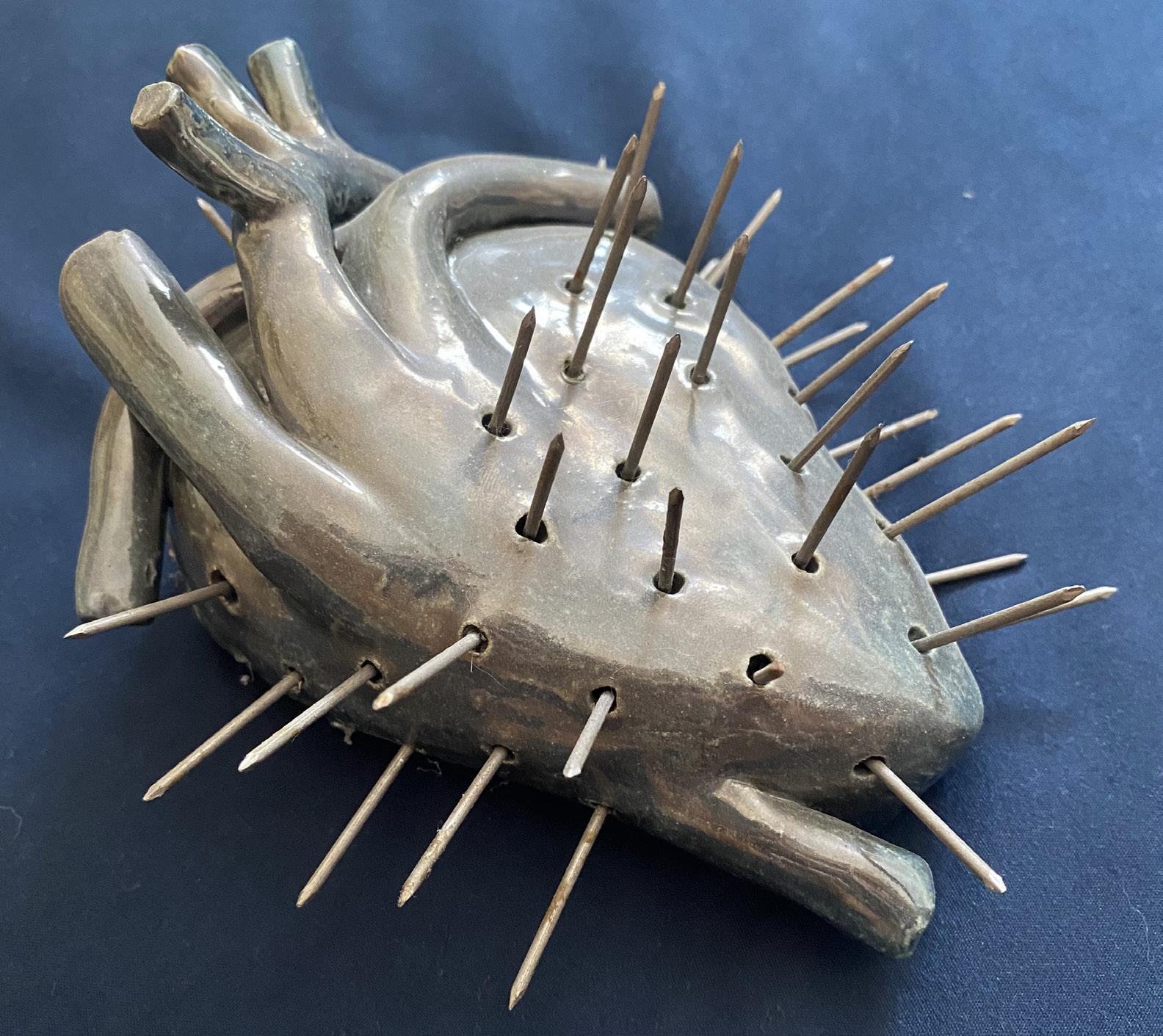
Case Report
De novo Familial Adenomatous Polyposis with undiagnosed von Willebrand’s Disease:
A Case Report
Brytani White, PA-S III, Sarah Neguse, MS, PA-C
Rocky Vista University
Patient consent was obtained via HIPAA
Authorization for the Release and Use of Private Health Information for Research.
IRB Approval #2023-021
1. ABSTRACT
This case details the presentation of a 29-year-old male that was found to have de novo familial adenomatous polyposis (FAP) in which treatment was complicated by undiagnosed von Willebrand’s disease (VWD). The patient presented to his primary care physician for routine labs, and the results were consistent with anemia. The patient was urgently referred to gastroenterology (GI) for further evaluation and diagnosed with FAP with no evidence of cancer. The patient had no known family history of GI cancer. FAP is a rare autosomal dominant disorder in which hundreds of adenomatous polyps develop in the colon with a nearly 100 percent chance of developing colon cancer by age 40 if untreated. De novo mutations with no family history account for only 10-30 percent of FAP cases (1-3 in 100,000 individuals). Additionally, de novo cases are usually not diagnosed until cancer has developed. This case is unique in that the patient was diagnosed at a young age (29-years-old) and had not yet developed cancer. The patient was diagnosed based on abnormalities from routine labs. This reinforces recommendations for routine lab work and the importance of completing full investigations into abnormalities. To treat his FAP, the patient underwent a total colectomy with mucosal proctectomy and ileoanal pull through. Pre-surgical labs revealed a minimally prolonged clotting time. However, since it was barely longer than the normal range, the surgeon decided to proceed without further investigation. Post-operative bleeding occurred, leading to the patient undergoing three additional unplanned surgeries and further evaluation for a bleeding disorder, after which, the patient was diagnosed with VWD. Von Willebrand’s disease, which affects about one percent of the population, is the deficiency of a protein leading to decreased ability to form a blood clot. A literature review revealed no reported case studies or known associations between FAP and VWD. After the delay for hematology work up, the patient returned three months later for surgical ileostomy reversal as planned, which proceeded without complication. This case report highlights the importance of routine screenings and presurgical labs and full investigation of abnormalities even if they are slight.
2. INTRODUCTION
Familial Adenomatous Polyposis (FAP) is a rare autosomal dominant colon cancer disorder associated with a mutation of the Adenomatous polyposis coli (APC) gene on chromosome 5 in which hundreds of adenomatous polyps develop in the colon.1 Individuals with FAP have a nearly 100 percent chance of developing colon cancer by age 40 if untreated and it accounts for about one percent of colon cancer cases.2 It occurs in 1 in 10,000 individuals.3 Of those, the majority have a known family history with only 10-30 percent of cases (1-3 in 100,000 individuals) being a de novo mutation.4 Most patients are asymptomatic until adenomas are large, numerous, cause bleeding, and/or become cancerous leading to later age of diagnosis for de novo cases.5 FAP has also been linked to a wide range of extracolonic features including upper gastrointestinal polyps, osteomas, congenital hypertrophy of the retinal pigment epithelium (CHRPE), epidermal cysts, fibromas, dental abnormalities and desmoid tumors.6 Prophylactic colectomy is recommended for all FAP patients to avoid risk of developing colon cancer.7
In 1991, mutations in the APC gene on chromosome 5, which encodes a tumor suppressor protein, were discovered as the cause of FAP.8 Since then, hundreds of mutations in the APC gene have been implicated in FAP and correlations have been identified between specific mutations and rate of extracolonic features, number of polyps developed, and age at which polyps develop have been identified.8 However, variation is still seen in individuals with identical mutations, alluding to the contribution of other factors in disease course such as environmental or other genetic factors.9
In addition to FAP, this case also presented with the complication of undiagnosed von Willebrand’s disease. Von Willebrand’s disease is the deficiency of the von Willebrand’s factor, which is a glycoprotein that functions as a carrier for factor VIII, helps with platelet adhesion, and binds to endothelium after a vascular injury.10 Von Willebrand’s disease can be inherited or acquired and affects about one percent of the population.10 Many mutations in the von Willebrand factor gene on chromosome 12 have been implicated in inherited von Willebrand’s disease and contribute to the variation in presentation and severity of symptoms.11 Acquired Von Willebrand’s disease occurs when another process leads to impairment
of the von Willbrand factor and is most commonly associated with malignancies (including lung cancer, Wilm’s tumor, gastric cancer, multiple myeloma, chronic lymphocytic leukemia, hairy cell leukemia, and lymphomas), systemic lupus erythematosus, autoimmune disorders, metabolic disorders, and adverse effects from drugs.10
The presentation of patient’s with von Willebrand’s disease can range from asymptomatic to prolonged bleeding from minor skin injury, recurrent/excessive bruising, heavy menstrual cycles, and epistaxis.10 Diagnosis is confirmed based on laboratory results, including prolonged partial thromboplastin time (PTT), normal prothrombin time (PT), decreased Von Willebrand factor activity (measured via Ristocetin cofactor activity), presence of Von Willbrand factor antigen, and decreased Factor VIII activity.10 Stable patients are treated with desmopressin.10 Von Willebrand factor replacement is often considered for severe presentations and episodes of serious bleeding such as with trauma or major surgery.10
3. CASE DESCRIPTION
A 29-year-old healthy male with no known medical conditions presented to his primary care physician for evaluation of cholesterol blood levels due to family history of high cholesterol. Routine labs were performed, including complete blood count, complete metabolic panel, and lipid panel, revealing low hemoglobin of 8.6 g/dL (normal range 13.3-18.2) and low hematocrit of 29.0 percent (normal range 38.0-53.0), indicating that the patient was anemic. The patient denied any recent trauma, a change in weight, a change in bowel habits, blood in the stools, or family history of GI disorders, including cancer. He reported recent fatigue but associated that with attending school full time, working a full-time job, and having a newborn baby at home. Due to the severity of the anemia and no known personal history of trauma or medical conditions, the patient was started on oral iron supplements and urgently referred to a gastroenterologist for further evaluation of a possible gastroenterological bleed the next day. A colonoscopy revealed friable polyps carpeting the entire colon and rectum and biopsy results were consistent with FAP with no evidence of cancer. The patient was referred to general surgery to discuss three types of surgeries to remove
the colon, which included: (1) proctocolectomy with end ileostomy, (2) total abdominal colectomy with ileoproctostomy or (3) total colectomy with mucosal proctectomy and ileoanal pull through. The patient elected to proceed with the third option as it allowed total removal of all colon tissue without the need for an ostomy for defecation. Pre-surgical labs showed improved hemoglobin levels of 12.3 g/dl and hematocrit of 39.8 percent as well as a slightly prolonged partial thromboplastin time (PTT), a measure of blood clotting time, of 35.3 seconds (normal range 25-35 seconds). The PTT normalized when the blood was mixed with control plasma, suggesting a possible blood clotting factor deficiency. However, since PTT was minimally out of normal range, the surgeon decided to proceed with the operation without further investigation of potential bleeding issues. The surgery was performed successfully with no visualized cancer, no complications, and an estimated blood loss (EBL) of 900mL (blood loss > 750 mL carries a risk of shock and inability to deliver blood to the whole body).12,13
During the night after the surgery, the patient’s heart rate increased, and blood began oozing from the surgical wound. The next morning, the patient was taken back to the operating room (OR) due to postoperative bleeding. During the procedure, no site of active bleeding was found, however 500mL of free blood was removed from the abdomen and pelvis. The patient was doing well after the second operation until that evening when his vitals revealed elevated heart rate again and a drop in blood pressure. The patient was given four units (1 unit is approximately 500mL) of blood for suspected blood loss. He was taken back to the OR and a site of mild active bleeding was found in the pelvis. During the operation, 2300mL of free blood was also removed from the abdomen and pelvis. A massive transfusion protocol was initiated, leading to the patient receiving an additional four units of red blood cells, platelets, and fresh frozen plasma. The patient was then packed with surgical gauze to provide pressure to control the bleeding with the intention of returning to the OR later that day to remove the packing. During the return to the OR, no sites of active bleeding were found, and packs were removed. The patient was discharged on postoperative day 12 with an ileostomy and referred to hematology/oncology for evaluation of a bleeding disorder.
Surgical ileostomy reversal was delayed
by a month while awaiting hematology work up to minimize bleeding risks and complications. Von Willebrand’s disease was eventually diagnosed by the hematologist. The patient returned three months after his first surgery for surgical ileostomy reversal which proceeded without complication. The patient began routine screening with endoscopies and pouchoscopies to monitor for polyps in the digestive tract. Since the initial presentation, the patient has been diagnosed with five desmoid tumors and undergoes regular CT scans for monitoring. The patient also has had three children, two of which have also been diagnosed with FAP and undergone total colectomies with mucosal proctectomies and ileoanal pull through.
4. DISCUSSION
Patients with FAP have an almost 100 percent risk of developing colon cancer, highlighting the importance of early diagnosis and intervention. However, with a lack of family history as is the case with de novo mutations and the lack of symptoms, de novo FAP is not usually identified until cancer has developed.14 This case is unique in that the patient was diagnosed at a young age (29-years-old) and no cancer was found at the time of diagnosis. The patient was diagnosed based on abnormalities from routine labs drawn by his primary care physician. This reinforces recommendations for routine lab work and the importance of providers completing full investigations into abnormalities.
Although the genetic basis of FAP is well known, genetic testing is mainly used for screening of at-risk family members and confirmation of diagnosis in clinically diagnosed patients.5 With the growing prevalence of genetic testing, the question is raised of whether mutations in the genes implicated in FAP should be routinely tested for mutations in the general population. Genetic counseling and testing are often offered to patients and their families after a clinical diagnosis is established. Currently, many genetic tests for cancer related mutations are only covered by insurance if the patient meets high risk criteria and the test is considered medically necessary, though these tests usually come with copays and deductibles.15 Out of pocket costs for genetic testing ranges depending on complexity of the testing, with the high end of the range costing thousands of dollars.15 However, the field of genomics is continuing to advance and
costs are expected to continue to decline for genetic testing and sequencing.16 Discussions and research have started to broach the idea of general population genetic testing. One study investigating breast cancer and BRCA mutations found that general population genetic testing may be beneficial to identify mutations and more cost effective compared to our current approach to genetic testing of only high-risk individuals.17 As this idea continues to develop, these trends could be translated to genetic mutations implicated in other malignancies, including FAP. As genetic testing becomes more available and affordable for patients, general population genetic screening should be further explored regarding risks and benefits to help increase early identification of the disease.
In addition to FAP, this case also presented the complication of undiagnosed von Willebrand’s disease. A literature review revealed no reported case studies or known associations between FAP and von Willebrand’s disease. Genetically, the mutations for these diseases are found in two different chromosomes.8,11 While no genetic connection was identified between FAP and von Willebrand’s disease, cancer (especially hematological malignancies) has been linked to acquired von Willebrand’s disease.18 However, while this patient was at high risk for cancer, pathology was consistent with FAP that had not yet developed into cancer. The additional discovery of von Willebrand’s disease added an extra layer of complication, highlighting the uniqueness of this case. Additionally, this warrants the question of whether further work up should have been performed given the patient’s slightly elevated PTT on pre-surgical labs, specifically checking for factor deficiencies, including von Willebrand factor. In this case, due to only being a slight increase in PTT (0.3 seconds above normal range), the surgeon proceeded without further investigation into a possible factor deficiency, which was suggested by the correction of PTT with mixing studies. While knowledge of the presence of von Willebrand’s disease would not have changed the treatment course for FAP, it would have allowed the surgeon to be better prepared for possible complications of postoperative bleeding. It would have also allowed for pre-operative prophylaxis with desmopressin or von Willebrand factor replacement. This preparation may have helped prevent the multiple returns to the operating room as well as the prolonged hospital stay and secondary monetary,
emotional, mental, and physical effects of the complications. Again, this highlights the importance of routine screenings and fully investigating any abnormalities that are uncovered.
5. REFERENCES
1. Galiatsatos P, Foulkes WD. Familial Adenomatous Polyposis. Off J Am Coll Gastroenterol ACG. 2006;101(2):385.
2. Carr S, Kasi A. Familial Adenomatous Polyposis. In: StatPearls. StatPearls Publishing; 2023. Accessed April 28, 2023. http://www.ncbi.nlm. nih.gov/books/NBK538233/
3. Kanth P, Grimmett J, Champine M, Burt R, Samadder JN. Hereditary Colorectal Polyposis and Cancer Syndromes: A Primer on Diagnosis and Management. Off J Am Coll Gastroenterol ACG. 2017;112(10):1509. doi:10.1038/ ajg.2017.212
4. Waller A, Findeis S, Lee MJ. Familial Adenomatous Polyposis. J Pediatr Genet. Published online March 15, 2016:78-83. doi:10.1055/s-0036-1579760
5. Half E, Bercovich D, Rozen P. Familial adenomatous polyposis. Orphanet J Rare Dis. 2009;4(1):22. doi:10.1186/1750-1172-4-22
6. Jasperson KW, Tuohy TM, Neklason DW, Burt RW. Hereditary and Familial Colon Cancer. Gastroenterology. 2010;138(6):2044-2058. doi:10.1053/j.gastro.2010.01.054
7. Beech D, Pontius A, Muni N, Long WP. Familial adenomatous polyposis: a case report and review of the literature. J Natl Med Assoc 2001;93(6):208-213.
8. Nieuwenhuis MH, Vasen HFA. Correlations between mutation site in APC and phenotype of familial adenomatous polyposis (FAP): A review of the literature. Crit Rev Oncol Hematol. 2007;61(2):153-161. doi:10.1016/j. critrevonc.2006.07.004
9. Familial Adenomatous Polyposis | VCU Massey Cancer Center. Accessed June 13, 2023. http:// www.masseycancercenter.org/patients-andfamilies/patient-resources-and-support-services/ inherited-cancers/the-genetics-of-colorectalcancer/familial-adenomatous-polyposis
10. Sabih A, Babiker HM. Von Willebrand Disease. In: StatPearls. StatPearls Publishing; 2023. Accessed June 23, 2023. http://www.ncbi.nlm.nih. gov/books/NBK459222/
11. Goodeve AC. The genetic basis of von Willebrand disease. Blood Rev. 2010;24(3):123-134. doi:10.1016/j.blre.2010.03.003
12. Hypovolemic Shock: Causes, Symptoms and Treatment. Cleveland Clinic. Accessed July 12, 2023. https://my.clevelandclinic.org/health/ diseases/22795-hypovolemic-shock
13. Johnson AB, Burns B. Hemorrhage. In: StatPearls. StatPearls Publishing; 2023. Accessed July 12, 2023. http://www.ncbi.nlm.nih.gov/ books/NBK542273/
14. Dalavi SB, Vedpalsingh TH, Bankar SS, Ahmed MHS, Bhosale DN. Familial Adenomatous Polyposis (FAP)—A Case Study and Review of Literature. J Clin Diagn Res JCDR. 2015;9(3):PD05-PD06. doi:10.7860/ JCDR/2015/11636.5696
15. What is the cost of genetic testing, and how long does it take to get the results?: MedlinePlus Genetics. Accessed August 14, 2023. https:// medlineplus.gov/genetics/understanding/testing/ costresults/
16. The Cost of Sequencing a Human Genome. Genome.gov. Accessed August 14, 2023. https:// www.genome.gov/about-genomics/fact-sheets/ Sequencing-Human-Genome-cost
17. Ficarazzi F, Vecchi M, Ferrari M, Pierotti MA. Towards population-based genetic screenings for breast and ovarian cancer: A comprehensive review from economic evaluations to patient perspectives. Breast Off J Eur Soc Mastology. 2021;58:121-129. doi:10.1016/j. breast.2021.04.011
18. Colonne CK, Favaloro EJ, Pasalic L. The Intriguing Connections between von Willebrand Factor, ADAMTS13 and Cancer. Healthcare. 2022;10(3):557. doi:10.3390/healthcare10030557
Delivery
By Anna Jacobs
moaning, screaming voice the dark red stench of blood my hands, shaking reaching out to grasp a tiny head
one contraction one howl one push one tiny human being clawing her way into the world
a large gasp a piercing cry
as two voices meld into one song a mother and child embracing for the first time
my hands, no longer shaking my eyes wet with tears
I’ll never forget the first baby I ever delivered
New Beginnings
By Gianna DeCosmo
New Beginnings is a painting born from the culmination of feelings I was having during a rough period of my life. I felt hopeless and lost because nothing seemed to be going to plan, and I didn’t know what I was doing. I turned to my art for inspiration and to find the meaning behind the chaos that is life, and I found the inspiration for this painting. I encountered a tale about people who would eat seeds right before they died so that trees or flowers would grow from their bodies. Their bodies would provide nutrients for something new and beautiful to grow. Through their deaths, something else could blossom—something new. This story reminded me that beautiful outcomes can be created from something dark, sad, or hopeless. The end of one chapter is the beginning of something new that is ready for exploration. New Beginnings explores the concepts of new life and fresh starts with its vibrant colors and personification of nature. It serves as a reminder that, like the cyclical style of nature, we can find beauty after pain and grow into someone new. The exploration of this piece gave me the space to let things that once felt hopeless begin to bloom.

t h i c s &
Tackling the Epidemic of Medical Misinformation: Nurturing Trustworthy Voices in
the Social Media Era
Arpit Danewalia, Second-year Medical Student. RVU College of Osteopathic Medicine, Ivins, UT arpit.danewalia@ut.rvu.edu
Maison Evensen-Martinez, Third-year Medical Student. Rocky Vista University College of Osteopathic Medicine, Ivins, UT, USA, maison. evensen@ut.rvu.edu
In the vast expanse of social media, platforms like TikTok and X (formerly Twitter) have evolved into bustling marketplaces where influencers wield significant power. However, this power, when misused, becomes a perilous conduit for the dissemination of misleading and harmful medical information. This brief piece delves into the state of medical misinformation, a vast area of growing concern, and explores potential solutions. For medical practitioners and students, the misuse of influence not only breeds fear but also challenges the sanctity of evidence-based healthcare practices. This fear stems from witnessing influential individuals peddling misinformation about health and medicine, often to bolster their following or pocket hefty sponsorships. Distorted information can instill doubts, sow confusion, and even lead to dangerous decisions for those seeking guidance amid the vast and overwhelming sea of online content. A 2021 study identified health misinformation topics such as vaccines (especially HPV), drugs or smoking, noncommunicable diseases, pandemics, eating disorders, and medical treatments to be the most prevalent topics online and expressed the importance of creating evidence-based digital policy action plans to combat them.1 Aside from misinformation from uniformed influencers and sources, studies have also investigated the insidious and targeted spread of misinformation, such as e-cigarette use,2–4 complementary and alternative medicine use,5 and harmful cancer information.6,7 Recently, the 2020 COVID-19 pandemic showed everyone just how prevalent misinformation could be. Misinformation regarding disease prevention, treatment, diagnostics, and vaccine safety8 was rampant with more spread of misinformation related to the novelty of information
or the social status of the sharer.9,10
The core of this issue lies in the power wielded by these influencers, reaching millions with a mere click. Nevertheless, there is a glimmer of hope amidst this chaos. Health professionals possess the knowledge, expertise, and ethical responsibility to counteract this wave of misinformation. Methods for counteracting misinformation have been classified into four pillars, including (broadly) information monitoring, education, knowledge refinement (e.g., fact checking), and accurate, timely, and clear knowledge distribution.11
One potential solution lies in amplifying the voices of credible health professionals on the same platforms used by influencers.12,13 Engaging, informative, and fact-based content can counteract misleading narratives; by harnessing the power of social media, medical professionals can disseminate accurate information, debunk myths, and bridge the gap between complex medical knowledge and the public. Returning to the 2020 pandemic, specific efforts at counteracting misinformation about the novel coronavirus in the social media space included educational campaigns, targeted communication strategies, and fact-checking initiatives.8 However, brand influencers, celebrities, politicians, and science communicators constituted most comments,14 identifying the need for increased engagement from the government and health officials.
To combat the allure of sensationalized but inaccurate content, health professionals must embrace the virtual realm. If there are officials moderating content and clarifying information, research shows that they can help dispel rumors when they arise.15,16 By engaging with audiences through informative videos, Q&A sessions, or simplified explanations of complex medical concepts,17 they can establish themselves as trusted pillars of knowledge and compassion.
Moreover, collaboration between social media platforms and healthcare organizations could help in the creation of vetted, verified channels for medical advice. Implementing robust fact-checking measures and highlighting verified health professionals can steer audiences toward reliable sources,16,18–20 creating a virtual haven for accurate medical information. Additionally, utilizing influencers’ reach to spread accurate information can have large impacts on the content received from viewers, as seen with the flu vaccine21 and COVID-19 pandemic-era.22
Furthermore, accurate and easy-to-understand information is key. Empowering the public with tools to discern credible information from misleading content is pivotal, in addition to providing clear and accurate information.17 Initiatives that promote health literacy and critical thinking skills regarding online medical information can serve as shields against misinformation.
The journey toward establishing credibility and trust in the digital space might be challenging, but the impact of disseminating accurate information is immeasurable. In the battle against medical misinformation, every like, share, and view of evidence-based content becomes a beacon of enlightenment amidst the noise. By harnessing the power of social media responsibly, health professionals can transcend the confines of their clinics and lecture halls, becoming beacons of accurate and trustworthy medical information in the virtual world.
1. Suarez-Lledo V, Alvarez-Galvez J. Prcovevalence of Health Misinformation on Social Media: Systematic Review. J Med Internet Res 2021;23(1):e17187. doi:10.2196/17187
2. Amin S, Dunn AG, Laranjo L. Social Influence in the Uptake and Use of Electronic Cigarettes: A Systematic Review. Am J Prev Med. 2020;58(1):129-141. doi:10.1016/j. amepre.2019.08.023
3. Smith MJ, Hilton S. Youth’s exposure to and engagement with e-cigarette marketing on social media: a UK focus group study. BMJ Open. 2023;13(8):e071270. doi:10.1136/ bmjopen-2022-071270
4. Smith MJ, Buckton C, Patterson C, Hilton S. User-generated content and influencer marketing involving e-cigarettes on social media: a scoping review and content analysis of YouTube and Instagram. BMC Public Health. 2023;23(1):530. doi:10.1186/s12889-023-15389-1
5. Gülpınar G, Uzun MB, Iqbal A, Anderson C, Syed W, Al-Rawi MBA. A model of purchase intention of complementary and alternative medicines: the role of social media influencers’ endorsements. BMC Complement Med Ther. 2023;23(1):439. doi:10.1186/s12906-023-04285-1
6. Kureyama N, Terada M, Kusudo M, et al. FactChecking Cancer Information on Social Media in Japan: Retrospective Study Using Twitter. JMIR Form Res. 2023;7:e49452. doi:10.2196/49452
7. Rahimy E, Sandhu NK, Giao DM, Pollom EL. #TrendingNow: Instagram Versus Twitter Activity Among Radiation Oncology Patients and Professionals. Pract Radiat Oncol. 2021;11(6):e506-e514. doi:10.1016/j. prro.2021.06.008
8. Smith R, Chen K, Winner D, Friedhoff S, Wardle C. A Systematic Review Of COVID-19 Misinformation Interventions: Lessons Learned. Health Aff Proj Hope. 2023;42(12):1738-1746. doi:10.1377/hlthaff.2023.00717
9. Photiou A, Nicolaides C, Dhillon PS. Social
status and novelty drove the spread of online information during the early stages of COVID-19. Sci Rep. 2021;11(1):20098. doi:10.1038/s41598021-99060-y
10. Wasike B. When the influencer says jump! How influencer signaling affects engagement with COVID-19 misinformation. Soc Sci Med 1982. 2022;315:115497. doi:10.1016/j. socscimed.2022.115497
11. Eysenbach G. How to Fight an Infodemic: The Four Pillars of Infodemic Management. J Med Internet Res. 2020;22(6):e21820. doi:10.2196/21820
12. Walter N, Brooks JJ, Saucier CJ, Suresh S. Evaluating the Impact of Attempts to Correct Health Misinformation on Social Media: A MetaAnalysis. Health Commun. 2021;36(13):17761784. doi:10.1080/10410236.2020.1794553
13. Xue H, Gong X, Stevens H. COVID-19 Vaccine Fact-Checking Posts on Facebook: Observational Study. J Med Internet Res. 2022;24(6):e38423. doi:10.2196/38423
14. MacKay M, Ford C, Colangeli T, Gillis D, McWhirter JE, Papadopoulos A. A content analysis of Canadian influencer crisis messages on Instagram and the public’s response during COVID-19. BMC Public Health. 2022;22(1):763. doi:10.1186/s12889-022-13129-5
15. Eckert S, Sopory P, Day A, et al. Health-Related Disaster Communication and Social Media: Mixed-Method Systematic Review. Health Commun. 2018;33(12):1389-1400. doi:10.1080/1 0410236.2017.1351278
16. Zhang J, Featherstone JD, Calabrese C, Wojcieszak M. Effects of fact-checking social media vaccine misinformation on attitudes toward vaccines. Prev Med. 2021;145:106408. doi:10.1016/j.ypmed.2020.106408
17. Bauder L, Giangobbe K, Asgary R. Barriers and Gaps in Effective Health Communication at Both Public Health and Healthcare Delivery Levels During Epidemics and Pandemics; Systematic Review. Disaster Med Public Health Prep.
2023;17:e395. doi:10.1017/dmp.2023.61
18. Bond RM, Garrett RK. Engagement with fact-checked posts on Reddit. PNAS Nexus 2023;2(3):pgad018. doi:10.1093/pnasnexus/ pgad018
19. Lanius C, Weber R, MacKenzie WI. Use of bot and content flags to limit the spread of misinformation among social networks: a behavior and attitude survey. Soc Netw Anal Min 2021;11(1):32. doi:10.1007/s13278-021-00739-x
20. Porter E, Wood TJ. The global effectiveness of fact-checking: Evidence from simultaneous experiments in Argentina, Nigeria, South Africa, and the United Kingdom. Proc Natl Acad Sci U S A. 2021;118(37):e2104235118. doi:10.1073/ pnas.2104235118
21. Bonnevie E, Smith SM, Kummeth C, Goldbarg J, Smyser J. Social media influencers can be used to deliver positive information about the flu vaccine: findings from a multi-year study. Health Educ Res. 2021;36(3):286-294. doi:10.1093/her/ cyab018
22. Li W, Ding H, Xu G, Yang J. The Impact of Fitness Influencers on a Social Media Platform on Exercise Intention during the COVID-19 Pandemic: The Role of Parasocial Relationships. Int J Environ Res Public Health. 2023;20(2):1113. doi:10.3390/ijerph20021113
Ephemeral Embrace: A Symphony of Lilies
By Anna L Megenhardt
In my oil painting, “Ephemeral Embrace: A Symphony of Lilies,” I sought to capture the essence of nature’s poetry through the delicate and timeless beauty of lilies. Like a conductor harmonizing a symphony, I aimed to evoke emotions and memories, drawing viewers into a world where the ethereal and the earthly intertwine.
Lilies, with their enchanting form and mesmerizing colors, have always held a profound fascination and adoration for me. They symbolize purity, rebirth, and the transient nature of existence, acting as a powerful reminder of life’s fleeting yet profound moments. These delicate blooms carry with them a subtle dance between fragility and resilience, embodying the essence of human experiences.
My intention was to portray lilies not merely as still objects on canvas but as vibrant, living entities, each petal an expression of profound emotion and every hue a reflection of nature’s brushstroke. Through the interplay of light and shadow, I endeavored to unveil the hidden mysteries within these flowers, allowing viewers to discover the subtle shifts in tones and textures that breathe life into the canvas.
In this piece, I have chosen to embrace a balance between realism and abstraction, allowing viewers to
interpret and engage with the artwork on a personal level. The lilies stand out as a detailed centerpiece to an abstract, dynamic backdrop. The fluidity of the oil medium has enabled me to create a mesmerizing dance of colors, evoking a sense of tranquility and contemplation. The textured brushstrokes carry a tactile quality that invites viewers to immerse themselves in the painting’s rich depths.
Moreover, I see “Ephemeral Embrace” as a reflection of our relationship with the natural world. In a rapidly changing world, it serves as a gentle reminder of the importance of cherishing the fleeting moments of beauty and connection with our environment. The lilies’ graceful presence serves as a call to pause, contemplate, and appreciate the subtle wonders that surround us daily.
Ultimately, “Ephemeral Embrace: A Symphony of Lilies” seeks to evoke an emotional response, stirring the soul and sparking introspection. It is my hope that this artwork transports viewers to a tranquil sanctuary where they can momentarily escape the chaos of the outside world and delve into the enchanting realm of the lilies’ ephemeral embrace

Research
ANALYSIS OF GUIDELINES REGARDING MENTAL HEALTH HOLD REQUIREMENTS AND DURATION OF ADULTS FROM MOUNTAIN STATES
Dallin Trout, PA-C
Darcy Solanyk, MS, PA-C
Rocky Vista University
1. ABSTRACT
Emergency commitments and involuntary holds are tools that medical providers possess to treat individuals with mental illness who may be a danger to themselves or others. State laws govern these holds in both requirement and length, and thus vary from state to state. We researched if the state laws of Arizona, Colorado, Idaho, Montana, Nevada, New Mexico, Utah, and Wyoming (Mountain States) follow recommendations set forth by the American Psychiatric Association (APA) and United Nations (UN) regarding these holds. In this evaluation, we compared the requirements recommended by the APA and the UN and noted what differences they had. We then examined Mountain States and the specific length and requirements for each of these states to enact emergency holds and involuntary commitments in the respective state statutes. We found that APA guidelines incorporated the UN guidelines and added two recommendations: an individual must be likely to suffer physical hardship due to being unable to satisfy their basic needs and that the state must present ‘clear and convincing’ evidence to enact involuntary holds. In total, the APA and UN present seven guidelines. Arizona is unique in that it is the only state that fulfills all guidelines by the APA while Wyoming and Colorado lack one and the remaining states lack at least two. All Mountain States require that a patient suffers from a mental disorder and is likely to cause harm to self or to others, as well as a judicial review to present evidence for continued care. Each state varies in who can provide evidence. Additionally, we find that all states vary in the length of their emergency holds and involuntary commitments. All states also allow the initiation of a hold by persons other than a medical professional. Moreover, neither the APA nor the UN recommend a minimum or maximum length of a hold. We theorize that this can allow the states flexibility in treating mental illness while simultaneously causing barriers to care due to lack of standardization. This analysis recommends further research-based guidelines from the APA to better standardize care across the United States.
2. INTRODUCTION
In a mental health crisis, individuals may find themselves in an altered mental state for hours or days despite prompt treatment.1 During this time, their mental status can put themselves or others at risk of injury or further mental deterioration.2 Thus, a solution is required to ensure a patient’s safety while maintaining treatment. This solution is found in emergency holds and involuntary commitments.3
Although emergency holds and involuntary, or civil, commitments are terms that are often used interchangeably, they refer to two distinct situations. Emergency holds are “a brief involuntary detention of a person presumed to have a mental illness in order to determine whether the individual meets criteria for involuntary civil commitment.”4 These contrast with involuntary commitments which can last for multiple days or weeks.
Emergency holds and involuntary, or civil, commitments are vital in treatment of mental illness.3 They are often necessary to maintain a patient’s safety.2 As utilizing them requires holding a person against their will, they must meet certain requirements5; for example, the state of Colorado requires that a patient suffers from a mental disorder, is likely to cause harm to themselves or others, suffers physical hardship due to being unable to satisfy their basic needs, and suffers substantial mental deterioration.5
It is here where we find a complication: (1) this is a legal process based on a medical determination and (2) each state can make its own statutes for the requirements of emergency holds, including who can make the determination and the length of the holds.4 This length, as previously noted, often varies.
While there are no strict federal guidelines, the American Psychiatric Association (APA) and United Nations (UN) have developed guidelines on the requirements these commitments should possess.6,7
Previous studies and books have examined the process of emergency holds and involuntary commitments,4 demographics,8 and ethical conflicts.9 However, previous studies have not compared these processes to the guidelines set by the UN or APA.
Here, we examine and compare those guidelines and the current practice of Mountain States (within in the United States) to determine if they align with these guidelines. We chose these states, as opposed to the whole of the United States, to ascertain whether there is variability in laws and procedures in a localized geographic area.
In addition, we compared the length of emergency holds and involuntary commitments, their requirements, and the requirements to lengthen such holds to ascertain if and how these guidelines are followed.
3. MATERIALS AND METHODS
We utilized the APA and UN guidelines6,7 and focused specifically on their requirements for emergency holds and involuntary commitments. We chose eight states (Arizona, Colorado, Idaho, Montana, Nevada, New Mexico, Utah, and Wyoming) that are within the Rocky Mountain range (termed Mountain States) to review their current laws regarding emergency holds of people dealing with an acute mental health crisis, such as a suicide attempt.
We detailed the specific recommendations that the APA puts forth; out of ten guideline recommendations we focused on recommendations 2 through 7, which dealt with the criteria for involuntary admission.6 Other guidelines were excluded from our review as they dealt with voluntary admission, denial of hospitalization, refusal of treatment, and transportation.6
After this, we compared these guidelines to the guidelines encouraged by the UN in their resolution. We focused on Principles 4, 16, and 17 which detailed criteria recommended for involuntary holds.7 We focused on the wording of the APA and the UN guidelines, as well as their content as it pertained solely to the criteria of emergency holds and involuntary commitments, their lengths, and processes.
We conducted a search of current emergency hold laws and requirements for involuntary commitments as of June 1, 2023. These laws were defined as statutes regarding the requirements, duration, and processes that regulated psychiatric holds due to mental illness as defined in the Diagnostic and Statistical Manual of Mental Disorders (DSM-V).5,11-17
We first conducted a Google search using the Google.com search engine. We focused on keywords of “current emergency hold laws” and “requirements for involuntary commitments.” We searched for databases and statutes that contained these phrases. This led us to state legislature websites which prompted us to use said state legislature websites in addition to Justia Law for their processes for emergency and involuntary holds. We then
compared the statutes that we found to the APA and UN recommended guidelines.6,7,11-17 We detailed the length of the holds, who could initiate holds, who could authorize involuntary holds, and the lengths of involuntary commitments.
4. RESULTS
We focused primarily on the guidelines dealing with the process and requirements of involuntary holds. Both the UN and the APA align in recommending judicial review of the case, having legal representation for the individual present during any review, and maintaining the rights of the individual.6,7 Both recommend that a person should only be held as long as necessary but do not specify any specific limit on that hold.6,7
In reviewing the recommendations for admission criteria of the UN and the APA, we found that they are nearly identical; the only area in which they differ is an additional recommendation by the APA that a person is likely to suffer physical hardship as a result of their mental illness.6 This could include but not be limited to being unable to provide for essential needs such as housing, food, or clothing.5
State statutes that discussed definitions, emergency mental health holds, extensions of treatment, court orders, and hearing procedures were included. Statutes that involved appeals, transportation, and voluntary applications were considered beyond the scope of this research and were not included.
We found that all states require individuals to suffer from a mental disorder and be likely to cause harm to self or others to be eligible for an involuntary hold. Additionally, all states require a judicial determination for continued care.5,11-17 A comparison of the recommendations of the UN and the APA and eight statutes of the eight states is summarized in Table 1.
Table 1. Recommended criteria for initial involuntary holds.
APA
-
UN Guidelines For Admission Suffering from mental disorder Evaluated by two physicians for initial assessment Likely to cause harm to self Suffer substantial mental deterioration
Arizona
Colorado
Idaho
Montana
Nevada
New Mexico
Utah
Likely to cause harm to others
Judicial determination for need for continued involuntary hospitalization
State Statutes
(2023)
(2022)
(2023)
(2021)
(2023)
(2021)
(2023)
(2022)
All Mountain States require that a patient suffering from a mental disorder who is likely to cause harm to self or others requires a judicial review for continued care. Of the Mountain States, Arizona11 fills all seven recommendations while Wyoming17 and Colorado5 lack one (evaluation by two physicians) and the remaining states lack at least two (see Table 1).12-16
We further explore the initiation of emergency holds in Table 2. Arizona is unique in the fact that mental health holds can be initiated by a far wider variety of people than the other states, but an initial evaluation must be made with two physicians.11 Otherwise, there is a common trend of requiring either an agent-of-the-law or a medical professional to initiate the hold,5,12-17 which deviates from UN and APA recommended guidelines of having two physicians initially evaluate a patient. While all states endeavor to have a medical professional initially
evaluate a patient as recommended, we see that they also allow this process to be initiated by someone other than a physician, most usually a peace officer (see Table 2).5, 12-17
Table 2. Initiation of emergency holds and length.
State Emergency commitment length
Who can perform initial evaluation for emergency hold State Statutes
Arizona 72 hours* Anyone from a ‘certified agency’, but is then recommended to be then certified by two physicians
Colorado 72 hours* A certified peace officer, including a police officer
A physician
A licensed psychologist
A licensed nurse with experience in psychiatric or mental health, A licensed marriage or family therapist with mental health experience
A professional counselor with mental health experience
An addiction counselor with experience evaluating mental health disorders
A licensed clinical social worker.
Idaho 24 hours* A certified peace officer, including a police officer
A physician
A physician assistant
An advanced practiced registered nurse
Montana 24 hours* A certified peace officer, including a police officer
A physician
A professional counselor with mental health experience
A licensed psychologist
A licensed clinical social worker
An advanced practiced registered nurse
Nevada 72 hours* A certified peace officer, including a police officer
A physician
A licensed psychologist
A licensed nurse with experience in psychiatric or mental health,
A licensed marriage or family therapist with mental health experience
A professional counselor with mental health experience
An addiction counselor with experience evaluating mental health disorders
A licensed clinical social worker.
New Mexico 24 hours* A certified peace officer, including a police officer
A physician
A licensed psychologist
Utah 48 hours* A certified peace officer, including a police officer
A physician
A physician assistant
A licensed mental health professional designated by the division as specially qualified by training and who has at least five years’ continual experience in the treatment of mental illness
Wyoming 72 hours* A physician
A physician assistant
A licensed marriage or family therapist with mental health experience
A licensed clinical social worker
An advanced practiced registered nurse
* denotes the statute specifying the maximum time allowed
§36-501, 530* (2023)
§27-65-102, 106* (2022)
§66-317, 326* (2023)
§53-21-102, 129* (2021)
§433A.018, .150* (2023)
§43-1-3, 10* (2021)
§62A-15-602, 629* (2023)
§25-10-101, 109* (2022)
UN and APA guidelines also stipulate that a judicial review should take place in the case of involuntary commitment.6,7 Here, the UN guidelines defer to the legal process of the country. The APA recommends that all states implement judicial review that brings “clear and convincing evidence”19 as their standard. Clear and convincing evidence is defined as evidence that is highly and substantially more likely to be true than untrue.20 For instance, evidence that a person is considered a danger to themselves could possibly include a recent suicide
attempt.5 This standard is not required in initiating a hold, but it is required to extend a hold. Those processes are shown in Table 3.
Table 3. Judicial review of Mountain States.
State Who may authorize Who must provide clear and convincing evidence
Arizona Superior court of the county*
Colorado District court of the county*
Idaho District court of the county*
Montana Judge or a justice of the peace of the county (may petition for a jury trial)
Nevada District court of the county*
New Mexico District court of the county
Utah District court of the county
Wyoming District court of the county
The evaluation of least two physicians, preferably psychiatrists
The evaluation of any professional per- son at the emergency detention facility (a person licensed to practice medicine or psychologist)
The evaluation of a professional per- son and a court ordered independent evaluation
The evaluation of a professional person (the same persons that can order an emergency hold)
The evaluation of least two additional physicians
The evaluation of an additional physi- cian, preferably with a mental health professional if available or needed
A designated examiner of the court (a licensed physician or other licensed mental health professional)
A designated examiner of the court (the same persons that can order an emer- gency hold)
* denotes the statute for jurisdiction; † denotes the statute for maximum length
Maximum Length of Commitment
365 days†
90 days
30 days
90 days
180 days
180 days
Up to 18 months
No limitation: initial review after 3 months, then every 6 months after
State Statutes
§36-533, 535*, 544† (2023)
§27-65-108111, (2022)113*
§66-328*, 337 (2023)
§53-21-128 (2021)
§433A.240, 310* (2023)
§43-1-12 (2021)
§62A-15-602 (2023)
§25-10-101 (2022)
All states require that a court in the original county where treatment began to authorize an extension of the hold.⁵,11-17 States differ in who presents evidence and how many persons presenting are required: three states (Idaho,12 Utah,16 Wyoming17) require a designated examiner of the court to independently verify and three states (Arizona,11 Idaho,12 Nevada14) require at least two examiners to extend a hold: Colorado,⁵ Montana,13 New Mexico,15 Utah,16 and Wyoming17 only require one.
With regards to additional examiners, both Nevada and New Mexico require additional evaluation. Nevada states that court will “promptly cause two or more physicians”14 to examine the patient. New Mexico also recommends an additional physician, one preferably with a background in mental health, to evaluate the patient. Variation in length of a maximum hold is also noted. Idaho can extend to a maximum of thirty days, while Wyoming can theoretically have no limit. All other states (Utah at 18 months)16 have maximums either at a year (Arizona)11 or less (Colorado and Montana at 90 days,5,13 Nevada and New Mexico at 180 days.)14,15
5. DISCUSSION
It is important to note that the APA and the UN guidelines are not binding resolutions. Strictly speaking, they possess no legal weight behind them. While their guidelines do rest upon established case law, such as O’Connor v. Donaldson18 and Colorado v. New Mexico19 as previously noted, states ultimately possess the power to write statutes for their territory and citizens. As seen in Table 1, not every state meets every guideline set forth. It is possible that states are primarily concerned with the possibility to harm self or others, the requirement for mental illness, and continued judiciary review which are ubiquitous throughout these states.
As we focused on a well-defined geographic location, it is possible that demographics may play a role in not only these differences, but in the differences seen in the subsequent tables. For instance, 77 percent of Colorado is considered rural territory.20
In Table 2, we see differences in how states approach the initiation of holds, particularly who may initiate holds. As many of these states have rural populations, it is unlikely that mental health professionals are available in every situation. This allows the states with areas without a mental health professional initially present to start the commitment process. This offers flexibility in providing treatment for mental illness; however, this also means that those that initiate the process may not necessarily have formal training in that area, which can lead to misdi-
agnoses, e.g., altered mental status due to ingestion of foreign substances or other medical etiologies can be mistaken for having a mental illness by someone not trained or experienced. This creates both possible barrier-to-care and a possibility of improper care.
Initiation of holds is not the only area in which we find variation. The length of holds, in both the emergency holds and involuntary commitments, vary from state to state. Despite this variation, neither the APA nor the UN make a recommendation for how long a commitment can last.
Previous research has been completed discussing the history of a 72-hour emergency hold,21 but research into the effectiveness of the length of the hold is not as forthcoming. For instance, one study shows that in a particular medical center, 88 percent of patients were discharged within 24 hours.22 Additional research should be encouraged to gauge the effectiveness of the emergency hold.
Thus, in the meantime, it is left to medical professionals and the courts to determine how long a person should be held and if that hold should be extended. This could lead to holds of differing lengths based upon physician preferences or experiences. Complications could also arise if a patient required transfer over state lines; as state maximums differ, this could lead to another barrier-to-care if a patient is transferred to a state that has a shorter maximum length.
This maximum length can also be affected by local courts and physicians. As the burden of proof of clear and convincing evidence is required to hold a person,18 it could become easier to maintain a hold if two people are able to present evidence. Conversely, requiring two people for a hold could result in fewer holds as having the review of two professionals could result in fewer misdiagnoses. However, even if the burden of proof is met, it may not necessarily translate to a hold. If there is no court available, the case may be dropped,23 which could create another barrier-to-care.
Despite these challenges, possible solutions exist. Ideally, federal statutes could determine how long a person can be held. They would codify the length of holds and make one standard for all states. However, legislative power is reserved by the states and a federal statute would not allow states to adjust their statutes best according to their own citizens.
A more feasible solution would be for the APA to review the current statutes and give additional guidelines regarding length of commitments according to best outcomes supported by research. This would most likely be the preferable solution as doing so would possibly help standardize care across not only the states examined here, but all states in the United States.
This study is limited by the scope of reviewing the guidelines of just the APA and the UN. Other factors, including hospital protocol, physician medical decision making, and judicial limitation can affect involuntary holds. Moreover, this study is qualitative and not quantitative in nature; further qualitative research could provide additional context to differences among states with regards to their holds.
6. CONCLUSION
In summation, all Mountain States appear to follow, to some extent, the guidelines of the APA and the UN for emergency holds and involuntary commitments, though that extent varies. Only Arizona follows all guidelines recommended, though all states appear to follow a similar judicial process for these holds. This analysis also highlights variation between the length of emergency holds, involuntary commitments, and who can initiate these holds. While limited in scope, this analysis illustrates the need for additional research-based guidelines to standardize care.
7. REFERENCES
1. Wilber ST, Ondrejka JE. Altered mental status and delirium. Emerg Med Clin North Am. 2016;34(3):649-665. doi:10.1016/j.emc.2016.04.012
2. Malouf N, Jackson BF, Borg K. 196 - Self-Harm and Danger to Others. In: Adams JG, ed. Emergency Medicine (Second Edition). W.B. Saunders; 2013:1639-1643.e1. doi:10.1016/B978-1-4377-35482.00196-8
3. Testa M, West SG. Civil commitment in the United States. Psychiatry (Edgmont). 2010;7(10):30-40.
4. Hedman LC, Petrila J, Fisher WH, Swanson JW, Dingman DA, Burris S. State laws on emergency holds for mental health stabilization. Psychiatr Serv. 2016;67(5):529-535. doi:10.1176/appi. ps.201500205
5. Care and Treatment of Persons with Mental Health Disorders, Behavioral Health Mental Health, Colorado Revised Stat. §§ 27-65-102, 106, 108-111, 113 (2016), accessed October 10, 2023 https://law.justia. com/codes/colorado/2016/title-27/mental-health/article-65/section-27-65-106
6. American Psychological Association. Position statement on voluntary and involuntary hospitalization of adults with mental illness. April 2020. Accessed October 10, 2023. https://www.psychiatry.org/ getattachment/46011d52-de5d-4738-a132-f5aaa249efb5/Position-Voluntary-Involuntary-Hospitalization-Adults.pdf
7. United Nations. Principles for the protection of persons with mental illness and the improvement of mental health care. December 17, 1991. Accessed October 10, 2023. https://www.ohchr.org/en/instruments-mechanisms/instruments/principles-protection-persons-mental-illness-and-improvement
8. Roy A, Lachner C, Dumitrascu A, et al. Patients on involuntary hold status in the emergency department. South Med J. 2019;112(5):265-270. doi:10.14423/SMJ.0000000000000968
9. Beauchamp TL, Childress JF. Principles of biomedical ethics. 8th ed. Oxford University Press; 2019.
10. American Psychiatric Association. Diagnostic and Statistical Manual of Mental Disorders. 5th ed. Washington D.C.: 2013. https://dsm.psychiatryonline.org/doi/book/10.1176/appi.books.9780890425787
11. Public Health and Safety, Arizona Revised Statues. §§ 36-501, 505, 520, 521, 524, 526, 530 (2023) Accessed October 10, 2023. https://www.azleg.gov/viewdocument/?docName=https%3A%2F%2Fwww.azleg.gov%2Fars%2F36%2F00501.htm
12. Hospitalization of Mentally Ill, Idaho Stat. §§ 66-317, 326, 328, 337 (2023). Accessed October 10, 2023. https://legislature.idaho.gov/statutesrules/idstat/title66/t66ch3/sect66-317/
13. Treatment of the Seriously Mentally Ill, Montana Stat. §§ 53-21-102, 118, 120, 128, 129 (2021). Accessed October 10, 2023. https://leg.mt.gov/bills/mca/title_0530/chapter_0210/part_0010/sections_index.html
14. Admission to mental health facilities or assisted outpatient treatment; hospitalization. Nevada Stat. §§ 433A.011, 0175, 018, 0195, 150, 240, 310 (2023). Accessed October 10, 2023. https://www.leg.state. nv.us/nrs/nrs-433a.html
15. Commitment Procedures, New Mexico Stat. §§ 43-1-3, 10, 11, 12 (2021). Accessed October 10, 2023. https://law.justia.com/codes/new-mexico/2021/chapter-43/article-1/section-43-1-11/#:~:text=Section%2043%2D1%2D11%20%2D,adults%20for%20thirty%2Dday%20period.&text=A.,waived%20 after%20consultation%20with%20counsel.
16. Involuntary commitment, Utah Stat. §§ 62A-15-602, 629, 631 (2022). Accessed October 10, 2023. https://law.justia.com/codes/utah/2010/title-62a/chapter-15/62a-15-628
17. Hospitalization of Mentally Ill Persons, Wyoming Stat. §§ 25-10-101, 109-110 (2022). Accessed October 10, 2023. https://law.justia.com/codes/wyoming/2011/title25/chapter10/
18. O’Connor v. Donaldson, 422 U.S. 563 (1975). Justia Law. Accessed October 10, 2023. https://supreme. justia.com/cases/federal/us/422/563/
19. Colorado v. New Mexico, 467 U.S. 310 (1984). Justia Law. Accessed October 10, 2023. https:// supreme.justia.com/cases/federal/us/467/310/
20. Colorado Rural Health Center. The state of health in rural Colorado; snapshot of rural health: 2022. Accessed October 10, 2023. https://coruralhealth.org/wp-content/uploads/2013/10/2022-Snapshot-of-Rural-Health-February-final-release.pdf
21. Morris NP. Reasonable or Random: 72-Hour limits to psychiatric holds. Psychiatr Serv. 2021;72(2):210-212. doi:10.1176/appi.ps.202000284
22. Francis E, Marchand W, Hart M, et al. Utilization and outcome in an overnight psychiatric observation program at a Veterans Affairs Medical Center. Psychiatr Serv. 2000;51(1):92-95. doi:10.1176/ps.51.1.92
23. Vernick JS, Gakh M, Rutkow L. Emergency detention of persons with certain mental disorders during public health disasters: legal and policy issues. Am J Disaster Med. 2012;7(4):295-302. doi:10.5055/ ajdm.2012.0102
Psychology, The Memory, Vagabond, & Masquerade
By Ben Graf
As a mental health therapist, one of my sole duties is to look at these small details and build a conceptualization of an entire person. This picture of a whole person contains thoughts, feelings, memories, successes, and pain. These four ink drawings are just a small glimpse at reflecting on this process. I started the series with my piece “Psychology” in 2018. While working in adult inpatient behavioral health, I saw some of the most powerful transformations that I may ever see in my entire career. When starting this work, I found great inspiration in the stories of actively psychotic patients finding themselves and all their little details amidst a fierce battle with mental health. In short, these details were recovered along their journeys back into themselves. In essence, healing entailed recalling the details that make up just who they are.
Something that stands out to me personally from each of these works is how each of these pieces were crafted during landmark events in my journey towards becoming a licensed therapist. After creating the first piece while working at a psychiatric hospital, the next hallmark of my journey was graduate school. To encapsulate the discovery of my own professional identity during this time, I completed “Masquerade.” At the start of my psychotherapy internship at a private practice in Pueblo, CO, I finished “Vagabond” to capture some of my feelings of drifting from site to site often with great distances between the site and home. Finally, prior to completing my thesis and graduating and starting the pursuit of licensure I completed “The Memory” to also represent how pieces of my educational journey were becoming a memory. Throughout the series, I focused on maintaining the central theme of the therapist’s role when looking at another person.
Each of these pieces is rife with symbolism from psychological literature. For example, the crumbling undercity and sewage outflow from the pipes in “Psychology” are representative of the forgetting curve. The second piece “Masquerade” depicts a woman putting up psychological defenses in the form of a glamourous mask to hide the vulnerable, wild-haired girl underneath. The third piece in the series, “Vagabond,” attempts to capture the sense of a man trying to fill a void of chronic and pervasive emptiness as time marches on outside of his control. Finally, the fourth piece in the series, “The Memory,” evokes an image of a young woman longing for a past that has all since flown away from her.
My hope was to capture the complexity of looking at an individual and seeing a new piece of them and their story every time, then taking that piece and wondering truly what it means about us as humans. At the same time, I also did not want to ascribe too many of my own interpretations of these symbols to the work. I, as the artist and therapist, may look at each of these four pieces and extrapolate my own meaning and stories from the portraits. However, I truly wanted the audience to look at the portraits and make their own determination based on the details in the drawings and who the silhouettes were. Perhaps some of those meanings and stories are relatable, while other are extremely alien and foreign to the viewer


Case Report
Investigation into Management Deficits in a Patient with Life-threatening GPA, and Future Management Course
Alyssa Funk, Rocky Vista University College of Osteopathic Medicine, Englewood, CO, USA
Patient consent was obtained via HIPPA Authorization for the Release and Use of Private Health Information for Research IRB #2023-030
1. ABSTRACT
A 49-year-old female presented to an Ear, Nose, and Throat (ENT) provider with progressive dyspnea. Pertinent past medical history includes childhood asthma, hemithyroidectomy, septoplasty for septal perforation, and nasal crusting. The patient reported her airway being narrowed to the point of requiring a pediatric endotracheal tube during surgeries. Additionally, subglottic stenosis was identified by ENT utilizing flexible tracheoscopy. Suspicion for granulomatosis polyangiitis (GPA) was high and antineutrophilic cytoplasmic antibody (ANCA) levels were obtained. Unexpectedly, perinuclear anti-neutrophil cytoplasmic antibody (P-ANCA) was positive and cytoplasmic anti-neutrophil cytoplasmic antibody (C-ANCA) negative. Typically, in GPA, C-ANCA is expected to be positive and P-ANCA is more associated with microscopic polyangiitis.1 Laryngeal biopsies
obtained during an incision and dilation procedure to open the airway were negative. What makes this case unique is that her clinical presentation correlated with GPA but was not serologically supported. Specifically, GPA is a rare autoimmune disease, and this case is an abnormal presentation of GPA.
In general, GPA is not well understood, although etiologic/pathogenic theories exist with no clear definition at this time.2 The manner in which GPA manifests varies widely (e.g., subglottic/bronchial stenosis, glomerulonephritis, and sensorineural hearing loss), as it affects nearly any tissue of the body.3 This makes it difficult to recognize especially when clinical manifestations appear over years such as in this case. Furthermore, this case depicts seronegative GPA, bringing attention to the increased difficulty in diagnosis and treatment. Currently, there is no singular test for GPA; however, combination of clinical manifestations, serology, biopsies, and diagnostic imaging are utilized. Although, positive and negative serologic or biopsy results neither include nor exclude a diagnosis of GPA.2 Treatment is dependent on the patient’s presenting symptoms, and often focuses on treating individual symptoms. Current studies have found success in utilizing immunologic suppression and steroid therapy, but guideline-directed management is limited.4 In this case, the treatments focused on the patient’s individual symptoms and included one failed trial of immunosuppression utilizing methotrexate. This case is ongoing, and investigation into what treatment possibilities exist is underway between the patient and medical team. Discussion includes risk stratification for trialing cyclophosphamide and glucocorticoids in attempt to place the patient in remission.
2. INTRODUCTION
Granulomatosis polyangiitis (GPA) is a rare autoimmune disorder that causes abnormal inflammation (vasculitis) of small and medium-sized vessels.2 Inflammation leads to inadequate blood flow in these vessels, resulting in oxygen deprivation and causing tissue injury called granulomatous inflammation.3 Nearly any organ or tissue can be affected by GPA, with upper and lower respiratory tracts and kidneys being the most commonly affected.5 Eyes, ears, nervous system, and musculoskeletal involvement has also been seen.6 Granulomatosis polyangiitis symptoms vary widely and may begin vaguely.7 Systemic symptoms often occur as fever, weight loss, malaise, polyarthralgia, decreased hearing, cough, and dyspnea.7 The diverse involvement and symptoms often make it difficult to recognize GPA as the underlying causative factor, delaying diagnosis and treatment.
Incidence of GPA is rare, and in the US is reported to occur in three per 1,000,000 (0.0003%) persons, and predominately reported in White peoples.3 To date, etiology of GPA is not well understood but may include genetic and/or environmental factors.7 Etiopathogenesis has been credited to high levels of antineutrophil cytoplasmic antibody (ANCA) in the patient’s blood. These high levels of ANCA are widely presumed as responsible for the cause of inflammation in GPA.3 There are two main types of ANCA: C-ANCA that targets a protein called proteinase 3 (PR3) and P-ANCA that targets a protein called myeloperoxidase (MPO).8 In serologic analysis, C-ANCA is expected to be positive with GPA and P-ANCA is more associated with microscopic polyangiitis (MPA), another rare autoimmune vasculitis.1 In comparison, MPA is reported to occur in 13-19 per 1,000,000, making GPA more rare.9 ANCAs cause vasculitis by attacking neutrophils, causing white blood cells to attack vessel walls creating swelling.10 To date, no singular test for GPA exists; thus, diagnosis is made from clinical and diagnostic findings. This includes signs and symptoms reported by the patient, physical exam findings, laboratory evaluation, imaging, and biopsy of involved tissue.7 Treatment is focused to the affected organs and/or tissues, severity of symptoms, and individual patient medical factors such as comorbidities (e.g., diabetes, chronic kidney disease, airway disease, etc.), age, and any previously attempted treatment.4
3. CASE DESCRIPTION
A 49-year-old female presented to ENT at the recommendation of her primary care provider after experiencing progressive dyspnea and learning from previous surgeries that she required a pediatric endotracheal tube for intubation. ENT employed a flexible tracheoscopy and found significant stenosis emanating from the infraglottic true vocal folds into the upper airway with a widely patent distal trachea. Due to her airway restriction, direct microlaryngoscopy bronchoscopy laser incision, balloon dilation, and steroid injection was done, as well as laryngeal biopsy during the procedure. With previously known nasal perforation and significant nasal crusting, granulomatosis with polyangiitis was highly suspect so ANCA levels were obtained post procedure. The patient was unexpectedly P-ANCA positive and C-ANCA negative, which is unique as patients with GPA are expected to be C-ANCA positive. Based on her abnormal serologic findings, the patient was sent to rheumatology where repeated serologic testing was done. The patient continued to be C-ANCA and PR3 negative. The rheumatologist reviewed the laryngeal biopsy results, which were negative, meaning there was not significant evidence of granulomatous necrosis to support the diagnosis of GPA. When comparing biopsy sites in respect to GPA, airway biopsies (e.g., laryngeal biopsies) are generally nonspecific and less sensitive than renal and pulmonary biopsies.1 This meant that the negative laryngeal biopsy did not exclude GPA as the diagnosis but was not supportive of GPA. At this time, it was determined that GPA was not the causative factor. Symptomatic treatment was recommended, including Pulmicort inhaler daily and mupirocin nasal rinses daily for 4-weeks due to the patient’s crusting rhinitis. Following the airway opening procedure, and combining the new daily regimen, the patient noticed an improvement in her ability to breathe and feel comfortable. The patient did not return to ENT until her symptoms of dyspnea returned around ten months later.
Prior to one year after the initial procedure to open the patient’s airway, she noted worsening dyspnea, new stridor, and now auditory fullness with hearing loss. At this time she returned to ENT for reevaluation, and testing showed narrowing of the patient’s airway had returned. A repeat incision and dilation procedure with steroid injection was done exactly one year after the first procedure. In attempt to reduce the rate at which abnormal subglottic tissue
developed in the patient’s airway, a trial of intratracheal steroid injections was also completed. The patient received a steroid injection at 4-week intervals over a three-month period. After completion of the steroid injections, the patient was evaluated for her auditory fullness and hearing loss. Audiometric evaluation revealed moderate to severe sensorineural hearing loss (SNHL) in the right ear, and moderate SNHL in the left ear. The patient was then given a 2-week course of oral prednisone, and audiometric evaluation was repeated after completing the steroid course. Results showed improvement to moderate SNHL of the right ear and mild SNHL of the left ear. The positive response to steroid treatment for the patient’s airway and hearing raised suspicion for GPA once again.
The combination of subglottic stenosis, P-ANCA positive, nasal crusting and septal perforation, new SNHL, and positive response to steroid treatment gave the clinical picture of GPA. Rheumatology reevaluated the patient five months after her second airway procedure and the diagnosis of seronegative GPA was given to the patient. The rheumatologist recommended immunosuppression therapy with methotrexate (MTX) once weekly. This was initiated but ultimately failed due to patient intolerance of side effects that lasted four days after the initial dose. Following the first dosing, the patient experienced nausea/vomiting, intermittent loss of consciousness, significant headaches, and weakness to the extent of being unable to get up from her bed. Patient did not continue taking the MTX and ultimately did not return for treatment until her symptoms of dyspnea and stridor returned 8 months later.
On follow up, the patient was seen by ENT again and presented with narrowing of her airway. The patient underwent another incision, dilation, and steroid injection procedure one month later, for a total of three. Following that, she is continuing daily Pulmicort inhaler, gluten free and low-sodium diet. As the writing of this report, the patient has not been seen for follow up post procedure.
4. DISCUSSION
This case presentation highlights the difficulties in diagnosing a patient with GPA, and furthermore the difficulties of diagnosing a unique presentation of GPA with negative serology. Healthcare providers determine diagnosis of GPA from clinical manifestation and physical exam, typically positive C-ANCA and PR3, elevated erythrocyte sedimentation rate (ESR)
and c-reactive protein (CRP) as they show inflammation occurring, radiologic evidence, and biopsies showing granulomatous inflammation.2 Although, negative ANCA markers and/or biopsy results do not exclude the diagnosis of GPA, positive markers serve as strong supporting evidence.2 At baseline, diagnosis of GPA is difficult, and this case specifically made it more difficult as the patient presented with seronegative GPA. The patient showed a pattern of receiving care and then not following up for extended periods of time or until her symptoms presented again. This likely added to the difficulty and length of time it took to determine GPA as her diagnosis. The patient’s unique presentation of multiple clinical manifestations, including upper airway, lower airway, and auditory, allowed her providers to come to the conclusion of seronegative GPA. Few other cases of seronegative GPA have been reported, and when investigating, it appeared that only one system was typically affected, making this case unique with multiple clinical manifestations. One case report described seronegative GPA with multiple cranial nerves palsies in a patient, but GPA was not described to be affecting multiple systems.11
In this case, treatments have been directed at individual symptoms such as her subglottic stenosis versus her sensorineural hearing loss, but no treatment has provided long-term relief. In 2021, the Vasculitis Foundation released a guideline to treating ANCA-associated vasculitis, and it directs providers to focus treatment on disease state (active vs. remission), severity, and treatments previously tried.2 Based on this guideline, the patient has attempted using oral glucocorticoids as monotherapy, intratracheal steroids, and immunosuppression without entering remission. The guideline has multiple recommendations on immunosuppressive agents, including cyclophosphamide and azathioprine, as well as monoclonal antibodies, specifically rituximab.2 The patient and her medical team are investigating how to utilize those alternative medications for possible treatment options. In researching, treatment from a 2017 study found success placing patients in remission by starting cyclophosphamide at 2mg/kg daily for 3 months in combination with a glucocorticoid at 1mg/kg for 1 month with taper and discontinuation after 6 months.12 If remission is achieved, then azathioprine at 2mg/kg daily is used for remission maintenance for a minimum of 2 years.12 The patient and her medical providers are currently considering trialing this treatment algorithm, in
hopes of placing her in remission. Due to the patient’s intolerance of MTX, it is difficult to determine if trialing another immunosuppressive agent such as cyclophosphamide would be beneficial to her or cause more harm. Risk stratification between the patient and her healthcare team is planned following her most recent incision, dilation, and steroid injection procedures. It is unknown if this suggested regimen will work for this individual patient, as her case is unique in presentation and does not follow anticipated diagnostic criteria.
This case report lays out the most up to date timeline of events and can serve as a regrouping mechanism during discussions between patient and medical team to adapt a management plan for the patient as previous treatments have not yielded long-term benefits.
5. REFERENCES
1. Csernok E, Gross WL. Current understanding of the pathogenesis of granulomatosis with polyangiitis (Wegener’s). Expert Review of Clinical Immunology. 2013;9(7):641-648. doi:10.1586/174 4666x.2013.811052
2. Chung, Langford, Gorelik, Guyatt, Archer, Conn. 2021 American College of Rheumatology/Vasculitis Foundation guideline for the Management of Antineutrophil Cytoplasmic Antibody– Associated Vasculitis. American College of Rheumatology. 2021;Vol. 0, No. 0(art.41773):1-18. doi:10.1002/ art.41773
3. StatPearls. Granulomatosis with polyangiitis. www.statpearls.com. Published online December 5, 2022. https://www.statpearls.com/ArticleLibrary/viewarticle/76416#:~:text=The%20 average%20life%20expectancy%20for,for%20 eight%20to%20nine%20years
4. Li W, Zhao F, Wang J. Granulomatosis with polyangiitis presenting with multisystem impairment. Medicine, Case Reports and Study Protocols. 2022;3(11):e0248. doi:10.1097/ md9.0000000000000248
5. Villa-Forte A. Granulomatosis with Polyangiitis (GPA). Merck Manuals Professional Edition. Published August 10, 2023. https://www. merckmanuals.com/professional/musculoskeletal-and-connective-tissue-disorders/vasculitis/ granulomatosis-with-polyangiitis-gpa
6. Johns Hopkins Vasculitis Center. Granulomatosis with Polyangiitis (GPA) - Johns Hopkins Vasculitis Center. Johns Hopkins Vasculitis Center. Published March 29, 2021. https://www.hopkinsvasculitis.org/types-vasculitis/granulomatosis-with-polyangiitis/
7. Granulomatosis with Polyangiitis - Vasculitis Foundation. Vasculitis Foundation. Published May 16, 2023. https://www.vasculitisfoundation. org/education/granulomatosis-with-polyangiitis-gpa-wegeners/#:~:text=other%20warning%20 signs.-,Diagnosis,required%20to%20confirm%20 the%20diagnosis
8. Professional CCM. ANCA test. Cleveland Clinic. https://my.clevelandclinic.org/health/diagnostics/22512-anca-test#:~:text=There%20are%20 two%20main%20types,protein%20called%20myeloperoxidase%20(MPO)
9. Professional CCM. Microscopic polyangiitis (MPA). Cleveland Clinic. https://my.clevelandclinic.org/health/diseases/13285-microscopic-polyangiitis-mpa#:~:text=Who%20is%20affected%20by%20microscopic,affect%20men%20 and%20women%20equally.
10. ANCA Vasculitis | UNC Kidney Center. UNC Kidney Center. Published September 26, 2018. https://unckidneycenter.org/kidneyhealthlibrary/ glomerular-disease/anca-vasculitis/#:~:text=ANCA%20vasculitis%20is%20caused%20when,vessel%20walls%2C%20which%20creates%20 swelling
11. Lee E, Park J, Choi SH, Park SH. Seronegative granulomatosis with polyangiitis presenting with multiple cranial nerve palsies. Neuropathology 2018;38(2):192-197. doi:10.1111/neup.12437
12. Naini AS. Otologic Manifestations and Progression in Patients with Wegener’s granulomatosis: A Survey in 55 Patients. PubMed Central (PMC). Published November 1,2017.https://www.ncbi. nlm.nih.gov/pmc/articles/PMC5785112/#:~:text=Granulomatosis%20in%20association%20 with%20polyangiitis,lower%20respiratory%20 tract%20(1)
Editor in Chief:
Nicole Michels, PhD
Co-managing Editors:
Hope Ruskaup, MFA
Alexis Marosi Horst, MA
Editorial Board:
Amanda Brooks, PhD
Lon Van Winkle, PhD
Mark Lee, MD, FACP
Jennifer Hellier, PhD
Brian Schwartz, PhD
Section Editors:
Case Reports: Mark Payton, PhD
Research: Rachel Linger, PhD
Ethics and Perspectives: Brad Thornock, PhD
Arts & Humanities: Hope Ruskaup, MFA; Alexis Marosi Horst, MA
Student Editors:
Arpit Danewalia (UT, OMSIII)
Rachell Chon (UT, OMSIII)
Maison Evenesen-Martinez (UT, OMSIV)
Julie Steinbeck (CO, OMSIV)
Graphic Design & Layout: Kyllie Mahoney
Cover Art: Jenna Buckleitner

Interested in contributing to the next issue of Articulate?
Explore our Submission Guidelines, Deadlines, and FAQs, and learn how to submit your work by scanning the QR code.