
VOLUME 5, AUGUST 2025
Journal of Organic Biochemistry at St. Andrew’s
Review articles researched and written by Upper School students.


VOLUME 5, AUGUST 2025
Journal of Organic Biochemistry at St. Andrew’s
Review articles researched and written by Upper School students.
Welcome to the fifth edition of the Journal for Organic Biochemistry at St. Andrew’s!
This project represents a culmination of work for Organic Biochemistry students as they prepare for collegiate work in science. We know that key literacy skills continue to build through the Upper School years. After fall and winter learning how to read and analyze current scientific research, students find and present an article to their peers. This year’s presentations included such topics as pediatric anesthesia, keratin modification of hair, and new methods for combined DNA/RNA extraction for use in forensics.
For a final project, each student takes their own turn to review the literature and write further about our current understanding of a topic. With the graduating senior class having experienced COVID during their middle school years, many are interested in how medical research is conducted and communicated. Students delved into many different subjects, including novel cancer treatment methods, iron deficiency, PFAS exposure, and more. Much like the true scientific process, students give peer review to iterate and improve their work. A sampling of their submissions is contained in this year’s journal. We hope you enjoy and find interest in their writing. May this be but their first writing as they pursue further studies at university.
William Ferriby Science Faculty
Radioligand Therapy for Prostate Cancer by Maya Chandra
CRISPR and its Applications in Cancer Screening, Diagnosis, and Treatment by Samina Bhatia
Human Exposure to Per- and Polyfluoroalkyl Substances (PFAS):
Kosette Koons-Perdikis
Bioprinted Vascular Tissues: Assessing Current Progress & Applications by Thomas Ludecke
Maya Chandra
Abstract: The five-year sur vival rate for patients with metastatic castration-resistant prostate cancer is approximately 30%.1 The poor outcomes demand better treatment options, and radioligand therapy helps improve those results This targeted inter vention offers an extended prognosis and enhanced quality of life while maintaining safety standards. At the same time, questions remain to optimize it.
Introduction:
Prostate cancer is the most common cancer among American men, causing the second most cancer-related deaths.2 Prostate cancer often grows slowly, and treating it before symptoms appear does not always improve outcomes.1 However, once it metastasizes by spreading to other parts of the body, it becomes much more lethal. Therefore, it is imperative to develop effective treatments with limited risks
A new approach to prostate cancer, radioligand therapy (RLT), employs
1 Xingyue Huo, Manish Kohli, and Joseph Finkelstein, "Predicting Sur vival in Metastatic Castration-Resistant Prostate Cancer Patients: Development of a Prognostic Nomogram," Studies in Health Technology and Informatics, April 8, 2025, 164, accessed April 30, 2025, https://doi.org/10.3233/shti250070.
2 "Prostate Cancer Patient Version," National Cancer Institute, accessed April 26, 2025, https://www.cancer.gov/types/prostate.
radioligands to target metastatic castration-resistant prostate cancer (mCRP C), which is prostate cancer that has spread to other parts of the body and continues to grow despite reduced testosterone levels.
A radioligand is made up of two main parts: a targeting ligand and a radioisotope 3 The targeting ligand is a molecule that identifies and attaches to specific indicators on cancer cells. They are often membrane receptors, and are specific to the cell type. A chelator will bind the radioisotope. The radioisotope then decays, delivering a targeted radiation to cancer cells.2 Similarly, it can tag tumors to aid in diagnostic imaging
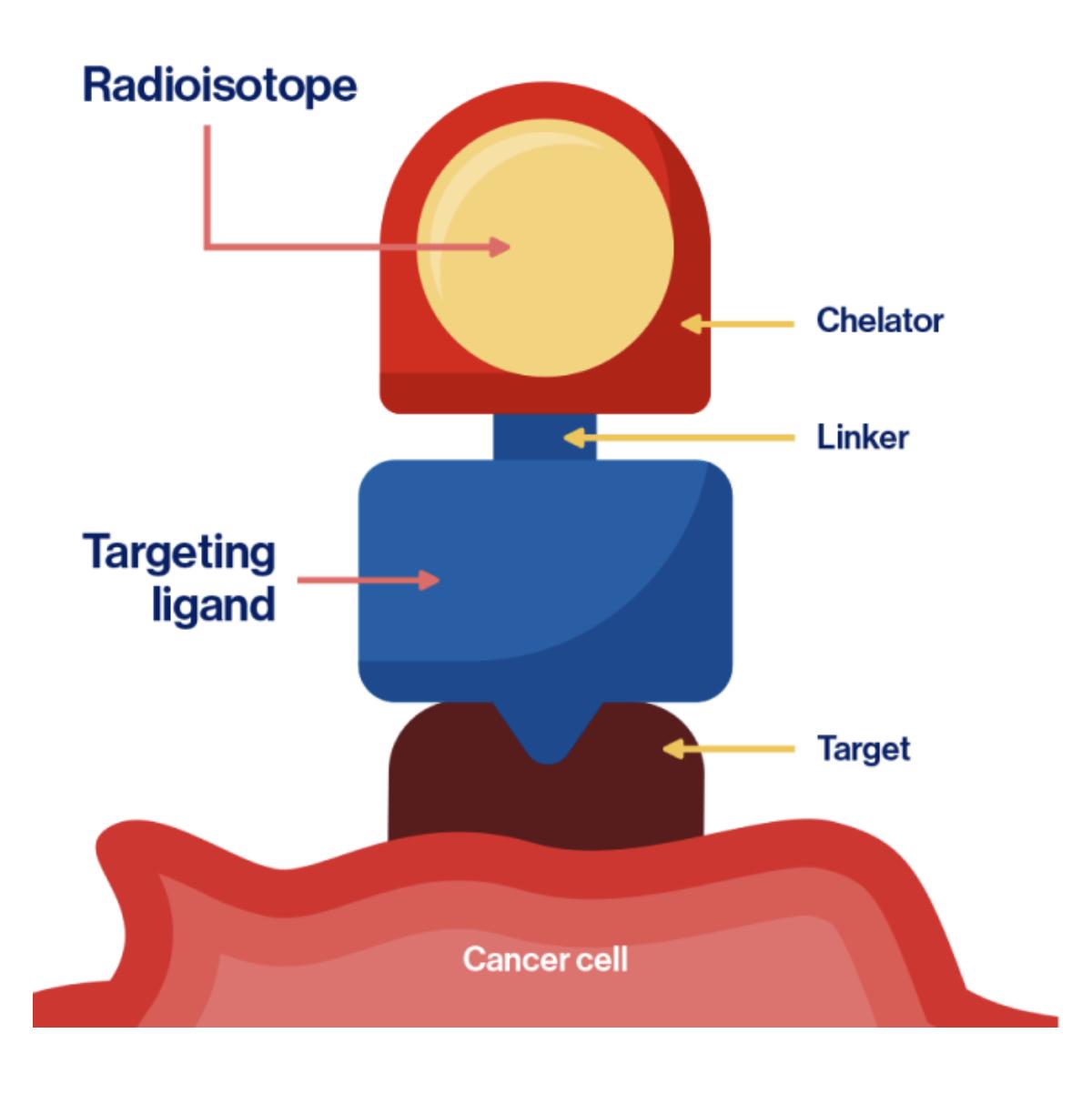
Figure 1: A radioligand binds to its target on a cancer cell.2
Prostate-specific membrane antigen (PSMA) is the receptor that is common on the cell membranes of prostate cancer cells, so the coordinating
3 "Foundations of theranostics," Novartis RLT Institute, accessed April 26, 2025, https://www rltinstitute novartis com/foundation s-of-theranostics/emerging-pillar/what-are-radiol igands/.
antibody ligand binds to it. The ligand is labeled with a radioisotope such as 177Lu. PSMA is a reliable target for the radioligand because it is expressed on the membranes of most metastases, regardless of location in the body since they are still prostate cancer cells.4
A 2017 study evaluated the efficacy of the 177Lu-PSMA radioligand to treat prostate cancer. It references 177Lu-PSMA-617 and 177Lu-PSMA I&T (imaging and therapy), two distinct radioligands, but summarized them as 177Lu-PSMA due to a lack of evidence for the benefit of one of them over the other.5 The study measured prostate-specific antigen, PSA, which becomes more abundant when there is a prostate issue such as cancer. The results were promising: in a follow-up with 99 of 145 patients (68%), at least 50% PSA level decline in 45% of patients and at least some PSA decline in 60% of patients.4 These positive outcomes gave credence to subsequent studies that continue to explore this innovative treatment.
More recently, the VISION trial has confirmed suspected benefits for patients. The trial was international, multi-centric, open-label (nonblinded),
4 Susan D. Sweat et al., "Prostate-specific Membrane Antigen Expression Is Greatest in Prostate Adenocarcinoma and Lymph Node Metastases," Urology 52, no. 4 (1998): 637, accessed April 26, 2025, https://doi org/10 1016/s0090-4295(98)00278-7
5 Wolfgang P. Fendler et al., "177Lu-PSMA Radioligand Therapy for Prostate Cancer," Journal of Nuclear Medicine 58, no 8 (2017): 1197, accessed April 29, 2025, https://doi.org/10.2967/jnumed.117.191023.
phase three (study that tests efficacy and safety as compared with a standard treatment), and randomized involving mCRP C patients who had progressed even after other inter ventions.6 Patients were randomly assigned to receive 177Lu-PSMA-617 with standard care or only standard care in a 2:1 ratio, meaning that 551 patients composed the experimental group while 280 were in the control group.5 The standard care for the VISION trial did not include chemotherapy.7 The overall sur vival (OS) of the patients receiving 177Lu-PSMA-617, 15.3 months, was significantly better than that of the other group, 11.3 months, and the image-based progression-free sur vival (IB-PFS), which is the duration when the cancer is controlled after treatment, was 8.7 months for the experimental group as compared to 3.4 months for the control group.5 Moreover, the experimental group demonstrated a meaningful improvement in PSA response rate. Approximately 46% of patients (versus 7.1% of the control group) had a greater than 50% reduction in PSA levels, and more than 33% of patients (versus 2% of the control group) had a greater than 80% reduction in PSA levels 5 The long
6 Gorrepati Rohith, "VISION Trial," Indian Journal of Urology 37, no 4 (2021): 372, accessed April 29, 2025, https://doi.org/10.4103/iju.iju 292 21.
7 Albert Jang, Ayse T Kendi, and Oliver Sartor, "Status of PSMA-targeted Radioligand Therapy in Prostate Cancer: Current Data and Future Trials," T herapeutic Advances in Medical Oncology 15 (Januar y 2023): 3, accessed April 29, 2025, https://doi.org/10.1177/17588359231157632.
follow-up (median of 20.9 months8) and the large sample size (831 participants) establish the validity of these results.5
Based on the success of this trial, the US Food and Drug Administration (FDA) approved Pluvicto, which is 177Lu-PSMA-617, in March 2022 for adults with PSMA-positive mCRP C that is metastatic.9 It is the first targeted RLT that the FDA has approved,8 so many questions and potential developments remain.
Patient care teams continued to monitor for additional adverse health effects of this novel therapy. The VISION trial found that 52.7% of the experimental group experienced adverse effects as compared to 38% of the control group.5 90% of mCRP C patients have bone metastasis, and almost half of those will have a critical skeletal event within two years.5 The length of the treatment (7.6 months versus 2.1 months) could have influenced this discrepancy 5
8 Oliver Sartor et al , "Lutetium-177–PSMA-617 for Metastatic Castration-Resistant Prostate Cancer," New England Journal of Medicine 385, no 12 (2021): 1091, https://doi org/10 1056/nejmoa2107322
9 Novartis Media Relations, "Novartis Pluvicto™ approved by FDA as first targeted radioligand therapy for treatment of progressive, PSMA positive metastatic castration-resistant prostate cancer, " Novartis, last modified March 23, 2022, accessed April 29, 2025, https://www.novartis.com/news/media-releases/n ovartis-pluvictotm-approved-fda-first-targeted-ra dioligand-therapy-treatment-progressive-psma-p ositive-metastatic-castration-resistant-prostate-ca ncer.
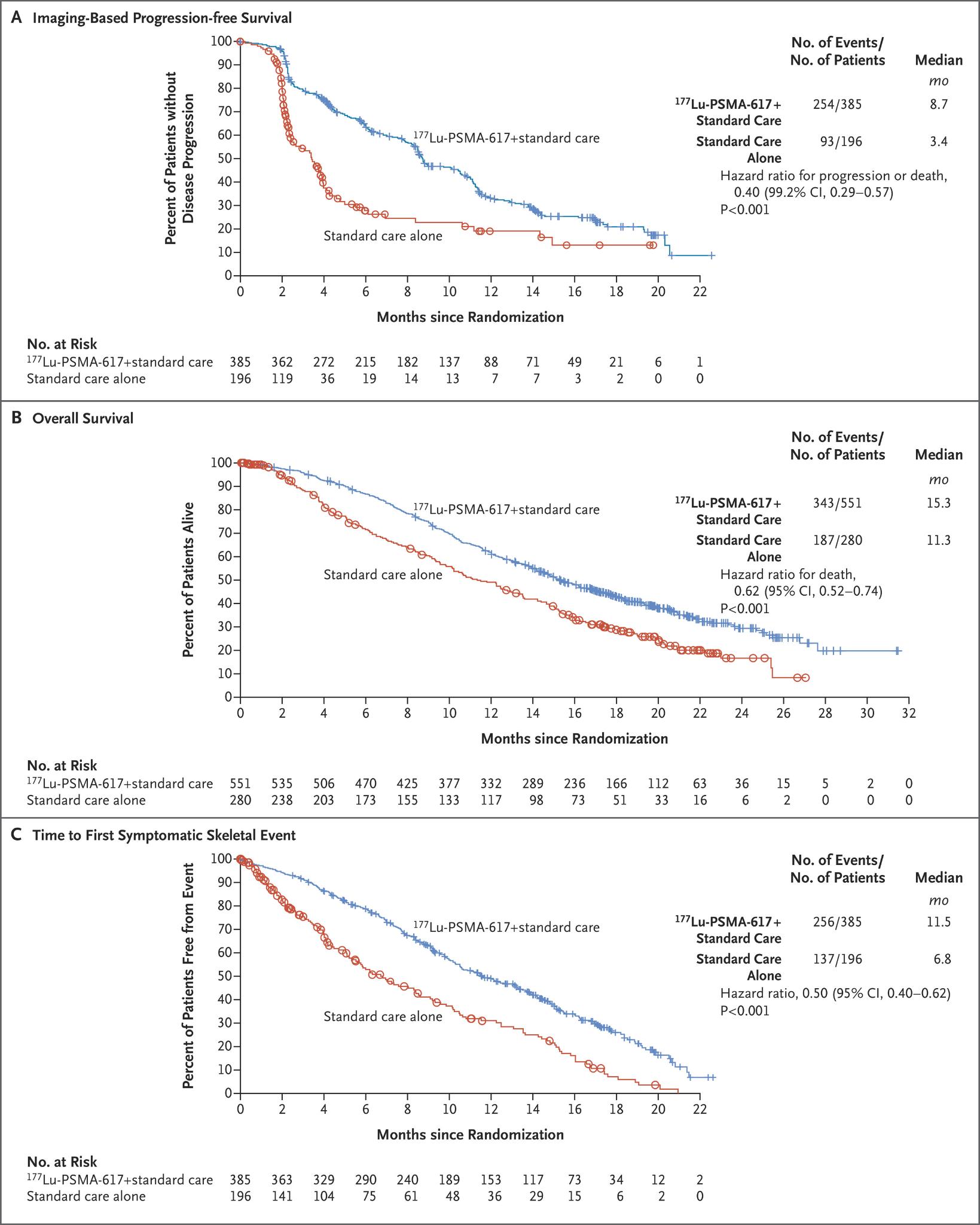
Figure 2: T he V ISION trial shows that 177Lu-PSMA-617 plus standard care (blue) is more effective than standard care alone (red) based on improved IB-PFS, OS, and time to first symptoms from a skeletal-related e vent 7
177Lu-PSMA-617 seemed to meaningfully delay the development of symptomatic bone lesions.5 Several trial participants also under went additional therapies to combat these effects, though overall combination therapy has not yet been fully assessed. A further analysis of the safety for this trial corroborates the relative insignificance of the risks compared to the benefits.10
Conclusion:
The recent information has solidified RLT as a compelling option for treating mCRP C. The FDA approval of Pluvicto is an important achievement, but more research is needed to determine the optimal dosing, timing, and treatment inter vals.11 Additionally, 2022 production supply shortages left questions about therapeutic access.10 Despite these unknowns, RLT already is improving lives and outcomes, and it provides exciting possibilities for future research and better treatment options for patients
10 Kim N Chi et al , "Safety Analyses of the Phase 3 VISION Trial of [177Lu]Lu-PSMA-617 in Patients with Metastatic Castration-resistant Prostate Cancer," European Urology 85, no 4 (2024): [Page #], accessed April 29, 2025, https://doi.org/10.1016/j.eururo.2023.12.004.
11 Karine Tawagi and Natalie Reizine, "PSMA-Targeted Therapy Is a Game Changer in Prostate Cancer Treatment, but Lingering Questions Remain," ed Fang Zhu, ASCO Daily News, last modified December 7, 2023, accessed April 29, 2025, https://dailynews ascopubs org/do/psma-targeted -therapy-game-changer-prostate-cancer-treatme nt-but-lingering-questions.
Samina Bhatia
Abstract: Cancer is one of the leading causes of death each year, with around 2 million new cancer cases occurring in 2021.1 Traditional methods of treatment, including surger y, chemotherapy, and radiation therapy, have their own side effects and challenges, such as failing to prevent disease recurrence or having a lack of specificity. Because of these limitations, scientists have been exploring new alternative methods including gene editing techniques such as CRISPR. The CRISPR technology, specifically Cas9, has been used to make cell models to understand diseases, and screen and treat cancer.
What is CRISPR
CRISPR, which stands for clustered regularly interspaced short palindromic repeats, is a naturally occurring repetitive DNA sequence in many bacteria that was first obser ved in Escherichia coli in 19872 It uses a Cas
1 Dimitrios Stefanoudakis et al., "The Potential Revolution of Cancer Treatment with CRISPR Technology," Cancers, March 17, 2023, accessed April 27, 2025, https://www.mdpi.com/2072-6694/15/6/1813.
2 Ishino, Yoshizumi, et al "Nucleotide sequence of the iap gene, responsible for alkaline phosphatase isozyme conversion in Escherichia
(CRISPR-associated protein) nuclease to cut a DNA sequence and either delete or insert extra DNA sequences3. The first successful use of CRISPR in vitro occurred in 2012 when it was used to cut DNA 4 . Since then, CRISPR has had multiple applications, including making cell models of diseases, modifying yeast cells, helping cure sickle cell anemia, and changing genes in certain tissues. Gene editing techniques are faster, cheaper, and more efficient ways to treat diseases because they mimic the natural DNA repair process3. Cas9 is the specific CRISPR-associated protein that is used for most of these processes. Cas9 contains guide RNA (gRNA), and the Cas9 protein with a bi-lobed structure consisting of the recognition lobe and the nuclease lobe. Cas9 recognizes the protospacer adjacent motif (PAM) sequence of the target DNA; the gRNA then pairs with the target sequence, activating sections of the Cas9 protein. The activated protein creates a blunt double-stranded break at a place near the PAM site, which triggers DNA repair mechanisms. Homology-directed repair (HDR) allows specific point mutations to be inserted, while nonhomologous end
coli, and identification of the gene product." Journal of bacteriology 169 12 (1987): 5429-5433
3 "Gene Editing – Digital Media Kit," National Institutes of Health, last modified November 5, 2020, accessed April 27, 2025, https://www.nih.gov/news-events/gene-editing-di gital-press-kit.
4 Mingxia Wang et al , "CRISPR applications in cancer diagnosis and treatment," Cellular & Molecular Biology Letters 28 (September 6, 2023) accessed April 27, 2025, https://link.springer.com/article/10.1186/s11658-02300483-4
joining (NHEJ) directly joins DNA fragments without needing external DNA. NHEJ is more commonly used; however it can cause accidental insertions or deletions that would change the function of the gene or make it non-functioning.3
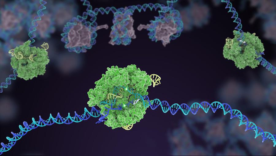
Figure 1: gRNA within the Cas9 enzyme (green) identifies a specific piece of DNA (blue), then cuts both strands in two places and removes the middle section 3
Scientists use CRISPR to screen for a variety of tumors. In a study researching hepatocellular carcinoma, a type of liver cancer, scientists in Hong Kong used Cas9 to identify phosphoglycerate dehydrogenase (PHGDH) as a form of resistance to a treatment, and then created an inhibitor for PHGDH. This inhibitor helped treat liver cancer5. In a study based in London, another group used Cas9 to discover the Haspin inhibitor CHR-6494 to reduce tumor
5 Lai Wei et al., "Genome-wide CRISPR/Cas9 librar y screening identified PHGDH as a critical driver for Sorafenib resistance in HCC," abstract, Nature Communications 10, accessed April 29, 2025, https://www.nature.com/articles/s41467-019-126067.
growth in breast cancer cells3. Different groups based in the US and China have studied using Cas12a to screen for breast cancer, because it is more sensitive and could cut single strands of DNA3. A separate German-based group used micelleplex to deliver Cas9 mRNA to lung cancer cells. They found no significant cytotoxicity and found that it generally led to tumor cell death.6 CRISPR has also been used to model cancers in order to learn more about them. Researchers have used Cas9 to create a knock-in mouse model by giving the mouse adenocarcinoma to alter certain genes. They then studied the mouse ’ s organs to learn more about cancer3. A different study generated a knockout of the tumor suppressor ARID1A in a human organoid. Another study created a librar y of Cas9 knockouts, and used this librar y to identify many genes that contribute to melanoma3.
A specific type of Cas9 cell, called dCas9 (endonuclease deficient Cas9), has the target recognition aspect but no nuclease activity. It has been used to regulate transcription. T-cells are a type of cell used in immunotherapy treatments of cancer, and are genetically modified cells that can kill cancer cells1. They can be programmed with Cas9 to target tumor cells, and new technology
6 Siyu Chen et al., "A novel micelleplex for tumour-targeted deliver y of CRISPR-Cas9 against KRAS-mutated lung cancer, " Royal Society of Chemistr y, Januar y 13, 2025, accessed April 27, 2025, https://pubs.rsc.org/en/content/articlehtml/2025/n r/d4nr03471f.
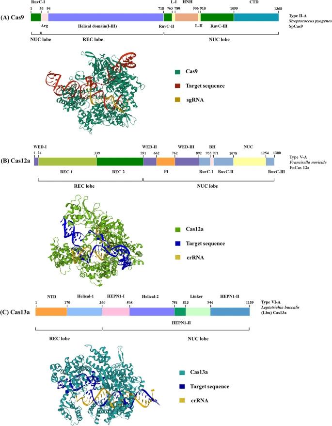
Figure 2: Cas9, 12a, and 13a have different structures and components that account for the differences in uses.3
with CRISPR makes the development of these cells cheaper and easier than before. A study placed Cas9-edited CART cells in patients with leukemia and lymphoma, and many improved or had a good long term response without side effects2. Another study applied the programmable aspects of Cas9 to telomerase, an enzyme that adds units onto cancer cells to prevent their death7. Editing this enzyme made cancer cells weaker and more likely to die in studies both in vitro and in vivo, demonstrating how it can be used for cancer treatment1. CRISPR is also useful post-transcription. A different form, called Cas13a, has a more diverse range of abilities for regulating genes. In a study done by American scientists, they found that Cas13a can be used to knockdown, or weaken, certain areas of genes in humans such as KRAS, CXCR4, and PPIB3.
Although CRISPR is generally considered a better method of cancer treatment, there are some downsides. Some studies have shown that it is not always as specific or efficient as intended. For example, one study had off-target editing in 16% of human embr yonic cells2. There are also sometimes unwanted byproducts such as accidental deletions or translocations that can be detrimental, so CRISPR currently has
7 R. Coleman Lindsley, "What are Telomeres and How Do They Play a Role in Cancer?," Dana-Farber Cancer Institute, last modified November 17, 2020, accessed April 29, 2025, https://blog dana-farber org/insight/2020/11/whatare-telomeres-and-how-do-they-play-a-role-in-ca ncer/.
limited uses in gene therapy2. A Chinese group did create a new base editing system that is more specific when editing and has fewer unwanted mutations, however it has not been as widely used3. In addition to problems with CRISPR uses, there are also some ethical concerns. The National Institutes of Health prohibits the use of genome editing on human embr yos because they could alter the future gene line without consent. The National Academies of Science, Engineering, and Medicine also recommended that gene editing trials using embr yos should be strictly regulated4. In November 2018, He Jiankui used Cas9 to create the first genomeedited babies. Jiankui was arrested, and his work raised the question of whether gene editing is necessar y, and at what age.3
Conclusion
Ultimately, multiple studies have shown that CRISPR gene editing technology is successful as a cancer treatment. CRISPR, particularly Cas9, can be used to screen for cancer, treat cancer, and create models to learn more about it. However, there are still obstacles to its widespread use, such as transcription errors and accidental deletions, as well as ethical concerns. In the future, new research will develop even better forms of CRISPR that are more accurate, so hopefully it will one day become a standard cancer treatment.
Dustin
Cheng
Abstract: PFAS are human-made chemicals used in products like cookware, clothing, and firefighting foam. They build up in human bodies through pathways like drinking water, food, and breast milk. PFAS exposure can harm the immune system, liver, thyroid, and metabolism This journal reviews how PFAS enters the body and why it is important to keep studying their health effects.
Introduction: Per- and polyfluoroalkyl substances (PFAS) are a large group of synthetic chemicals characterized by strong carbon-fluorine bonds, making them resistant to mixing with water or oil, or degrading with heat.
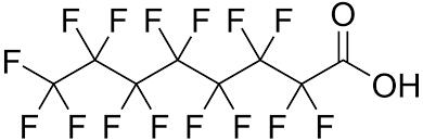
Figure 1: T he structure of perfluorooctanoic acid(PF OA), a legacy compound of PFAS.1
1 Environmental Exposomics Laborator y. "Polyfluoroalkyl Acid." Polyfluoroalkyl
These unique properties have led to their various use in both industrial and consumer products, including non-stick cookware, food packaging, and firefighting foams.2 Additionally, PFAS are commonly used as the fundamental materials in outdoor gear such as Gore-Tex, which offers durable waterproofing and breathability.3 Due to their chemical stability, PFAS do not easily degrade in the environment, leading to persistent existence and accumulation in ecosystems As a result, these substances end up in environmental pollutants, detected in air, water, soil, and within living organisms globally.4 Many studies focus on a few of the most commonly used historical PFAS compounds, such as perfluorooctanoic acid(PFOA) or perfluorooctanesulfonic acid (PFOS), collectively known as legacy PFAS compounds.
Substances (PFAS), Duke University, https://exposomics duke edu/ser vices/pfas
2 National Institute of Environmental Health Sciences. "Perfluoroalkyl and Polyfluoroalkyl Substances (PFAS) " National Institute of Environmental Health Sciences, U S Department of Health and Human Ser vices, https://www.niehs.nih.gov/health/topics/agents/p fc
3 W. L. Gore & Associates. "PFAS and Gore’s Structural Firefighting Products." GORE-TEX Professional, 2 Apr 2021, https://www.goretexprofessional.com
4 Hansen, S.F., Bunde, C.T.H., Roy, M. A. et al. Late lessons from early warnings on PFAS Nat Water 2, 1157–1165 (2024). https://doi.org/10.1038/s44221-023-00168-4
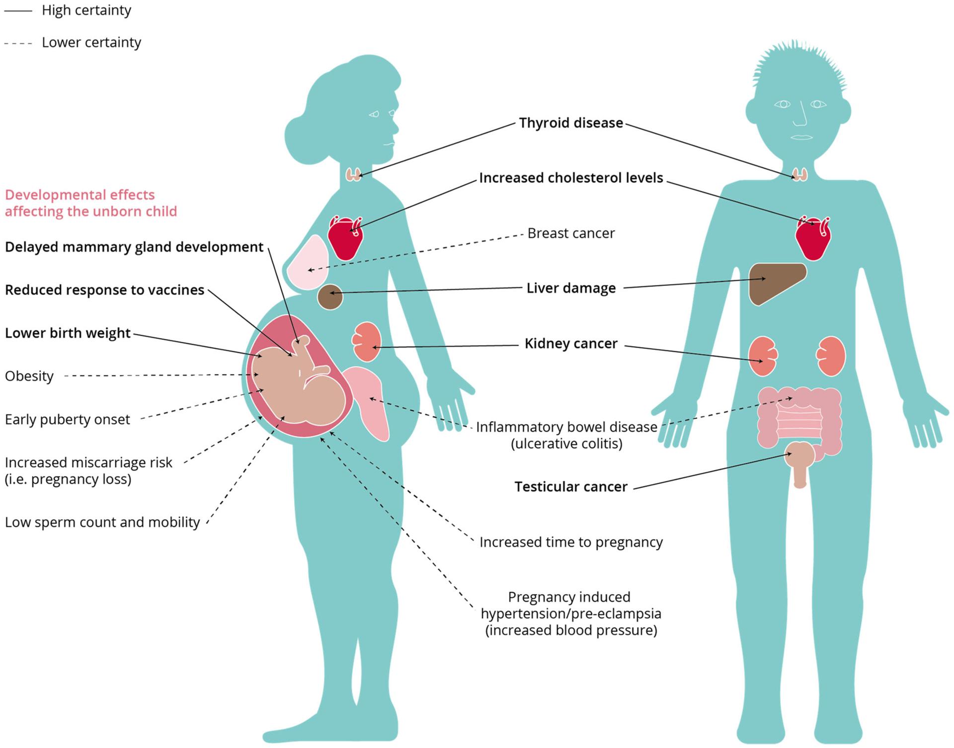
2: Effects of per- and polyfluoroalkyl substances on human health5
Current knowledge of PFAS health impact on humans focuses on the risk of damaging the immune system, thyroid function, liver health, and metabolic regulation. Epidemiological studies have shown that PFAS exposure, especially its legacy compounds such as perfluorooctanesulfonic acid (PFOS) and perfluorooctanoic acid (PFOA), can significantly suppress immune function.5 For instance, higher PFAS levels have been linked to reduced antibody responses to childhood vaccinations and
5 Fenton, Suzanne E et al “Per- and Polyfluoroalkyl Substance Toxicity and Human Health Review: Current State of Knowledge and Strategies for Informing Future Research ” Environmental toxicology and chemistr y vol. 40,3 (2021): 606-630. doi:10.1002/etc.4890
an increased risk of infections in infants and children.6 Thyroid dysfunction is another major concern. PFAS exposure is often associated with both hyperthyroid disease in women and hypothyroid disease in men7, affecting those with existing thyroid autoantibodies more severely. In terms of liver health, PFAS exposure has been connected with elevated liver enzymes,
6 Grandjean P, Andersen EW, Budtz-Jorgensen E, Nielsen F, Molbak K, Weihe P, Heilmann C 2012. Serum vaccine antibody concentrations in children exposed to perfluorinated compounds. JAMA 307:391–397
7 C8 Science Panel. 2012. Probable Link Evaluation of Thyroid disease.[cited 2025 Apr. 24] Available from: http://www.c8sciencepanel.org/pdfs/Probable Li nk C8 Thyroid 30Jul2012.pdf
fatty liver disease, and in some cases, liver cancer, which happens particularly among highly exposed populations.8 Furthermore, PFAS could also cause metabolic effects, including disruptions in lipid and insulin regulation, while some evidence also links PFAS to increased insulin resistance9. Overall, these findings underscore the significant and wide-ranging health risks of PFAS exposure, highlighting the need for continued monitoring and regulator y action.
One key pathway for early-life PFAS exposure is through breast milk, causing significant risks to newborns A recent study analyzing breast milk samples from U.S. mothers revealed the presence of both legacy PFAS, PFOS and PFOA, and current-use short-chain PFAS, including perfluoro-n-hexanoic acid (PFHxA) and perfluoro-nheptanoic acid (PFHpA).10 Researchers collected breast milk samples from 50 primiparous women living in or near Seattle, Washington, in 2019. The result showed that concentrations of total
8 Massoud O, Charlton M. 2018. Nonalcoholic fatty liver disease/nonalcoholic steatohepatitis and hepatocellular carcinoma Clin Liver Dis 22:201–211.
9 Nelson JW, Hatch EE, Webster TF. 2010. Exposure to polyfluoroalkyl chemicals and cholesterol, body weight, and insulin resistance in the general U.S. population. Environ Health Perspect 118:197–202
10 Guomao Zheng, Erika Schreder, Jennifer C. Dempsey, Nancy Uding, Valerie Chu, Gabriel Andres, Sheela Sathyanarayana, and Amina Salamova. 2021. Per- and Polyfluoroalkyl Substances (PFAS) in Breast Milk: Concerning
PFAS ranged from 52.0 to 1850 pg/mL, with PFOS and PFOA detected in all samples at median levels of 30.4 and 13.9 pg/mL, respectively The study also found that current-use PFAS are increasingly prevalent, with detection frequencies growing and a doubling time of about 4.1 years. These findings indicate that while regulator y actions have reduced the levels of phased-out compounds, newer PFAS continue to be a risk.
Beyond infancy, dietar y habits become another major factor influencing PFAS accumulation, particularly among teenagers. PFAS are chemicals used in many ever yday products like food packaging, fast food wrappers, and microwave popcorn bags because of their resistance to oil and water.11 One study focused on 193 adolescents in Cincinnati and found that diet plays a big role in PFAS exposure.12 Foods that are packaged, processed, or come from animals, like fish and dair y, are more likely to contain PFAS.11 Susmann et. al. used 24-hour food recalls and tested the teens’ blood to measure PFAS levels. They found that teens who ate more
11 Susmann, H P , Schaider, L A , Rodgers, K M , Rudel, R. A., 2019. Dietar y habits related to food packaging and population exposure to PFAS. Environ Health Perspect 127 (10), 107003 https://doi.org/10.1289/EHP4092
12 Harr y Sultan, Jessie P. Buckley, Heidi J. Kalkwarf, Kim M Cecil, Aimin Chen, Bruce P Lanphear, Kimberly Yolton, Joseph M. Braun, Dietar y per- and polyfluoroalkyl substance (PFAS) exposure in adolescents: The HOME study, Environmental Research, Volume 231, Part 1, 2023, https://doi.org/10.1016/j.envres.2023.115953.
packaged and processed foods had higher PFAS levels, while those who ate more fruits and fiber had lower levels. This suggests that PFAS can transfer from packaging materials into food, and then enter the body when the food is eaten. The study also noted that fast food, take-out, and certain types of meat were linked to higher exposure Common food habits, especially those involving packaged or convenience foods, are a major source of PFAS for teenagers, making diet an important area to focus on when looking for ways to reduce chemical exposure.
Drinking water is another major exposure pathway, especially for those living near airports, militar y bases, or industrial sites 13 PFAS are chemicals heavily used in firefighting foams known as aqueous film-forming foams (AFFF) to put out fuel fires. When these foams were sprayed, PFAS seeped into the ground and contaminated the water supply.14 Because of PFAS’s persistance, they can stay in groundwater and drinking water sources for years. Studies have shown that people living near places where AFFF was used often have
13 Hu XC, Andrews DQ, Lindstrom AB, Bruton TA, Schaider LA, Grandjean P et al. Detection of Poly- and Perfluoroalkyl Substances (PFASs) in U S Drinking Water Linked to Industrial Sites, Militar y Fire Training Areas, and Wastewater Treatment Plants. Environmental Science & Technology Letters 2016; 3: 344–350
14 Banzhaf S, Filipovic M, Lewis J, Sparrenbom C, Barthel R A review of contamination of surface-, ground, and drinking water in Sweden by perfluoroalkyl and polyfluoroalkyl substances (PFASs). Ambio 2017; 46: 335–346.
higher levels of PFAS in their blood.15 Even if someone has no direct contact with PFAS-containing products, they can still be exposed just by drinking water from a contaminated source. The contamination can spread over large areas, making it difficult to remove and leading to long-term exposure risks for entire communities
PFAS exposure remains a serious issue through sources like breast milk, diet, and water. Although efforts to phase out older PFAS have helped, newer PFAS are still a growing problem. Continued research and stronger safety rules are needed to protect people from health risks.
15 Health Canada Health Canada’s Drinking Water Screening Values for Perfluoroalkylated Substances (PFASs). Februar y 2016, Ottawa, ON.
Daniel Lobsenz
Abstract: Alpha-gal syndrome (AGS) is an allergic reaction to galactose-α-1,3-galactose (alpha-gal) that happens after consuming mammalian meat or other animal-based products. Evidence suggests that bites from certain tick species can lead to the development of this syndrome.1 From 2017 to 2021, there was a spike in positive test results for AGS in the United States.2 This article ser ves as a review of the current evidence surrounding AGS and its transmission; however, it does not address the treatment of the allergy.
Introduction: Galactose-α-1,3-galactose (alpha-gal) is an oligosaccharide that was previously known for its role as a barrier in xenotransplantation (see Figure 1). As a result of a loss of functional 1,3 galactosyltransferase, humans and other catarrhine primates
1 Young, Ian, et al "Tick Exposures and Alpha-gal Syndrome: A Systematic Review of the Evidence." Ticks and Tick-borne Diseases, vol. 12, no 3, May 2021, p 101674, https://doi.org/10.1016/j.ttbdis.2021.101674.
2 Thompson, Julie M., et al. "Geographic Distribution of Suspected Alpha-gal Syndrome Cases United States, Januar y 2017–December 2022. " MMW R. Morbidity and Mortality Weekly Report, vol 72, no 30, 28 July 2023, pp 815-20, https://doi.org/10.15585/mmwr.mm7230a2.
distinguish themselves from other mammals due to their inability to generate alpha-gal.3

Figure 1: Galactose-α-1,3-galactose molecule4
The earliest symptoms of AGS began to emerge around 20 years ago. After the FDA approved a monoclonal antibody treatment for metastatic colorectal cancer, the number of immunoglobulin E(IgE) mediated reactions appeared to have significant geographical variation, with higher rates in the southeast U.S. A 2008 study identified the specific antigen galactose-α-1,3-galactose and confirmed the geographical variation. In control subjects, 20.8% had the IgE antibodies in Tennessee but only 0.6% in Boston. At this time, the association between tick bites and alpha-gal was obser vser ved.5
In 2009, Australian allergists found 25 patients who had allergic reactions to red meat; all but one had a large local reaction to tick bites, and the outlier had
3 Wilson, Jeffrey M , et al "Galactose-α-1,3-Galactose: Atypical Food Allergen or Model IgE Hypersensitivity?" Current Allergy and Asthma Reports, vol 17, no 1, Jan. 2017, https://doi.org/10.1007/s11882-017-0672-7.
4 "Galactose-α-1,3-galactose." American Chemical Society, 1 July 2024, www.acs.org/molecule-of-the-week/archive/g/gal actose-alpha-1-3-galactose.html.
been bitten recently5. This report connected alpha-gal to the allergic reaction, but did not connect it to the fact that the allergen was present in tick saliva.
Allergists in 2009 identified 24 patients with IgE antibodies to alpha-gal with similar histories of allergic reactions after ingesting meat6 These antibodies reacted against beef, pork, lamb, cow ’ s milk, cat, and dog, but not turkey or chicken.
Three lines of evidence connect the allergy to red meat and the bites of the Lone Star tick (see Figure 2). These included that there was a strong correlation between the histor y of tick bites and levels of IgE to alpha-gal, these antibodies tended to exist in states where the Lone Star tick is common, and there was a significant correlation between the antibodies and Lone Star tick-derived substances.
Researchers theorized that tick bites were the only cause of alpha-gal in the United States It has been reported in the United States, Japan, the Middle East, Europe, South Africa, and Australia, and it occurs in both adults and children.7
5 Nunen, Sher yl A. Van, et al. "An association between tick bite reactions and red meat allergy in humans " Medical journal of Australia 190 9 (2009).
6 Commins, Scott P., et al. "Delayed anaphylaxis, angioedema, or urticaria after consumption of red meat in patients with IgE antibodies specific for galactose-α-1, 3-galactose." Journal of Allergy and Clinical Immunology 123 2 (2009): 426-433
7 Edlow, Jonathan A. "Alpha-Gal Syndrome: A Novel and Increasingly Common Cause of Anaphylaxis " Annals of Emergency Medicine, vol 83, no. 4, Apr. 2024, pp. 380-84, https://doi.org/10.1016/j.annemergmed.2023.08.491.
While initial reports indicated it was most common in the southeastern states, the range of the Lone Star tick has begun to spread, as seen in Figure 2 In recent years, the number of cases has increased.2
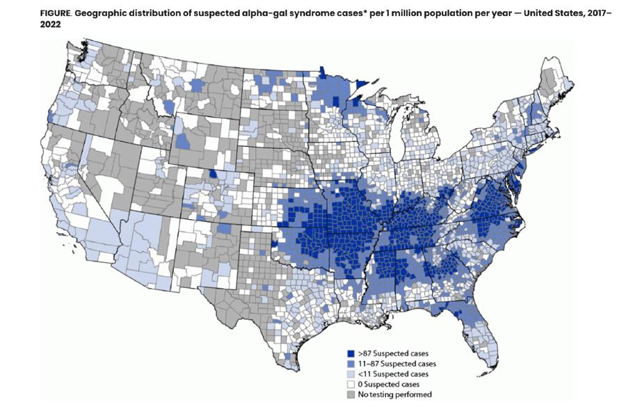
Figure 2: Geographic distribution of suspected alpha-gal syndrome cases per 1 million population per year - United States, 2017-20222
Discussion: Human immune response to alpha-gal may have evolved with the natural levels of alpha-gal found in tick saliva; however, this does not explain why IgE is being expressed at these levels. Exposure to alpha-gal through the gut is not sufficient to promote IgE to alpha-gal. This indicates that there is something unique either about transmission via the skin or, potentially, there is another feature of tick saliva that favors IgE promotion.
In studies with mice, guinea pigs, and cattle, it appears that after a primar y tick infestation, there is an immune response that diminishes the effects of following tick infestations, including transmission of tick-borne pathogens. Type 2 immune cells and mediators play a crucial role in protecting mammals against ticks.
Basophils, eosinophils, IgE, histamine, and mast cells all play a part in this
acquired tick resistance.8 One study reported that subcutaneous inoculation of lar val-stage lone star tick extracts in mice resulted in the promotion of IgE class switch that was dependent on MyD88 signalling in B cells. Exposure via the skin seems to be important to the transmission of alpha-gal, since when introduced intraperitoneally, the extracts did not result in IgE (see Figure 3).9
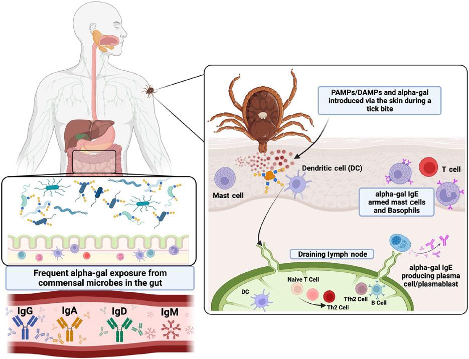
Figure 3: Diagram demonstrating the exposure to alpha-gal via tick and the formation of alpha-gal-specific IgE antibodies7
While the presence of alpha-gal in tick saliva is likely why ticks induce AGS, the reason why ticks have alpha-gal in their saliva is debated. While some argue that the presence comes as a result of residual
8 Wilson, Jeffrey M , et al "Tick Bites, IgE to Galactose‐alpha‐1,3‐galactose and Urticarial or Anaphylactic Reactions to Mammalian Meat: The Alpha‐gal Syndrome." Allergy, vol. 79, no 6, 9 Jan 2024, pp 1440-54, https://doi.org/10.1111/all.16003.
9 Chandrasekhar, Jessica L., et al. "Cutaneous Exposure to Clinically Relevant Lone Star Ticks Promotes IgE Production and Hypersensitivity through CD4+ TCell– and MyD88-Dependent Pathways in Mice " T he Journal of Immunology, vol. 203, no. 4, Aug. 2019, pp. 813-24, https://doi.org/10.4049/jimmunol.1801156.
amounts of alpha-gal in tick saliva from prior meals, one study seems to indicate that ticks have alpha-gal as a result of both a mammalian blood meal and endogenous production. One found that alpha-gal is present in both fed and unfed ticks. Alpha-gal epitopes were found in the midgut, hemolymph, and salivar y glands, and in the immunofluorescence. Through immunoelectron microscopy, these epitopes were found in endosomes of the digestive gut cells of the ticks.10
In terms of the allergic reaction, AGS is the first known reaction where a carbohydrate present in mammalian food is the cause of IgE-mediated reactions. The symptoms can range from gastrointestinal issues to anaphylactic shock. However, unlike typical allergic reactions, where people usually react within minutes, there tends to be hours in between when people consume foods containing alpha-gal and when they develop symptoms. The diagnosis is based on the measurement of IgE antibody levels to alpha-gal, with an IgE level to alpha-gal >5.5 kUA/L giving a 95% probability of AGS. Besides humans and old-world monkeys, the alpha-gal disaccharide is common in all mammals. However, the origin of the meat, the cut of the meat, and the type of processing all affect the alpha-gal levels, with innards like pork and beef
10 Fischer, Jörg, et al. "Spatial Distribution of Alpha-gal in Ixodes Ricinus – a Histological Study " Ticks and Tick-borne Diseases, vol 11, no 5, Sept. 2020, p. 101506, https://doi.org/10.1016/j.ttbdis.2020.101506.
kidney having the highest levels, as seen in Figure 4.11
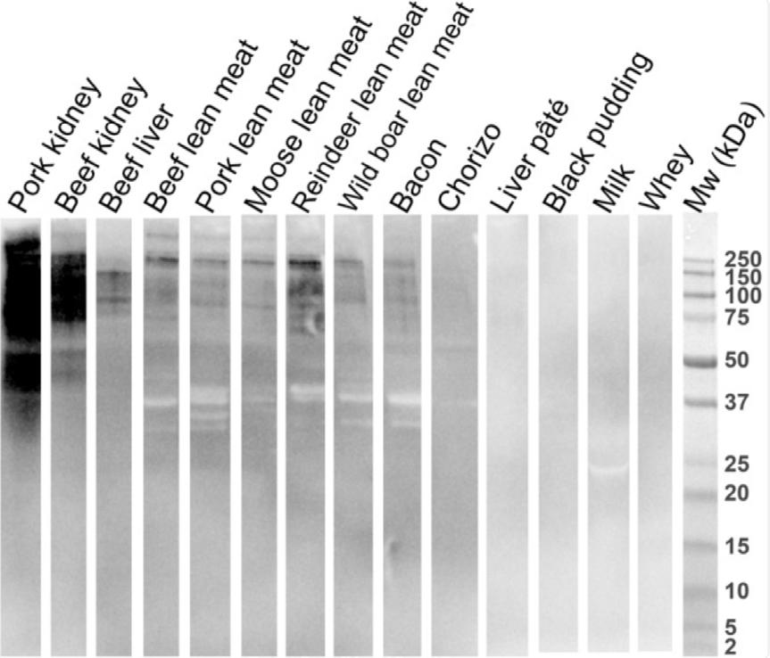
Figure 4: Immunoblot analysis of food extracts. Detection of alpha‐gal‐bearing proteins using the murine anti‐α‐Gal antibody (M86) 10
In the intestine, alpha-gal particles bind to lipids and are packaged into chylomicrons. These appear in the systemic circulation about two to three hours after consuming a fatty meal. The reason behind the delayed symptoms of alpha-gal is theorized, but not proven, to stem from the idea that it takes a larger amount of time to digest lipid particles. Over the next couple of hours, the lipids then transition into smaller lipoproteins that are small enough to pass through the endothelial walls and enter interstitial tissue, where mast cells that have the IgE antibodies reside, leading to the reaction This potentially explains the delayed reaction.12
11 Perusko, Marija, et al. "Allergenic Potency of Various Foods of Mammalian Origin in Patients with α‐Gal Syndrome." Allergy, vol. 80, no. 1, 15 July 2024, pp. 181-92, https://doi org/10 1111/all 16235
12 Wilson, Jeffrey M., et al. "Tick Bites, IgE to Galactose‐alpha‐1,3‐galactose and Urticarial or
Conclusion: With the emergence of this novel allergy, scientists have begun studying potential causes behind the transmission and development of this allergy. This has led to theories about the development of alpha-gal in ticks, the transition of alpha-gal from ticks to humans, the var ying reactions to differing animal products, and the explanation behind the delayed allergic response. While there has been sufficient research, many of the explanations still remain hypothetical and lack sufficient information to give a concrete explanation. More research regarding these topics is needed to fully understand the allergy.
Anaphylactic Reactions to Mammalian Meat: The Alpha‐gal Syndrome " Allergy, vol 79, no 6, 9 Jan. 2024, pp. 1440-54, https://doi.org/10.1111/all.16003.
Chrissy Graves
Abstract: The latest report from the World Health Organization (WHO) indicates that iron deficiency remains the most common single-nutrient deficiency worldwide and disproportionately affects women of reproductive age between the ages of 18 and 45 years.1 This paper evaluates some of the potential effects of iron deficiency as it relates to premenopausal women.
Introduction: Iron deficiency (ID) is the body's response to insufficient iron levels. Iron is mostly stored in hemoglobin (2500 mg), and additional amounts in myoglobin (130 mg), and the remaining in the liver.2 Iron is essential for the production of hemoglobin, which carries oxygen through the body. With a lack of iron, a human's body will struggle to make healthy red blood cells, leading to iron deficiency anemia (IDA).2
Women are at a much higher risk of ID due to menstruation, pregnancy, and a lack of iron in the diet.3 Although these
1 Ciulei, Mihaela A , et al "Iron Deficiency is Related to Depressive Symptoms in United States Nonpregnant Women of Reproductive Age: A Cross-Sectional Analysis of NHANES 2005–2010 " T he Journal of Nutrition, vol 153, no 12, 2023, pp 3521–3528
LANCET REVIEW pdf Accessed 28 Apr 2025
3 Mayo Clinis.“Iron Deficiency Anemia.” Mayo Clinic, 15 Jan. 2022, 2 Pasricha, Sant-Rayn, et al "Iron Deficiency " T he Lancet, vol. 397, no. 10270, 4 Dec. 2020, pp. 233–248. T he Blood P roject, www thebloodproject com/wp-content/uploads/2022/07/IDA
are the main causes of ID in women, it is important to know the various mental and physical effects that it can have on a premenopausal woman ID and IDA remain under-researched despite roughly 33% of premenopausal women experiencing the deficiency.4 Common symptoms often include lightheadedness, shortness of breath, fatigue, and weakness5, with some studies showing even larger effects. Here, we will discuss several additional symptoms that merit further consideration.
Sleep Disturbance: Although ID can cause fatigue, it can also negatively affect one ’ s sleep. Dopamine signals the body that time to wind down. If the lack of oxygen to the brain from low iron is causing a person ' s dopamine levels to get unbalanced, it can cause restless feelings, trouble falling asleep, or not being able to stay asleep during the night.6 In a study conducted by T he Journal of Nutrition, researchers took data from two groups of nonpregnant women aged 20-45 years from the National Health and Nutrition Examination Sur vey (NHANES). One group was from 2005-2008 (n=2497) and the other was
https://www mayoclinic org/diseases-conditions/iron-deficien cy-anemia/symptoms-causes/syc-20355034 Accessed 28 Apr 2025
4 Weyand, Angela C , et al "Prevalence of Iron Deficiency and Iron-Deficiency Anemia in US Females Aged 12–21 Years, 2003–2020 " JAMA, vol 329, no 24, 27 June 2023, p 2191 https://doi org/10 1001/jama 2023 8020
5 Benson, C. S., et al. "The Effect of Iron Deficiency and Anaemia on Women's Health " Anaesthesia, vol 76, no S4, 7 Mar 2021, pp 84–95 https://doi org/10 1111/anae 15405
6 Allen, Richard. "Dopamine and Iron in the Pathophysiology of Restless Legs Syndrome (RLS)." Sleep Medicine, vol. 5, no. 4, July 2004, pp. 385–91. https://doi.org/10.1016/j.sleep.2004.01.012.
from 2005-2010 and 2015-2018 (n=6731).7
NHANES asked questions that associated ID and IDA with sleep duration, latency, and quality The subjects were asked various questions about sleep quality and latency, and NHANES used differing hours for sleep duration. Results showed that 12.8% of females had ID and 6 0% had IDA 7 For those with ID or IDA, more than half reported poor sleep quality. They could not find a correlation between sleep latency and ID, but they did find that the adjusted odds ratio of short sleep duration was 1.22 times higher among those with ID compared with those who did not have ID (95% Confidence Inter val).7 The results from this study can show how ID can correlate with sleep disturbance, especially since iron also ties in with a person ' s dopamine levels.
Depression: Iron is essential for neurotransmitter synthesis. It is a cofactor for tyrosine hydroxylase, which converts tyrosine to dopamine, a neurotransmitter and hormone that is responsible for influencing motivation, pleasure, learning, and movement.8 (See figure 1).
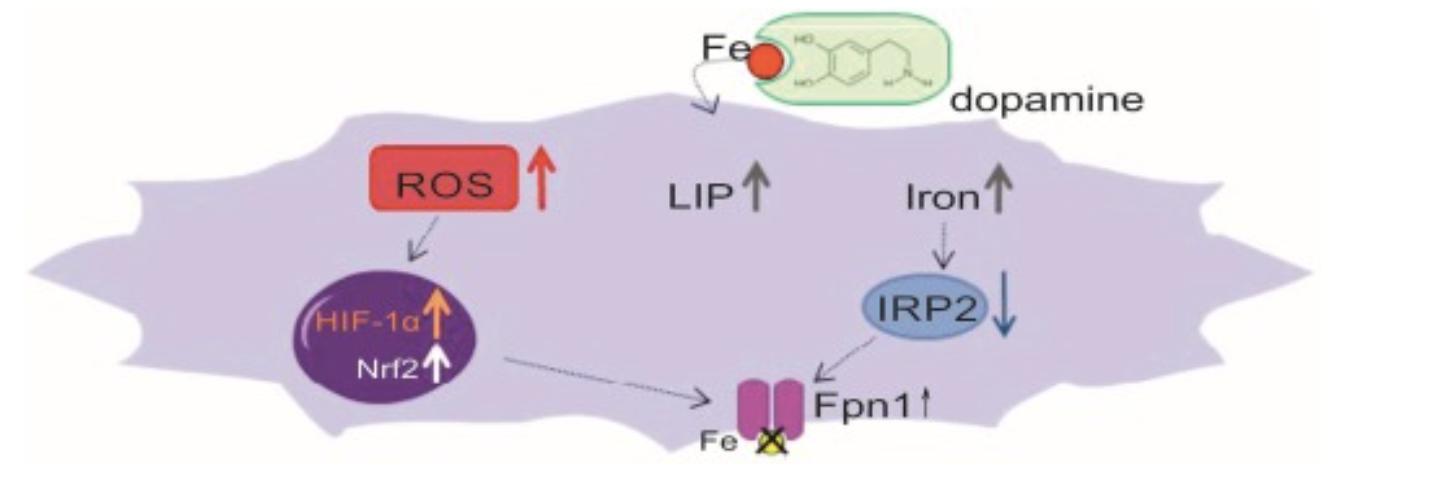
Figure 1: T he pathway where changes in iron le vels and dopamine metabolism can lead to stress (via ROS) 8
7 Al Hinai, Maymona, et al. "Iron Deficiency and Vitamin D Deficiency Are Associated with Sleep in Females of Reproductive Age: An Analysis of NHANES 2005–2018 Data." T he Journal of Nutrition, vol. 154, no. 2, Feb. 2024, pp. 648–57. https://doi.org/10.1016/j.tjnut.2023.11.030.
8 Dichtl, Stefanie, et al. "Dopamine Promotes Cellular Iron Accumulation and Oxidative Stress Responses in Macrophages." Biochemical P harmacology, vol. 148, Feb. 2018, pp. 193–201. https://doi.org/10.1016/j.bcp.2017.12.001.
If a female has low iron levels, this can cause dopamine levels to be lowered, possibly resulting in depressive symptoms. T he Journal of Nutrition conducted a study where they inter viewed and did physical examinations on 2216 nonpregnant women between the ages of 20 to 44 years old.1 They used a PHQ-9 scale via inter views to assess the depressive symptoms of these women. The study categorized depressive symptoms into different sections ranging from depressed mood to suicidal ideation and its possible severity. Their results showed that about 9.6% (n=296) of the women had depressive symptoms.1 The results found that women with ID were 1.6 to 1.8 times more likely to have depressive symptoms than those with healthy iron levels.1 They could not find a correlation between people with IDA and depression, as there were too few people in the study who had anemia. This study suggested that ID may lead to depressive symptoms because of the smaller production of dopamine in a person ' s body, resulting in a weaker motivation for pleasurable activities and movement
: Iron supplements are normally administered during pregnancy because ID and IDA in
pregnant women suggest that there could be risk factors for preterm deliver y, subsequent low birth weight, and inadequate prenatal health9 After looking at multiple studies, it can be concluded that there is a U-shaped link that ID can negatively affect a baby's birth weight.9 Abnormally high hemoglobin concentration can correlate with poor plasma volume expansions.10 In contrast, low hemoglobin levels can result in lower birth weight, as they can restrict fetal growth and development due to reduced oxygen transportation and availability. Specifically, in a study conducted in rural Nepal with 681 women, those who gave birth while having low hemoglobin levels had babies that were 38 to 187 grams less than the normal,11 demonstrating that the lower the mother's hemoglobin levels, the greater the risk for the baby being under weight at birth. Additionally, iron is essential for the growth and development of the fetus, including the develop of the brain and ner vous system, so if the mother’s blood is not carr ying enough oxygen because of ID, which is then transferred across the placenta to the fetus, then that can impair the fetus’s
9 Allen, Lindsay H "Anemia and Iron Deficiency: Effects on Pregnancy Outcome " T he American Journal of Clinical Nutrition, vol 71, no 5, May 2000, pp 1280S–1284S https://doi org/10 1093/ajcn/71 5 1280s Accessed 28 Apr 2025
10 Rahman, Syed Moshfiqur, et al "Association between Maternal Plasma Ferritin Level and Infants' Size at Birth: A Prospective Cohort Study in Rural Bangladesh " Global Health Action, vol 14, no 1, 1 Jan 2021 https://doi org/10 1080/16549716 2020 1870421
11 Dreyfuss, Michele Lynn Anemia and Iron Deficiency during P regnancy: Etiologies and Effects on Birth Outcomes in Nepal The Johns Hopkins University, 1998 P roQuest Dissertations and T heses. ProQuest, www.proquest.com/openview/a0fcf7a63441ac46d66d642aeeba df81/1?cbl=18750&diss=y&pq-origsite=gscholar. Accessed 28 Apr. 2025.
development and lead to lower birth weights9 (See figure 2).
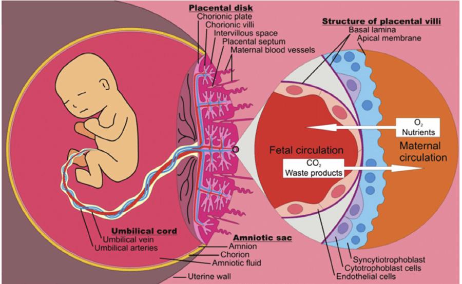
Figure 2: T he relationship between the mother and her fetus as it relates to the transmission of oxygen from the mother through the placenta.12 Furthermore, scientists have also found that low ferritin (iron storage) levels in the mother can lead to earlier births.11 Because of the reduced oxygen-carr ying capacity in the blood, the fetus and mother could experience hypoxia, which is a condition where the body does not receive enough oxygen to function properly. This can trigger a stress response in the fetus and mother and potentially lead to preterm births.9. Conclusion: While ID has many potential effects, treatments like oral supplements and increasing iron-rich food intake (eg, red meat, poultr y, dark leafy greens) have shown some benefits.3 Increasing iron intake may relieve some symptoms, but further research must be conducted to fully understand and address the condition, as well as understand how it affects different groups of women.
12 Elad, David, et al "Have We Neglected the Role of Fetal Endothelium in Transplacental Transport? Role of Fetal Endothelium Placental Transport " Traffic, vol 15, 2013, https://doi org/10 1111/tra 12130
Kosette Koons-Perdikis
Abstract: In 2023, the United States Food and Drug Administration reported that 60% of all packaged food and beverages contained artificial sweeteners.1 Due to their low caloric content and high sweetening intensity, artificial sweeteners have become a common substitute for sugar in food and beverage products. However, recent research has propagated growing concern around the adverse health effects of artificial sugars, specifically on the gut microbiome This paper investigates the impacts of aspartame, sucralose, and stevia extracts, three of the most popular artificial sweeteners, on the function of intestinal microbiota.
Introduction
Amidst the health craze that characterizes the modern era, one commodity in particular has fostered significant attention: artificial sweeteners. Also referred to as nonnutritive sweeteners, or NNSs, artificial sweeteners have a higher
1 "Artificial Sweetener Market Size, Share and Trends Analysis Report by Type, by Form by Application and by Region Forecasts, 2024-2032, " Straits Research, accessed April 29, 2025, https://straitsresearch com/report/artificial-sweete ner-market
sweetening intensity but lower caloric content per gram in comparison with nutritive sweeteners, such as sucrose.2 While natural sugars are part of a healthy diet, consuming excess sugars contributes to obesity, diabetes, and heart disease.3 This makes NNSs especially marketable to individuals who seek to balance consuming low-calorie food, which they deem healthier, with fulfilling their cravings. The Food and Drug Administration has set Acceptable Daily Intake (ADI) recommendations for various artificial sweeteners, the most common of which are aspartame, sucralose, and, though a naturally occurring source, stevia-derived substances.3 Nonetheless, the consumption of artificial sweeteners has generated much concern surrounding their negative health effects. Research conveys that the consumption of NNSs contributes to metabolic derangements, such as glucose intolerance, and alterations to the gut microbiome.2
The gut microbiome, located in the intestine, houses trillions of microorganisms such as bacteria,
2 Alexandra R Lobach, Ashley Roberts, and Ian R Rowland, "Assessing the in Vivo Data on Low/no-calorie Sweeteners and the Gut Microbiota," Food and Chemical Toxicology 124 (Februar y 2019): https://www.sciencedirect.com/science/article/pii/ S0278691518308780?via%3Dihub#bib65.
3 Mayo Clinic Staff, "Artificial Sweeteners and Other Sugar Substitutes," Mayo Clinic, last modified Januar y 2023, accessed April 23, 2025, https://www mayoclinic org/healthy-lifestyle/nutr ition-and-healthy-eating/in-depth/artificial-sweet eners/art-20046936.
viruses, fungi, and parasites.4 The function of the gut microbiome is significant to human health as it plays a pivotal role in the immune, digestive, and ner vous systems, and in metabolic processes.2 It depends on the mutualistic symbiotic relationship that an individual shares with the microbes in the gut: the intestine provides housing and nutrients to beneficial microorganisms that regulate bodily processes. The functionality of the gut microbiome is entirely dependent upon an individual’s diet and different environmental stimuli. An unbalanced diet may lead to “dysbiosis”, or a loss of beneficial bacteria paired with an overgrowth of negative bacteria in the gut.4 Because these bacteria play an essential role in human health, a loss of bacterial diversity is detrimental. Therefore, links between the consumption of artificial sweeteners and a disruption to intestinal microbiota pose a severe threat and are essential to explore.
Aspartame (C₁₄H₁₈N₂O₅) is a dipeptide of aspartic acid and phenylalanine joined by methyl ester bonds.

4 "Gut Microbiome," Cleveland Clinic, last modified August 18, 2023, accessed April 23, 2025, https://my clevelandclinic org/health/body/25201-g ut-microbiome
It is about 200 times sweeter than sucrose with an ADI of 40 mg/kg of body weight.5 The body rapidly metabolizes aspartame through presystemic hydrolysis, making it difficult to determine its concentration levels in the blood.6 Even bolus doses of aspartame that exceed 200 mg/kg are not able to be detected because of how rapidly it is broken down by the human body.6
Nonetheless, effective studies have been conducted with rats to illustrate the health implications of aspartame. An 8-week experiment on the effects of chronic low-dose aspartame on Male Sprague-Dawley rats linked aspartame ingestion to less weight gain and a more favorable body composition. In the intestinal microbiome, aspartame ingestion increased short-chain fatty acid production and the total bacterial abundance of Enterobacteriaceae and Clostridium leptum.7
5 Francisco Javier Ruiz-Ojeda et al., "Effects of Sweeteners on the Gut Microbiota: A Review of Experimental Studies and Clinical Trials," Advances in Nutrition 10 (Januar y 2019): https://doi org/10 1093/advances/nmy037
6 B A Magnuson et al , "Aspartame: A Safety Evaluation Based on Current Use Levels, Regulations, and Toxicological and Epidemiological Studies," Critical Re views in Toxicology 37, no. 8 (2007): accessed April 23, 2025, https://doi.org/10.1080/10408440701516184.
7 Marie S A Palmnäs et al , "Low-Dose Aspartame Consumption Differentially Affects Gut Microbiota-Host Metabolic Interactions in the Diet-Induced Obese Rat," PLoS ONE 9, no 10 (2014): https://doi.org/10.1371/journal.pone.0109841.
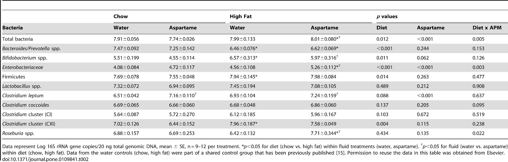
Sprague-Dawley Rats 7
Additionally, rats that were treated with aspartame for 11 weeks developed glucose intolerance, or a state of decreased efficiency of glucose metabolism that leads to elevated blood sugar levels.5 In a human trial, participants who were treated with 400 mg of aspartame experienced declines in plasma glucose and peak insulin concentrations while the gut microbiota was not affected.8 Because aspartame is so rapidly broken down by the human body, it is rare that any amount is able to reach the large bowel, minimizing its intestinal effects.5 Thus, although aspartame may have minimal effects on the human microbiome, evidence shows that aspartame threatens glucose tolerance and gut microbiota in rats
Sucralose
Sucralose (C₁₂H₂₂O₁₁) is synthesized by the substitution of the three hydroxyl groups in sucrose Sucralose is 320-1000 times sweeter than sucrose, with an ADI of 5 mg/kg of body weight.5
It has proven to have a major bacteriostatic effect in which it inhibits the growth of bacteria Sucralose has proven to cause a dysbiosis of the intestinal microbiome in rats and metabolic dysfunction in mice.9 In a 12-week study done with Sprague-Dawley rats, sucralose consumption decreased the amount of anaerobic and aerobic bacteria, bifidobacteria, lactobacilli, Bacteroides, and Clostridium in the gut.10 Furthermore, sucralose, especially when paired with a high-fat diet, displayed a synergistic effect with Firmicutes, a
9 Wang, Qiao-Ping, Qiao-Ping Wang, Duncan T Browman, Herbert Herzog and G. Gregor y Neely. "Non-nutritive sweeteners possess a bacteriostatic effect and alter gut microbiota in mice." PLoS ONE 13 (July 2018): https://doi.org/10.1371/journal.pone.0199080
8 David L. Hor witz, Michael McLane, and Peter Kobe, "Response to Single Dose of Aspartame or Saccharin by NIDDM Patients," Diabetes Care 11, no. 3 (1988): https://doi.org/10.2337/diacare.11.3.230.
10 Mohamed B Abou-Donia et al , "Splenda Alters Gut Microflora and Increases Intestinal P-Glycoprotein and Cytochrome P-450 in Male Rats," Journal of Toxicology and Environmental Health, Part a 71, no. 21 (2008): accessed April 23, 2025, https://doi.org/10.1080/15287390802328630.
negative bacterium.9 Recent studies correlating high Firmicute levels with obesity in humans bring into question the effectiveness of consuming sucralose as part of a “weight loss diet”.
Additionally, a bacteriological analysis of sucralose in rats displayed disruption to the regulation of amino acids, resulting in inflammation in the host 5 Thus, it is evident that sucralose has many adverse impacts on the functions and populations of gut microbiota in rats and mice.
Stevia rebaudiana standardized extracts (SSEs) are derived from the stevia rebaudiana shrub, which leaves contain compounds such as stevioside, steviolbioside, and rebaudioside A, B, C, D, E, F, and M SSEs are about 250 times sweeter than sucrose with an ADI of 4 mg/kg of body weight.
In humans, bacterial strains from fecal materials were treated with stevioside and rebaudioside A While both sweeteners were completely hydrolyzed into steviol within 10 to 24 hours, the human intestinal microflora was not able to degrade steviol.11 In comparison with that treated with glucose, the mixed fecal bacteria treated with stevioside and rebaudioside slightly altered the gut microbiome. Stevioside inhibited the function of anaerobic bacteria, and rebaudioside A weakly inhibited aerobic
11 Claudio Gardana et al., "Metabolism of Stevioside and Rebaudioside a from Stevia Rebaudiana Extracts by Human Microflora, " Journal of Agricultural and Food Chemistr y 51, no. 22 (2003): https://doi.org/10.1021/jf0303619.
bacteria.5 Although SSEs were not found to impact glucose intolerance, select compounds did have bacteriostatic effects on gut microbiota
Although NNSs are perceived to be healthier alternatives to refined sugars, they pose their own unique challenges Studies conducted on the most common NNSs -aspartame, sucralose, and SSEsare indicative of the adverse effects that artificial sweeteners have on metabolic function and dysbiosis. NNS consumption functionally and compositionally alters intestinal microbiota, therefore contributing to glucose intolerance.12 Although the rapid hydrolysis of certain artificial sweeteners, such as aspartame, makes it difficult to conduct research on humans in vitro, studies conducted on rats and mice illustrate the potential effects of NNSs. Overall, the mass consumption of NNSs calls for a major reassessment of how “ healthy” they are, and much more research needs to be done on the direct impact that artificial sweeteners have on the human intestinal microbiome.
12 Jotham Suez et al., "Artificial Sweeteners Induce Glucose Intolerance by Altering the Gut Microbiota," Nature 514, no. 7521 (2014): https://doi.org/10.1038/nature13793.
Thomas Ludecke
Abstract: As research investigations and grant funding compound in the field of bioengineering, the pursuit of bio-printed vascular tissues has emerged as the most sought after goal. Perhaps the most consequential effect of bio-printed vascular tissues would be on the field of bio-printing itself, as vascularity is foundational to the formation of more complex organoids.1 Not only this, but vascular tissue engineering would also allow for more accurate in-vivo models to address vascular diseases along with vascular grafts needed in surger y and other intensive procedures. This review will delve into the current methods of bioengineering vascular tissue along with the applications and avenues for future research into the technology.
Introduction:
A majority of living cells within the human body live within 100-200 μm of capillaries that allow for the essential process of gas exchange wherein CO2 is
1 Jiang, H., Li, X., Chen, T., Liu, Y., Wang, Q., Wang, Z., & Jia, J. (2023). Bioprinted vascular tissue: Assessing functions from cellular, tissue to organ levels. Materials Today Bio, 23, 100846. https://doi.org/10.1016/ j.mtbio.2023.100846
expelled and O2 is taken in. 2 The prevalence of these networks of capillaries in vivo emphasizes the acute necessity of capillaries in human health and how transformative bioengineered vascular tissue could be in medicine. In order to meet the demands of bioengineered vascular tissueresearchers have to overcome significant hurdles including the ability to produce complex and defined architectures of blood vessels, rapid manufacturing and printing of these tissues, and control of mechanical properties for application in clinical settings.3 If all of these requirements are met among others, complex processes such as arterial bypasses could be simplified and a slew of vascular-based diseases could be better studied in vitro.
Arteries and veins, which act as the larger routes of the human vascular system, are made up of three layers: the tunica intima, tunica media, and tunica adventitia. Capillaries (smaller routes) are made up of just tunica intima.1 Tunica intima are formed by endothelial cells (ECs) which line the capillaries and are responsible for a host of functions such as preventing clotting and inducing angiogenesis (new blood vessel formation).3 Although larger diameter
2 Jafarkhani M, Salehi Z, Aidun A, Shokrgozar MA Bioprinting in Vascularization Strategies Iran Biomed J. 2019 Jan;23(1):9-20. doi: 10.29252/.23.1.9. PMID: 30458600; PMCID: PMC6305822
3 Li, W., Li, J., Pan, C., Lee, J.-S., Kim, B. S., & Gao, G. (2024). Light-based 3D bioprinting
blood vessels are made up of more layers and thus have greater cellular diversity, their increased size makes their replication in a lab setting more straightfor ward.3 The challenge - from the bioengineering perspective - is the smaller capillaries. To meet the challenge of engineered smaller-capillaries a host of methods have been put forth - the most prominent of which are the top-down and bottom-up methods of vascular tissue engineering (VTE).1
The bottom-up approach to VTE is characterized by the stimulation of blood vessel formation through cellular and extracellular cues.1 Through this process, scientists use microfluidic devices to exploit cells into angiogenesis resulting in EC sprouting, tubular formation, and secretion of matrix proteins.4 Select studies have shown the role of proangiogenic factors (signals that allow for blood-vessel formation and growth) in the proliferation of microvasculature and EC sprouting allowing for researchers to replicate capillar y structures in a lab setting. The self assembly of these dense capillar y
networks - via microfluidic devices5 and fibrin gels containing endothelial cellshas advanced progress considerably with the ultimate goal of the networks of engineered vasculature being implanted in vivo.4 Although this strategy of self-assembly of microvessels has shown promise, its efficacy is sizably curbed by its use in large-scale tissues as several cells die through hypoxia.1
The top-down VTE approach is considered to be traditional1 and employs a strategy whereby cells are placed onto bioprinted scaffolds. This method has its advantages in the fact that the same vascular tissue is easily replicatedunlike the bottom up approach (as induced angiogenesis cannot be controlled).1 Scientists employing this strategy typically bioprint hollow, circular 3-d scaffolds made up of biocompatible synthetic polymers and line these with a variety of human endothelial cells such as human umbilical vein endothelial cells (HUVECs) to construct the three layers of vasculature described earlier (intima, media, adventitia) - (method layed out in Figure 1)6 A study from Xu et al
techniques for illuminating the advances of vascular tissue engineering. Materials Today Bio, 29,101286.
https://doi org/10 1016/j mtbio 2024 101286
4 Fleischer, S., Tavakol, D. N., & Vunjak‐Novakovic, G. (2020). From arteries to capillaries: Approaches to engineering human vasculature. Advanced Functional Materials, 30(37).https://doi.org/10.1002/adfm.201910811
5 Onoe, H., Okitsu, T., Itou, A. et al. Metre-long cell-laden microfibres exhibit tissue morphologies and functions. Nature Mater 12, 584–590 (2013). https://doi.org/10.1038/nmat3606
6 Xu, Y , Hu, Y , Liu, C , Yao, H , Liu, B , & Mi, S (2018) A novel strategy for creating tissue-engineered biomimetic blood vessels using 3D bioprinting technology Materials, 11(9), 1581 https://doi org/10 3390/ma11091581
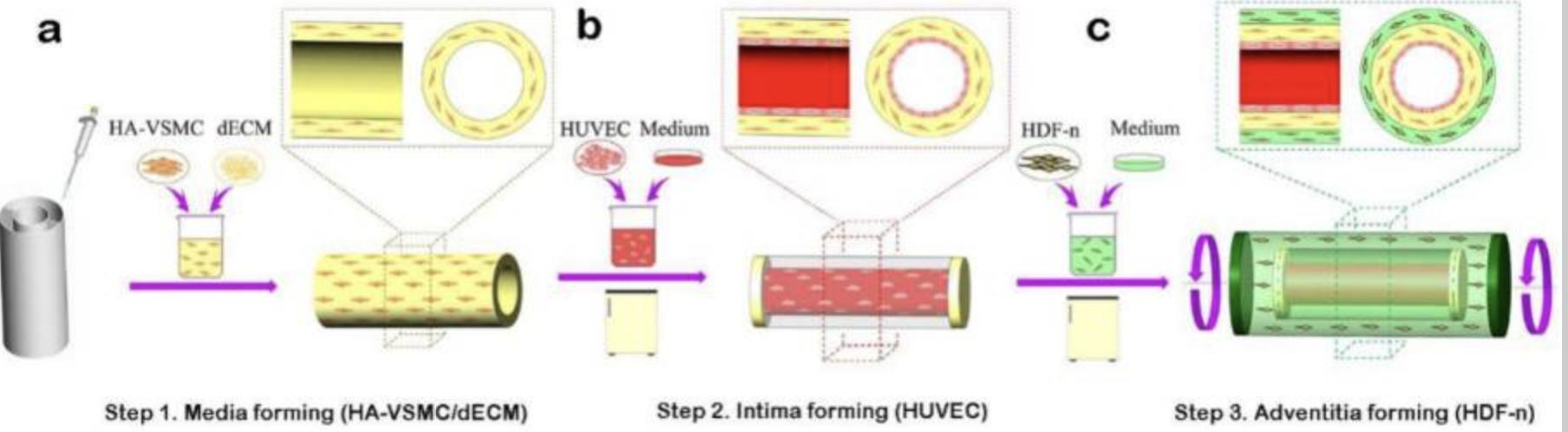
Figure 1: Displays the schematic process behind Xu et. al. top-down approach characterized by implementation of all three layers of the blood vessel and synthetic polymer scaffolding to hold it in place.6
showed the efficacy of this method asafter 48h of culture in vivo - the cells making up the vasculature were still viable and the structure of the tissue was intact.5 Similar to the bottom-up approach, top-down has its disadvantages as the method can be characterized by an uneven spread of cells on the scaffolding and misalignment.1 Although both of the prominent methods of bioengineering vascular tissue have their flaws, it is worth mentioning that a combination of the two methodologies is likely the key to an effective practice for VTE.1
A final, more innovative approach to VTE has been the use of extrusion 3-D printing technology to formulate vasculature. With this method, researchers can deposit bioink - blends of biomaterials and living cells - into a nozzle similar to that of plastic 3-d printer and use software to construct 3-d vasculature 1 While this method is just as promising as it is innovative, more research is needed into the correct formulation of bioink - to best replicate in-vivo vasculature.1
Cardiovascular disease is the leading cause of death in the developed world often leading to bypass procedures wherein significant vascular tissue is required for grafting.4 Not only would engineered vascular tissues meet the demands of grafting, but researchers could better test the efficacy of certain therapeutics meant to treat vascular diseases through using engineered tissue as in-vitro models.3 This could bring cures to devastating ailments such as atherosclerosis, intimal hyperplasia, and arterial stenosis.1 For this to be achieved, however, additional research needs to be done into hypoxia in relation to bottom-up methods, cell organization within top-down methods, a synthesis of both top-down and bottom-up methods, and more effective bioink. Progress in the formulation of bioengineered vascular tissue has made leaps and bounds and the conversations surrounding the topic will only be amplified as more discoveries are made.
