International Journal of Healthcare Sciences ISSN 2348-5728 (Online)
Vol. 10, Issue 1, pp: (315-319), Month: April 2022 - September 2022, Available at: www.researchpublish.com

International Journal of Healthcare Sciences ISSN 2348-5728 (Online)
Vol. 10, Issue 1, pp: (315-319), Month: April 2022 - September 2022, Available at: www.researchpublish.com
Department of Family Medicne, Chi-Mei Medical Center, Tainan, Taiwan
DOI: https://doi.org/10.5281/zenodo.7083070
Published Date: 15-September-2022
Abstract: Nasogastric tubes are frequently used in hospital and home care, however most medical and nursing students or postgraduates are not aware of the possible dangers that may be involved. It is important to have full medical education before starting this procedure, so as to prevent complications resulted from nasogastric tube insertion. The use of nasogastric tubes may bring with many kinds of complications. The goal of this article is to reevaluate the application, care and possible dangers of nasogastric tube insertion. If an incomplete NG is noted upon removal of NG, endoscopic removal should be considered since the retainment of a broken NG is not likely to be passed out easily and may induce ulcer, bleeding and other complication. We stress on insertion with great care and confirming correct placement, strategies to improve safety and timely management to avoid from further complications by giving detailed medical education in advance before commencing this procedure.
Keywords: nasogastric (NG) tubes, complications, endoscopy removal.
Since its first introduction, the nasogastric (NG) tube has become a method used to relieve gastrointestinal symptoms. NG tubes are used in the management of patients who need decompression of the gastrointestinal tract, diagnosis and assessment, nutrition support and medication administration. The use of NG tubes may bring with complications. The tube size selection, assessment of tube position and the method of securing NG tubes are important parts of care of NG tube to reduce the risks of NG tube-related complications and provide for patient safety. The aim of this article is to highlight the uses, care and hazards of NG tube.
The use of nasogastric (NG) tubes is a procedure often performed by medical and nursing students or young postgraduates. There are many reasons why people may need a nasogastric tube. Certain illness or cancers can prevent patients from eating or drinling normally, such as head injury, cerebrovascular accidents and neurological disorders like dementia or pakinsonism. Patients who can not eat well with choking and malnutrition are also candidates for nasogastric tube insertion
Care givers of patients who have dementia are often asked to consider the use of nasogastric tube due to more chances of choking or aspiration when swallowing which may lead to pneumonia, a serious condition that may lead to respiratory failure. The placement of nasogastric (NG) tubes is a very commonly used procedures. They are inserted both by nurses and doctors, although the insertion may be uncomfortable for the patient, it is usually thought of as a relatively benign procedure. Besides reviewing the literature related with complications from the insertion of nasogastric tubes. We also report a patient who developed a rare incident caused after nasogastric tube insertion by a home care nurse.
Erosin of nasal cavity: Erosion of nasal cavity could be serious enough to cause epistaxis and severe pain especially when proper lubrication and gentle insertion not applied in the early training .
International Journal of Healthcare Sciences ISSN 2348-5728 (Online)
Vol. 10, Issue 1, pp: (315-319), Month: April 2022 - September 2022, Available at: www.researchpublish.com
Pharyngeal aspect: If the patient complains of pharygeal pain after nasogastric tube insertion, it is possible to have ulceration and infection in the posterior cricoid area which may compromise the patients’airway.
Malpositioning of the NG tube: NG tube inserted into the trachea is sometimes noted even performed by experienced medical or nursing staff.
Tube kinking and impaction:The NG tube could be knotted in the esophagus and also coiled in the posterior nasopharynx causing obstruction to both the esophagus or airway. If the kinking of nasogastric tube is severe, the tube may be blocked.
Bronchialplacement:Bronchialplacementmay causeairway obstructionwhichmay lead topneumoniaandmortalitywhich indicate this simple procedure may be dangerous.
Intracranial penetration: This may occur follwing facial trauma or sugery.
Tube rupture: Tube rupturing may happen during feeding when the pressure of the feeding syringe is too high or forcefully pushed.
Tube breakage: This may happen as in our case.
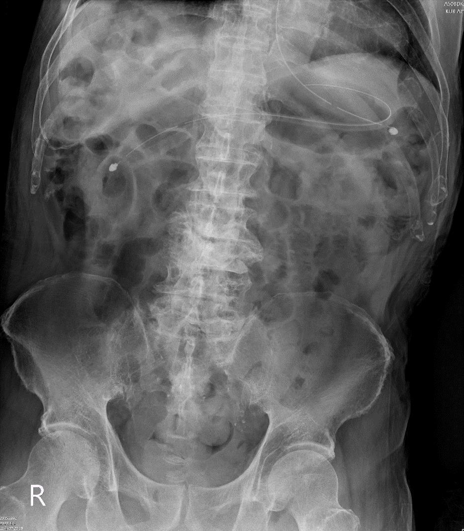
A76 year old male with history of old CVAwith bed ridden, dementia, parkinsonism was sent to ER due to coffee ground noted from NG. Besides, right foot woud with swelling and mild fever was noted for one day, he was brought to a local clinicl where augmentin was prescribed. Initial laboratory exam showed only mild hyponatremia Na[126.0 mmol/L] and findings compatible with UTI.
According to his family, he was used to grinding teeth for a long time. An oral ulcer over left soft palate has been noted recently. A broken part of NG was noted 40 days ago when changing NG by a home care nurse, but no intervention was made with only observation to see if the broken NG can be passed out along with stool.
International Journal of Healthcare Sciences ISSN 2348-5728 (Online)
Vol. 10, Issue 1, pp: (315-319), Month: April 2022 - September 2022, Available at: www.researchpublish.com
Endoscope was performed at ER with the following findings
Esophagus: perifocal hyperemia with small mucosal breaks <5mm in EG junction.
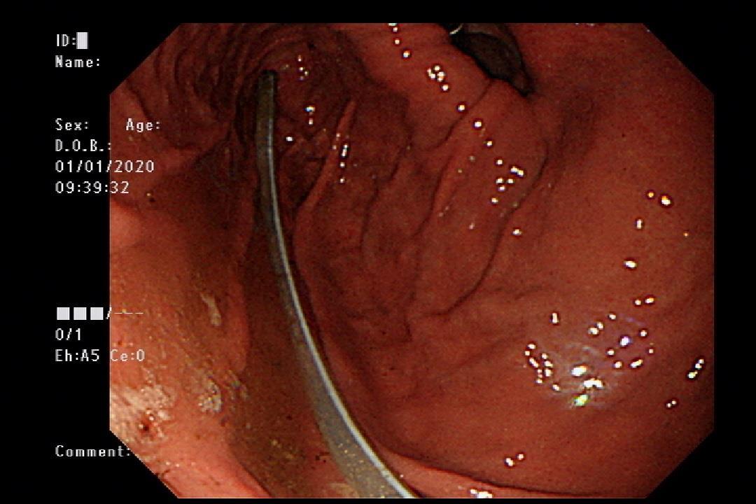
Upward displacement of EG junction over diaphragmatic-esophageal hiatus.
Stomach: chronic hyperemia noted with erosions in antrum. Rapid Hp urease test was done at antrum.Abroken NG tube about 20 cm was noted, removed with a snare catheter.
Duodenum: some small shallow ulcers in bulb. Negative to 2nd portion.
Diagnosis:
1. Reflux esophagitis, LAGr.A.
2. Sliding hiatal hernia
3. Chronic erosive gastritis.
4. Duodenal small shallow ulcers in bulb.
5. Broken NG tube s/p removal.
After admission, empirical antibiotic with augemtin has prescribed for UTI and bronchopneumonia. Dentist was consulted for r/o odontogenic infection and left facial cellulitis. No intraoral finding and no intraoral I&D. ENT has also consulted for stridor, and bilateral vocal cord palsy was impressed. We has informed family the risk of intubation if respiratory failure or distress. However, after days of treatment, symptoms improved. Madopa 0.5tab QD has prescribed for parkinsonism. Speech and swallowing therapist may be needed for swallowing rehabilitation. He was discharged under stable condition.
We often see patients with foreign body impaction in the larynx or in the gastrointestinal tract in the emergency room. Certain people are more likely to be complicated with foreign body impaction, for example patients who have mental disorder, dementia or aged. Those who have foreign body impaction may develop dysphagia, foreign body sensation, and vomiting. Most foreign bodies can go down into the digestive system without causing any discomfort, or need further
International Journal of Healthcare Sciences ISSN 2348-5728 (Online)
Vol. 10, Issue 1, pp: (315-319), Month: April 2022 - September 2022, Available at: www.researchpublish.com
management, but some are not including a broken NG as in our case. A careful review of medical history and physical examination is essential in making the diagnosis. Intervention by endoscope is an effective and safe to remove foreign bodies.
If the foreign bodies are small blunt objects, such as beads or coins, about half of them will pass within 24 hours. Usually objects that are less than 2 cm in size can pass through the entire GI tract without causing any problems. If the objects do not pass out after 3 weeks, they should be referred to receive endoscopic removal.
For long objects, larger than 6 cm, such as fork, spoon, toothbrush or a piece of broken NG as in our case, endoscopic removal is suggested, as these are unlikely to pass through the duodenum.
This case made us realize a rare but possible complication associated with NG tube insertion. Reviews of the literature point out the importance of putting the patient in the right position prior to insertion; understanding the possible inaccuracy of beside tests in checking the position of the NG tube; using x-ray to see if tube placement is correct.[8] If a broken NG is noted upon removal of NG, endoscopic removal should be considered since the retainment of a piece of broken NG is not likely to be passed out easily and may induce ulcer, bleeding as in our case.
Taking a X-ray to see whether the nasogastric tube is in the right position can be useful. But, even this is not fool proof as some tube may be blurred on x-ray and a tube look like in the stomach may be actually in the pleural space and taking a chest x-ray is not possible during a home care visit.
There are many complications after the insertion of NG tubes.
Some may have a complication such as aspiration pneumonitis which may require intubation and ventilator; tachycardia which in severe situation mayrequire electrical cardioversion; or pneumothorax if a NG tube being inserted into the pleural area.
Complications connected with NG tube insertion were reported in the literature for a very long time. The complication rate was between 0.3-15%. Complications in the insertion process could be just minor problem such as slight epistaxis to the potentially life threatening complications including tracheobronchial complications, vascular penetration, gastrointestinal complications and intracranial insertion.
Complications can also happen after long term use including ulceration or bleeding from the nasopharynx, or gastrointestinal tract. Insertion of a NG tube into the bronchial tree had been also reported. The risk of malpositioned tube is that feeding milk or other foods or medicine may go straight into the lungs. It is of utmost importance to confirm the correct position of the tube prior to starting feeding. Measures used to confirm the right position include: a very smooth entry; no cough or respiratory distress on insertion; aspiration of gastric content such as milk and listening via stethscope by injected air over the epigastrium
However, all of the above measures are not reliable and Rassius et al. pointed that incorrect placed tubes were difficult to be found out from bedside tests. The smooth insertion is particularly unreliable as patients who are requiring a NG tube often have poor gag reflex and may not show any signs of the tube being inserted incorrectly.
Patients who have a NG tube inserted into the lungs must be monitored carefully and have follw up x-rays after removal of the tube. Correct positioning of the patient can be helpful for NG insertion, sitting up the patient if clear or supine with the neck flexed if not clear with no contraindication.
Esophageal obstruction has been reported from a coiled NG tube. Perforation and dissectionof the esophagus by nasogastric tube were also reported. Cardiac arrhythmias with NG tube insertion are rare, but also possible.
Other complications may result from long term use such as pressure sore over nose area or bleeding from the nasopharynx or gastrointestinal tract.
NG tube feeding is frequently used to maintain the fluid and nutrition support of patients who can not eat or drink . However, this simple tube may be associated with various kinds of complications. Regular follow up of the condition of NG tube is needed.
In summary, we review the complications related with NG tube insertion with a case report to enhance medical education on the risks involved with this procedure.We highlight the importance of taking x-ray to confirm tube placement and follow up patients closely after the removal of NG tubes. Instructions should be given to patients and their family as when a follow
International Journal of Healthcare Sciences ISSN 2348-5728 (Online)
Vol. 10, Issue 1, pp: (315-319), Month: April 2022 - September 2022, Available at: www.researchpublish.com
up or emergency visit may be needed in certain condition.We stress on insertion with great care and confirming correct placement, strategies to improve safety and timely management to avoid from further complications by giving detailed medical education in advance before commencing this procedure.
[1] Yardley IE, Donaldson LJ: Patient safety matters: reducing the risks of nasogastric tubes. Clin Med 2010;10:228-30.
[2] Mohan Chandra Mandal, et al.: Nasogastric Tube Placement-ASimple Yet Difficult Procedure-AReview · Journal of Medical and Dental Sciences 2017;6(31):25 72-2576
[3] Nicholas L. Smith, Michael Park, Ross Freebairn: Case report and review – Nasogastric tube complications.Crit Care & Shock.2012;15:36-42
[4] Metheny, N, Titler, M:Assessing placement of feeding tubes.Am J Nurs 2001; 101: 36–46.
[5] Ginsberg G. G: Management of ingested foreign objects and food bolus impactions. Gastrointestinal Endoscopy. 1995;41(1):33–38.
[6] Sugawa C, Ono H, Taleb M, et al.: Endoscopic management of foreign bodies in the upper gastrointestinal tract: A review. World J Gastrointest Endosc. 2014;6(10):475-481.
[7] Smith M. T., Wong R. K. Foreign bodies: Gastrointestinal Endoscopy Clinics of NorthAmerica. 2007;17(2):361–382.
[8] Taylor SJ: Confirming nasogastric feeding tube position versus the need to feed. Intensive and Critical Care Nursing 2013; 29: 59-69
[9] American Association of Critical Care Nurses(AACN). AACN practice alert: Initial and ongoing verification of feeding tube placementin adults. Crit Care Nurse. 2017;37(5):100.
[10] O’Neil R, Krishnananthan R: Intrapleural nasogastric tube insertion.Australas Radiol
[11] Aroni F, Pourzitaki C, Logotheti H,et al.: Ventricular tachycardia and cardiac arrest during nasogastric tube insertion. South Med J 2010;103:1192-3.
[12] Wang PC, Tseng GY, Yang HB, Chou KC,Chen CH: Inadvertent tracheobronchial placement of feeding tube in a mechanically ventilated patient. J Chin MedAssoc 2008;71:365-7.
[13] RassiasAJ, Ball PA, Corwin HL:Aprospective study of tracheopulmonary
[14] complications associated with the placement of narrow-bore enteral feeding tubes. Crit Care 1998;2:25-8.
[15] Hutchinson R, Ahmed AR, Menzies D: A case of intramural oesophageal dissection secondary to nasogastric tube insertion.Ann R Coll Surg Engl 2008;90:W4-7.
[16] Shlamovitz GZ. Nasogastric Intubation [updated Apr 21 2020; cited 2021 Feb 21]. Available from: http://emedicine. medscape.com/article/80925-overview#a1.