International Journal of Healthcare Sciences ISSN 2348-5728 (Online)
Vol. 9, Issue 1, pp: (133-138), Month: April 2021 - September 2021, Available at: www.researchpublish.com

International Journal of Healthcare Sciences ISSN 2348-5728 (Online)
Vol. 9, Issue 1, pp: (133-138), Month: April 2021 - September 2021, Available at: www.researchpublish.com
Megha Mishra1, Karuna Raghuwanshi2
1 Swami Vivekanand college of Pharmacy Bhopal, India 2 Department of Pharmacy, Barkatullah University Bhopal, India
Corresponding Author- megham.pharma@gmail.com
Abstract: Mucormycosis or black fungus is a serious type of fungal infection caused by mucormycetes which are made up of group of molds. In this paper, we studied about what is fungus, mucormycosis, types of fungal infection and preventions, and amphotericin B is a choice of drug and other drugs like posconazole, isavuconazole are used as a salvage therapy. Surgical debridement is also one of the treatment for mucormycosis by removal of necrotic tissues. Deferasirox also used in mucormycosis treatment but further studies also required for better results. Hyperbaric oxygen also has been used in some patients of mucormycosis but research to be continue for more benefits.
Keywords: Black Fungus, Mucormycosis, Infections, Amphotericin B, Salvage therapy.
Fungus- A plant which are not green and does not have leaves or flowers or like a wet powder and grows on old wood or food, walls etc. A fungus is a group of eukaryotic organisms like yeasts, molds[1]
Fungus is a microbe that causes infections to human beings. ‗Mycology‘ is the study of fungi. Fungus, in plural called as fungi. Though fungi are the infectious agents, that producing infection in the human body, there are few fungi such as edible fungi. Examples for edible fungi is mushrooms. And some fungi are used in fermentation. Antifungal drugs are used for the treatment of fungal infections. Candidiasis is caused by fungus yeast[2]
Three types of fungus such as white fungus, black fungus and yellow fungus.
1. White fungus like Aspergeilosis infection caused by Aspergillis, it is a type of fungus that lives in outdoor as well as indoor. This type of infection includes allergic infection, lung infection and also seen in other body organs. Candidians caused by candida which lives in skin and inside the body like throat,mouth,gut,vagina without causes any serious health problems,
2. Whereas black fungus is white in color but it causes tissue damage which appears black in color. It mainly affects the sinus, brain and lungs
3. Apart from white and black fungus yellow fungus is very rare and less found in human beings[3]
In present pandemic or in Covid-19 crises, patients have high risk of black fungus. Blackfungus infection mainly occurs in post Covid-19 patients due to intake of high dose of steroids. This infection also seen in patients having diabetes (Diabetes mellitus) [4]
Mucormycosis- It is a type of fungal infections. Mucormycetes is a group of molds which causes fungal infection. These molds live throughout the atmosphere. Mucormycosis is being increasingly diagnosed worldwide after Covid-19 pandemic, particularly in India [5]. The recent trend is due to the increased awareness, advances in diagnostic techniques, and the increase in the pervasiveness of predisposing factors [6, 7]. Mucormycosis affects mainly the Human being, who have health problems or take medicines which lower the body's ability to fight germs and sickness. Commonly it affects on the lungs after inhaling fungal spores from the air. It is also present on the skin after a cut, wound and burn, or other
International Journal of Healthcare Sciences ISSN 2348-5728 (Online)
Vol. 9, Issue 1, pp: (133-138), Month: April 2021 - September 2021, Available at: www.researchpublish.com
type of skin injury [8] People with haematological malignancies (collective term for neoplastic disease), transplant recipients and in people with uncontrolled diabetes mellitus [9,10,11].
What are the symptoms of mucormycosis?[12,13,14]
• cough
• fever
• headache
• nasal congestion
• sinus pain
• blackened skin tissue
• blisters
• fever
• redness
• swelling
• tenderness
Mucormycosis is caused by exposure to mucormycetes molds. These organisms occur in:[15]
• leaves
• piles of compost
• soil
• rotting wood
Diagnosis:
1. Culture Microscopy or direct and histopathology, microscopic examination and culture of various clinical specimens are the cornerstones of diagnosing mucormycosis[15,16]
2. Serology (Enzyme-linked immunosorbent assays[17], immunoblots[18], and immunodiffusion tests[19]
3. Molecular based assays include conventional polymerase chain reaction (PCR),restriction fragment length polymorphism analyses (RFLP), DNA sequencing of defined gene regions, and melt curve analysis of PCR products[20,21,22]
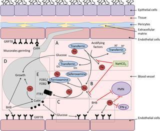
Chemistry of mucormycosis:

International Journal of Healthcare Sciences ISSN 2348-5728 (Online)
Vol. 9, Issue 1, pp: (133-138), Month: April 2021 - September 2021, Available at: www.researchpublish.com
The patients got recover from Covid-19 diseases but already suffering from diabetes, then those patients have high risk to get mucormycosis, because those patients have less immunity, so they should regularly check their sugar level and get injections of steroidsas per their doctor‘s advice to keep the risk of mucormycosis low.
Avoid going to the dust area, wear mask specially N95 mask, clean skin injuries with warm water and antiseptic solution to reduce the risk of infection, also maintain personal hygiene, wear shoes, long sleeves shirt, long trousers, gloves to reduce the risk of infection, followed all these preventions specially while handling soil and gardening[23].
Control high blood sugar, Monitor blood glucose level time to time after Covid-19 recovery. Use clean sterile water for humidifiers during oxygen therapy[24]
In other aspects,Antifungal intravenous injections are also very effective for preventing mucormycosis. A patient has advised to take two to three doses per day depending on the severity of the infection[25]
Usually antifungal drug like amphotericin B, posaconazole, or isavuconazole, these drugs administered by Intravenous route and posaconazole, isavuconazole by oral route. Other medicines like fluconazole, voriconazole and echinocandins cannot work against fungi that cause mucormycosis. Generally, surgery is the only well-known way, in that the infected tissues usually cutted away from the body. Usually antifungal drugs like amphotericin B, posaconazole, or isavuconazole, these drugs administered by IV route and posaconazole, isavuconazole by oral route[26]
1. Surgery- Surgical debridement is one of the treatment for mucormycosis. Debridement means to remove all necrotic tissues, whereas endoscopic debridement means to remove limited necrotic tissues. If patient have rhinocerebral infection, debridement to remove all necrotic tissuescan often be disfiguring, requiring removal of the palate, nasal cartilage, and the orbit[27]
2. Antifungal drugs Antifungal therapy improves the spreading of Infections with mucormycosis. Amphotericin B antifungal therapy is best for mucormycosis, posaconazole or an echinocandin are also useful for mucormycosis.
Starting dose is 5mg/kg of liposomal or lipid complex amphotericin B daily, then increase the dose up to 10mg/kg is an attempt to control these infections. If we can use Amphotericin B with Posaconazole or echinocandin combination therapy but there is no convincing data to supportthesekindsof combination therapy is beneficial to reduce mucormycosis. If patients having isolated renal mucormycosis prescribed Amphotericin B deoxycholate alone.
3. Step down therapy-Broad-spectrum azoles like Posaconazole and isavuconazole are active in-vitro against mucormycosis agents and these drugs available in parenteral as well as oral route. Amphotericin B, posaconazole or isavuconazole can be used for oral step-down therapy, if patients have responded to lipid formulations[28]
4. Salvage therapy- Posaconazole or Isavuconazole are used as a salvage therapy when patients not respond to Amphotericin B or shows tolerance with Amphotericin B, then we used these drugs.
300mg loading dose of posaconazole is required for patient every 12 hours on the first day by Intravenous route and then maintain the dose for every 24hours thereafter.
Isavuconazole loading dose is 200mg, given by Intravenous route or orally every 8 hours for the 1st six doses, followed by 200 mg Intravenous route or orally every 24 hours thereafter[29]
5. Combination antifungal therapy Unfortunately, the combination antifungal therapy is not very much recommended as the treatment of mucormycosis, because at present no such studies are there to satisfy the medical science that the combination antifungal therapy is also can be used[30].
6. Deferasirox It is a chelating agent,and beneficial for the treatment of mucormycosis (experiments based on mice has not been adequately studied in humans) small studies has been evaluated with mixed results. Studies says if deferasirox using within combination of antifungal drugs, seven of eight patients survived. liposomal amphotericin B plus deferasirox or placebo for the first 14 days therapy, reduces the risk of mucormycosis.
Further study is also very necessary to determine the possible benefits or harms of deferasirox[31].
7. Hyperbaric oxygen Hyperbaric oxygen has been also used in some patients with mucormycosis, but the benefit of this therapy has not been established [32,33,34].
International Journal of Healthcare Sciences ISSN 2348-5728 (Online)
Vol. 9, Issue 1, pp: (133-138), Month: April 2021 - September 2021, Available at: www.researchpublish.com
In complete study of fungus,types of fungus,we have found black fungus (mucormycosis) is a serious infection which is caused by group of molds called as ―mucormycetes‖ and other fungal infection like whiteinfection and yellow are not as serious as black fungus. To prevent or to care these funguses, we have found that post covid-19 patients have high risk of mucormycosis or black fungus whose patients already suffering from diabetes mellitus (Hyperglycemia), if those patients have given the high doses of steroids.
To control hyperglycemia, one should monitor glucose level, use clear sterile water for treatment. As we found, the patient should take care of himself post Covid-19,it will minimize the risk of mucormycosis like monitor the glucose level, avoid going to the dusty area,wear full sleeve clothes and use the gloves specially while handling soil,gardening, maintain personal hygiene, and use warm water with antiseptic solutions for washing skin injuries. Ifwe move to treatment aspects, antifungal drugs are widely used for black fungus or mucormycosis, Amphotericin humidifiers are used during oxygen therapy and use of antifungal drugs like posconazole and isavuconazole are also useful for treatment of mucormycosis.
Article highlights:
Mucormycosis is a serious type of life-threatening infection with some available treatments is available.
The medications for mucormycosis have been published in fewer review articles
The treatments like Amphotericin humidifiers are used during oxygen therapy and use of antifungal drugs like posconazole and isavuconazole are also useful for mucormycosis.
Prevention is also an important factor for minimizing the risk of mucormycosis.
Prevention aspects like personal hygiene is also essential factor that patient should care after Covid-19.
Monitor glucose level, avoid going to the dusty areas,wear full sleeve clothes and wear gloves specially while handling soil and gardening.
Use warm water with antiseptic solutions for washing skin injuries.
Some treatments are under clinical development for mucormycosis.
[1] Ramanan P, Wengenack NL, Theel ES (2017) Laboratory Diagnostics for Fungal Infections: A Review of Current and Future Diagnostics Assays.Clin Chest Med 38(3):535-554.
[2] Magill SS, Edwards JR, Bamberg W, Beldavs ZG, Dumyati G, KainerMA,et al (2017)Emerging Infections Program Healthcare-Associated Infectionsand Antimicrobial Use Prevalence Survey Team. Multistate pointprevalencesurvey of health care-associated infections. N Engl J Med 370:1198–1208.
[3] Bloos F, Bayer O, Sachse S, Straube E, Reinhart K, Kortgen A (2013) Attributable costs of patients with candidemia and potentialimplications of polymerase chain reaction-based pathogendetection on antifungal therapy in patients with sepsis. J Crit Care 28:2–8.
[4] Chakrabarti A, Das A, Mandal J, Shivaprakash MR, George VK, Tarai B, et al (2006) The rising trend of invasive zygomycosis in patients with uncontrolled diabetes mellitus. Med Mycol 44:335-42.
[5] Prakash H, Ghosh AK, Rudramurthy SM, Singh P, Xess I, Savio J, et al (2019) A prospective multicenter study on mucormycosis in India: epidemiology, diagnosis, and treatment. Med Mycol 57:395-402.
[6] Farmakiotis D, Kontoyiannis DP (2016) Mucormycoses. Infect Dis Clin North Am 30:143-63.
[7] Dioverti MV, Cawcutt KA, Abidi M, Sohail MR, Walker RC, Osmon DR (2015) Gastrointestinal mucormycosis in immunocompromised hosts. Mycoses 58(12):714-718.
[8] Wysong DR, Waldorf AR (1987) Electrophoretic and immunoblot analyses of Rhizopus arrhizus antigens. J Clin Microbiol 25: 358–363.
International Journal of Healthcare Sciences ISSN 2348-5728 (Online)
Vol. 9, Issue 1, pp: (133-138), Month: April 2021 - September 2021, Available at: www.researchpublish.com
[9] Jones KW, Kaufman L (1987) Development and evaluation of an immunodiffusion test for diagnosis of systemic zygomycosis (mucormycosis): preliminary report. Clin Microbiol 7: 97–101.
[10] Hsiao CR, Huang L, Bouchara J-P, Barton R, Li HC, Chang TC (2005) Identification of medically important molds by an oligonucleotide array. J Clin Microbiol 43: 3760–3768.
[11] Nagao K, Ota T, Tanikawa A et al (2005) Genetic identification and detection of human pathogenic Rhizopus species, a major mucormycosis agent, by multiplex PCR based on internal transcribed spacer region of rRNA gene. J Dermatol Sci. 39: 23–31.
[12] Petrikkos G, Skiada A, Lortholary O, Roilides E, Walsh TJ, Kontoyiannis DP (2005) Epidemiology and clinical manifestations of mucormycosisexternal icon. Clin Infect Dis 54(1):23-34.
[13] Lewis RE, Kontoyiannis DP (2013) Epidemiology and treatment of mucormycosisexternal icon; Future Microbiol 8(9):1163-75.
[14] Spellberg B, Edwards Jr. J, Ibrahim A (2005) Novel perspectives on mucormycosis: pathophysiology, presentation, and managementexternal icon. Clin Microbiol Rev 18(3):556-69.
[15] Frater JL, Hall GS, Procop GW (2001) Histologic features of zygomycosis: emphasis on perineural invasion and fungal morphology. Arch Pathol Lab Med 125: 375–378.
[16] Lass-Florl C (2009) Zygomycosis: conventional laboratory diagnosis. Clin Microbiol Infect 5: 60–65.
[17] Sandven PER, Eduard W (1992) Detection and quantitation of antibodies against Rhizopus by enzyme-linked immunosorbent assay. APMIS 100:981–987.
[18] Wysong DR, Waldorf AR (1987) Electrophoretic and immunoblot analyses of Rhizopus arrhizus antigens. J Clin Microbiol 25: 358–363.
[19] Jones KW, Kaufman L (1987) Development and evaluation of an immunodiffusion test for diagnosis of systemic zygomycosis (mucormycosis) preliminary report. Clin Microbiol 7: 97–101.
[20] Nagao K, Ota T (2005) Tanikawa A et al. Genetic identification and detection of human pathogenic Rhizopus species, a major mucormycosis agent, by multiplex PCR based on internal transcribed spacer region of rRNA gene. J Dermatol Sci 39: 23–31.
[21] Larche J, Machouart M, Burton K et al (2005) Diagnosis of cutaneous mucormycosis due to Rhizopus microsporus by an innovative PCR-restriction fragment-length polymorphism method. Clin Infect Dis 41: 1362– 1365.
[22] Machouart M, Larche J, Burton K et al (2006) Genetic identification of the ´ main opportunistic mucorales by PCRrestriction fragment length polymorphism. J Clin Microbiol 44: 805–10.
[23] Imran M, Alshrari SA, Tauseef M, Khan SA, Hudu SA, Abida (2021) Mucormycosis medications: a patent review. Expert OpinTher Pat 1-16.
[24] Jeong W, Keighley C, Wolfe R, Lee WL, Slavin MA, Chen SC, Kong DCM (2019) Contemporary management and clinical outcomes of mucormycosis- A systematic review and meta-analysis of case reports. Int J Antimicrob Agents. 53:589-97.
[25] Manesh A, John AO, Mathew B, Varghese L, Rupa V, Zachariah A, Varghese GM (2016) Posaconazole: an emerging therapeutic option for invasive rhino-orbitocerebralmucormycosis. Mycoses 59:765-72.
[26] Natesan SK, Chandrasekar PH (2016) Isavuconazole for the treatment of invasive aspergillosis and mucormycosis: current evidence, safety, efficacy, and clinical recommendations; Infect Drug Resist 9: 291-300.
[27] Roilides E, Antachopoulos C (2014) Simitsopoulou M. Pathogenesis and host defence against Mucorales: the role of cytokines and interaction with antifungal drugs. Mycoses 57: 40–47.
[28] Lanternier F, Poiree S, Elie C et al (2015) Prospective pilot study of high-dose (10 mg/kg/day) liposomal amphotericin B (L-Amb) for the initial treatment of mucormycosis. J Antimicrob Chemother 70: 3116–23.
[29] Wiederhold NP (2016) Pharmacokinetics and safety of posaconazoledelayedrelease tablets for invasive fungal infections. Clin Pharmacol 8: 1–8.
International Journal of Healthcare Sciences ISSN 2348-5728 (Online)
Vol. 9, Issue 1, pp: (133-138), Month: April 2021 - September 2021, Available at: www.researchpublish.com
[30] Spellberg B, Ibrahim A, Roilides E, et al (2012) Combination therapy for mucormycosis: why, what, and how?.Clin Infect Dis 54(1):73-78.
[31] Yohai RA, Bullock JD, Aziz AA, Markert RJ (1994) Survival factors in rhino-orbital-cerebral mucormycosis. SurvOphthalmol 39:3.
[32] Ferguson BJ, Mitchell TG, Moon R, et al (1998) Adjunctive hyperbaric oxygen for treatment of rhinocerebralmucormycosis. Rev Infect Dis 10:551.
[33] Bentur Y, Shupak A, Ramon Y, et al (1998) Hyperbaric oxygen therapy for cutaneous/soft-tissue zygomycosis complicating diabetes mellitus.PlastReconstr Surg 102:822.
[34] Strasser MD, Kennedy RJ, Adam RD (1996) Rhinocerebralmucormycosis. Therapy with amphotericin B lipid complex. Arch Intern Med 156:337.
[35] Baldin C, Ibrahim S (2017)Molecular mechanisms of mucormycosis-The bitter and the sweet. PlosPathog 13:8.