International Journal of Healthcare Sciences ISSN 2348-5728 (Online)
Vol. 8, Issue 2, pp: (347-352), Month: October 2020 - March 2021, Available at: www.researchpublish.com

International Journal of Healthcare Sciences ISSN 2348-5728 (Online)
Vol. 8, Issue 2, pp: (347-352), Month: October 2020 - March 2021, Available at: www.researchpublish.com
1Ms. Nisha Waghmare, 2Ms. Divya Sharma, 3Mr. Shishir Shukla 1(Author), 2,3 (Co- Author) 1,2,3 DEPARTMENT OF OPTOMETRY, SHRI RAWATPURA SARKAR UNIVERSITY, RAIPUR, CHATTISGARH.
Abstract: The aim was to study the Visual outcome of keratoconus (kc) with a corneal transplantation technique Deep anterior lamellar keratoplasty (DALK).
Material and Methods: This Retrospective cross sectional study included 40 patients eyes with moderate to advanced keratoconus underwent Deep anterior lamellar keratoplasty. Data were obtained by reviewing the records of 40 patients operated at National institute of Ophthalmology, Pune between June 2019 – June 2020 For Deep anterior lamellar keratoplasty Surgery. Patient at least completed 6 months follow up. Medical records were reviewed for Demographic Details , Risk Factors , Surgical procedure, size of donor , post operative complication and Visual outcome. According to the patient Record the Data was Measured and analyzed Statistically.
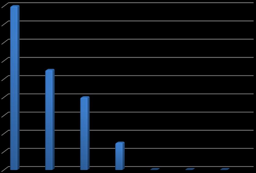
Result: Out of 40 Operated Patients with aged between 10 – 30 years were enrolled in this study. wherein Preoperatively , corrected distance visual acuity was >6/60( < 1.00 in log mar) in 18 eyes, 6/60(1.00 in log mar) in 11eyes, 6/36(0.8 in log mar) in 8 eyes, 6/24( 0.6 in log mar ) in 3 eyes. After performing the Deep anterior lamellar keratoplasty . the corrected distance visual acuity was after 6 month 6/12 ( 0.3 in log mar ) in 6 eyes , 6/18( 0.5 in log mar ) in 11 eyes, 6/24( 0.6 in log ma r) in 9 eyes, 6/36 ( 0.8 in log mar) in 11 eyes , 6/60 ( 1.0 in log mar ) in 3 eye.Similarly , Pre operatively 18 patients (45 %) were in blindness categories according to WHO ; After performing the Deep anterior lamellar keratoplasty only 3 patients (7.5 %) were in blindness categories.
Conclusion: Deep anterior lamellar keratoplasty should be procedure of choice in patient with keratoplasty induced by keratoconus. This technique causes minimal refractive change and provide rapid visual recovery for patient with keratoconus.
Keywords: Keratoconus , DALK Surgery, visual outcome.
Cornea is an avascular, transparent tissue that is an important component of the ocular refractive system. It is one of the most densely stimulated tissues in the body. Corneal stroma is composed of cellular and acellular components . Cornea is an immune-rich tissue due to the absence of blood and lymphatic vessels. Although normal cornea is without blood vessels, many conditions can cause neovascularization, scarring, and corneal blindness. It consist of 5 layers (possibly six , if the Dua’s layer is included). From the anterior to posterior the layers of the cornea are:
KERATOCONUS It is the most common primary ectasia. It usually occurs in the second decade of life and affects both genders and all ethnicities. Keratoconus (conical cornea) is a non – inflammatory biletral ( 85%) ectatic condition of cornea in its axial part. It usually starts at youth and Progresses slowly.
Ocular may have signs and symptoms vary depending on disease severity. Corneal thinning frequently precedes IIO ectasia. In moderate and advance cases, placido disc examination shows irregularity of the circle. And in keratometry reading Normal average keratometric values are 45 D . In keratoconus the keratometric values are increased and based on the severity of keratoconus.
International Journal of Healthcare Sciences ISSN 2348-5728 (Online)
Vol. 8, Issue 2, pp: (347-352), Month: October 2020 - March 2021, Available at: www.researchpublish.com
Morphological classification Depending on the size and shape of the cone , keratoconus is of three types:
• Nipple cone(<5mm)steep
• Oval cone(5-6mm)
• Globus cone(>6mm)
Treatment usually starts with spectacle correction . may improve vision in early cases.However , later in the course of diseases the falling vision may not be corrected by glasses due to irregular astigmatism. Contact lens ,usually Rigid permeable or scleral contact lens improves the vision in early cases.In early to moderate cases a specially designed scleral contact lens(Rose – K ) may be useful. The Intracorneal ring segments are reported to be useful in early to moderate cases. A treatment called C3R( Corneal collagen cross linking with riboflavin) to help prevent worsening and slow the progression.
Keratoplasty may be required in later stage, DALK or PKP may be performed.
DALK Deep anterior lamellar keratoplasty (DALK) is a surgical procedure for removing the corneal stroma down to Descemet’s membrane.it is most useful for the treatment of corneal diseases in the setting of a normally functioning endothelium .Traditionally , penetrating keratoplasty ,which involves a full thickness corneal graft, has been the treatment of choice for corneal stromal diseases . But PK can be complicated by graft rejection , irregular astigmatism and corneal opacification , thus resulting in visual impairment . DALK offers an alternative procedure that may lessen those risks because the recipient Descemet’s membrane and endothelium are preserved . At the same time , DALK carries the potential danger of decreased visual acuity due to possible opacification at the interface layer
As early as the 1950s, jose Barraquer and colleagues in Colombia applied new techniques of lamellar keratoplasty , dissecting the corneal stromal down to two thirds of its thickness in both the donor and the recipient tissue. Yet the procedure failed to gain favor because of poor visual result related to asymmetry of the dissected surfaces and scarring in the tissue interfaces .
Although exposure of descemet’s membrane in corneal dissection was performed in the 1970s, the term “ deep lamellar keratoplasty” as it is used today , was not employed until 1984 by Eduardo Arenas Archila, MD with the use of intrastromal air injection to smooth host tissues removal. By the late 1990s , studies indicated that DALK was associated with visual outcomes similar to PK without the risk of immunological rejection. In spite of positive reports in the literature , the classic technique of layer-by-layer stromal tissues removal was tedious and required great surgical experinces , thereby limiting its use around the world . Recent advances in technique present by many surgeons have started to familiarize the procedure.
Surgical Techniques:-
1. Direct open dissection 2. Dissection with hydrodelamination 3. Closed Dissection 4. Dissection with Big-Bubble Technique
Big-Bubble Techinique combined with Femto Second Laser technique.
Participants:
The study design was conceived and planned at the National Institute of Ophthalmology Hospital, Pune, Maharashtra, a comprehensive eye care center.
This was a Retrospective study involving 40 eyes of 40 patients.The patients who is suffering from severe keratoconus who attended the OPD at National Institute of ophthalmology, who have difficulty in vision due to keratoconus. The duration of the study was from june 2019 to june 2020
An informed assent, detailed ocular and methodical history was taken and following examination were done.
Inclusion criteria:
• Patient of Keratoconus who are not improving with spectacles and specialized contact lenses
• Patient with severe keratoconus
International Journal of Healthcare Sciences ISSN 2348-5728 (Online)
Vol. 8, Issue 2, pp: (347-352), Month: October 2020 - March 2021, Available at: www.researchpublish.com
• Patient with no media opacity other than cataract
• Age – young age between 10 to 30
Exclusion criteria:
• Patient with vernal keratoconjuntivitis , corneal dystrophies, posterior segment anomaly,secondary glaucoma and ocular surface diseases
• Patient with cicatrical pemphigoid and chemiacal / thermal burns
Outcome measurement:
Follow up will be performed the 6 months of the surgery.
The follow up visits will include Visual acuity testing (BCVA) Slit lamp examination: For signs of graft rejection, inflammation, uveitis, neovascularization and wound integrity. Intraocular pressure.
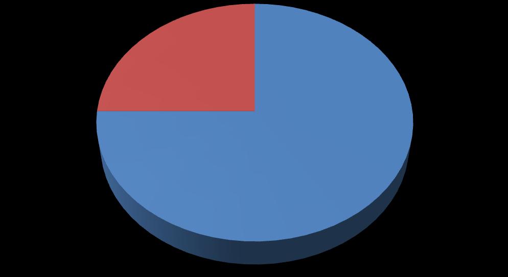
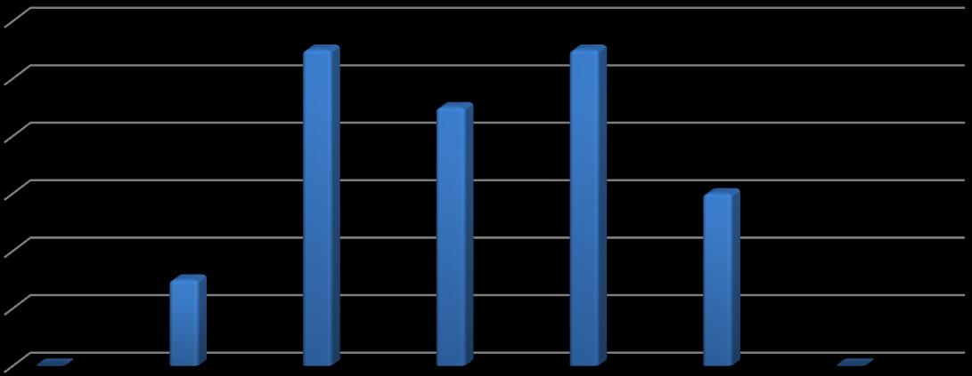
Out of 40 patients ,The mean age of the patient was 20.46 ±4.98 years, min 10 years, and maximum 30 yrs, range between 10 – 30 years. . In gender distribution 75% are males , 25% are female. Preoperatively the corrected visual acuity was <1.00 logMar in 14 eyes, 1.00 logMar in 9 eyes, 0.8 logMar in 5 eyes, 0.6 logMar in 2 eyes and, after performing the DALK the postoperatively distance corrected visual acuity was after 6 months 0.3 logMar in 4 eyes , 0.5 logMar in 8 eyes, 0.6 logMar in 7 eyes, 0.8 logMar in9 eyes, 1.0 logmar in 2 eyes.
Similarly , preoperatively 40 patients were in visual impairment categories according to WHO; After performing the DALK only 3 patients were in visual impairment categories.
TABLE 1: Table showing sex distribution of cases operated


International Journal of Healthcare Sciences ISSN 2348-5728 (Online)
Vol. 8, Issue 2, pp: (347-352), Month: October 2020 - March 2021, Available at: www.researchpublish.com
The major advantage of the lamellar keratoplasty techniques as compared to PK which can have unpredictable visual outcome with prolonged visual instability, the comparatively stable and predictable visual outcome obtained with lamellar keratoplasty are beneficial to most of the patient. The present study evaluated Visual Acuity after performing DALK surgery by 6 months postoperatively 0.3 logMar in 4 eyes , 0.5 logMar in 8 eyes, 0.6 logMar in 7 eyes, 0.8 logMar in 9 eyes, 1.0 logmar in 2 eyes. In fact, the DALK visual outcomes at 6 months were comparable to the best outcome obtained after PK even when compared with some longer PK postoperative times.Postoperative BCVA scores were improved over preoperative values in all patients. A DALK procedure strip Descemet’s membrane is considered the surgery of choice in eyes with a healthy endothelium. In general, even tough the outstanding result of PKP ,DALK may be preferred in patients with kc because of the absence of risk of endothelial rejection, earlier tapering of steroids, decreased risk of secondary glaucoma, and increased wound strength.
The advantage of DALK is even more noticeable in patients with mental retardation in which PK has a higher prevelance of postoperative complications such as globe rupture, corneal ulceration and graft rejection, and corneas with significant peripheral thinning.
Mainly this study was focused on the Visual outcome which includes Visual acuity before and after surgery was noticed it was improved after the DALK surgery and helps to stop the progression of Keratoconus. Several study reports have indicated that visual acuity continues to improve in DALK eyes for up to 1 years after surgery.clear advantages of DALK in terms of surgical risk, graft survival, complication rates,visual acuity and post operative astigmatism which in turn,has better than PKP, and being a safe refractive treatment for patient having keratoconus.
Treatment of keratoconus has experienced great advances in the last two decades. From being limited only to RGP contact lens wear and PK for the most moderate cases, to having different curative alternatives currently to treat not only the cone and avoid the necessity of a corneal transplant, but also being able to halt the progression of the disease with a very high rate of efficacy and safety. Also, the advances in refractive surgery including surface corneal ablation treatment and Phakic intraocular lenses have allowed a better management and visual rehabilitation of these patients after a corneal transplant is required, being able to achieve, in many cases, a 20/20 unaided vision.
Visual recapture after deep anterior lamellar Keratoplasty is effected, among the other optical variables, by the transparency of the corneal tissue. unusually than PKP In DALK the presence of a donor-host link may effected the visual result of the patient. Limitation of our study were as follows: Less sample size , Short follow up period.
After assessing the demographic profile of patients undergoing Deep anterior lamellar keratoplasty , it was found that most of the patients were males. The commonest indication for Deep anterior lamellar keratoplasty was Keratoconus because the patients benefit the most from preserving their own endothelium. Therefore DALK have expanded to other corneal ectasia ( Pellucid marginal degeneration and post lasik ectatia) , stromal opacities and scar. Vascularized corneal opacity has high chance of going into rejection. Causes for failure of graft was rejection due to inflammation and infection of graft. And there is less chance of astigmatism because both the layers form the support layer for the donor tissue; there is also less danger of rejection and more chance that the graft remains transparent. Final Visual Acuity improved From preoperative vision in majority of the cases. after performing DALK surgery without removing the donor endothelium. Thus Deep anterior lamellar keratoplasty done in ideal condition has huge success rate and can bring back light into the lives of many who have corneal blindness.
The author would like to acknowledge participant for their patience and coordination.
[1] Alió JL, Vega-Estrada A, Sanz-Díez P, Peña-García P, Durán-García ML Maldonado M. Keratoconus management guidelines. Intern J Keratoconus Ectatic Corneal Dis. 2015;4(1):1–39. doi: 10.5005/jp-journals-10025-1095. [Cross Ref].
[2] Parker JS, van Dijk K, Melles GR. Treatment options for advanced keratoconus A review. Surv Ophthalmol. 2015;60(5):459–80. doi: 10.1016/j.survophthal.2015.02.004. [PubMed] [Cross Ref]
International Journal of Healthcare Sciences ISSN 2348-5728 (Online)
Vol. 8, Issue 2, pp: (347-352), Month: October 2020 - March 2021, Available at: www.researchpublish.com
[3] van Dijk K, Parker J, Tong CM, Ham L, Lie JT, Groeneveld-van Beek EA, et al. Midstromal isolated Bowman layer graft for reduction of advanced keratoconus: a technique to postpone penetrating or deep anterior lamellar keratoplasty. JAMA Ophthalmol. 2014;132(4):495–501. doi: 10.1001/jamaophthalmol.2013.5841. [PubMed] [Cross Ref]
[4] Reinhart WJ, Musch DC, Jacobs DS, Lee WB, Kaufman SC, Shtein RM. Deep anterior lamellar keratoplasty as an alternative to penetrating keratoplasty a report by the american academy of ophthalmology. Ophthalmology. 2011;118(1):20918. doi: 10.1016/j.ophtha.2010.11.002. [PubMed][Cross Ref]
[5] El-Agha MS, El Sayed YM, Harhara RM, Essam HM. Correlation of corneal endothelial changes with different stages of keratoconus. Cornea. 2014;33(7):707–11. doi: 10.1097/ICO.0000000000000134.[PubMed] [Cross Ref]
[6] Castroviejo R. Keratoplasty for the treatment of keratoconus. Trans Am Ophthalmol Soc. 1948;46:127–53. [PMC free article] [PubMed]
[7] Appelbaum A. Keratoconus. Arch Ophthalmol. 1936;15(5):900–21. doi: 10.1001/archopht.1936.0084017011 2011. [Cross Ref]
[8] Paufique L, Charleux J. Lamellar keratoplasty. In: Casey T, editor. Corneal grafting. London: Butterworth; 1972. pp. 121–176.
[9] John T, et al.History. In: John T , et al., editors. Corneal endothelial transplant.New Delhi: jaypee- Highlights; 2010. Pp. 143- 157.
[10] A.K Khurana book “Diseases of eye’, diseases of lens{179}.