ISSN 2348-313X (Print)
International Journal of Life Sciences Research ISSN 2348-3148 (online) Vol. 10, Issue 4, pp: (27-34), Month: October - December 2022, Available at: www.researchpublish.com

ISSN 2348-313X (Print)
International Journal of Life Sciences Research ISSN 2348-3148 (online) Vol. 10, Issue 4, pp: (27-34), Month: October - December 2022, Available at: www.researchpublish.com
1Department of Chemistry, Merchant Science College, Basna, Gujarat, India
2 Department of Chemistry, Adarsh Science College, Patan, India
DOI: https://doi.org/10.5281/zenodo.7276919
Published Date: 03-November-2022
Abstract: The grouping of some rare metal ions with an significant 2-(1,3-benzoxazole -2-yl - sulfanyl )-N-phenyl acetamide (BSPA) ligand to form coordination compounds is an important area of present research. Less explored biologically important 2-(1,3-benzoxazole -2-yl - sulfanyl )-N-phenyl acetamide ligand is allowed to react with solution of some rare metal perchlorates and attempt has been made to synthesize solid 2-(1,3-benzoxazole -2-ylsulfanyl )-N-phenyl acetamide complexes. These 2-(1,3-benzoxazole-2-yl-sulfanyl )-N-phenyl acetamide complex are subjected to U.V Visible Spectroscopy, IR Spectroscopy,TGA Analysis, elemental analysis of these complex has been evaluated by standard methods and attempts have been made to correlate structural characteristics with properties of these 2-(1,3-benzoxazole -2-yl - sulfanyl )-N-phenyl acetamide complex.
Keywords: Spectroscopic analysis, characterization, 2-(1,3-Benzoxazole-2-yl-sulfanyl)-N-phenyl acetamide(BSPA) complex.
Thе rare–earth metals are bу definition, thе group IIIb elements Sc, Y, Lа аnd thе 14 lanthanides Ce–Lu. [1,2,3]
A definite volume of 70% acid was diluted with water to obtain 0.2M perchloric acid solution in 500 ml flask. The exact strength was determined by pH metric titration against 0.2M NaOH solution (standardized with 0.2N oxalic acid previously prepared). 75 ml 0.2M perchloric acid was taken and excess solid metal carbonate was added. The solution was stirred for 30 minutes and filtered (this way 0.133 M lanthanide was obtained).
The formation of complexes was carried out by mixing 75 ml 0.133 M metal perchlorate solution and 50ml 0.2 M BSPA ligand in DMSO solution. The mole ratio of ligand and metal was (1:1). The reaction mixture was refluxed for 2.5 to 3.0 hours at 95 0C temperature. After 3 hours the reaction mixture was cooled. There was no immediate precipitation. The pH of the above solution was then raised up to 6.5 using 0.1M sodium hydroxide solution which resulted in the precipitation of the semi solid sticky material. Then, this sticky product was dissolved in methanol to remove stickiness. This mixture with methanol was slightly heated for total dissolution and after that cooled. Then after around 30 ml of cold water was added for precipitation of the complex in non sticky form. The complex thus obtained was washed well with double distilled water to remove unreacted metal perchlorate and ligand. All the complexes were dried in oven at 40oC to 50oC.
ISSN 2348-313X (Print)
International Journal of Life Sciences Research ISSN 2348-3148 (online) Vol. 10, Issue 4, pp: (27-34), Month: October - December 2022, Available at: www.researchpublish.com
Sr. No. Ligand or Complexes
Brief name 1 Ligand BSPA BSPA 2 Gd(III)-BSPA Gd-BSPA
M P and TLC were taken with usual apparatus[solvent system for TLC 70% toluene + 30% methanol] TLC indicated single spot confirming complex formation. Elemental analyses wеrе performed with a Vario-MICRO CUBE C, H, N, S analyzer. The metal content was determined by titration with a solution of standardized disodium salt of EDTA [4].Magnetic susceptibilities were measured by the Gouy’s method [5], at room temperature using Hg[Co(CNS)4]as calibrant. Thе IR spectra wеrе recorded оn a BRUKER ALPHA FT-IR 400 – 4000 cm-1 spectrophotometer. Thе UV – visible spectra wеrе measured оn a UV-1800 Shimadzu (Double beam) spectrophotometer. Thermal measurements wеrе performed using a METTLER TOLEDO STARe system TGA/DSC1(11500C) thermal analyzer. The mass spectra analyses were performed with a model QDA of Waters and Alliance 2690 analyzer.
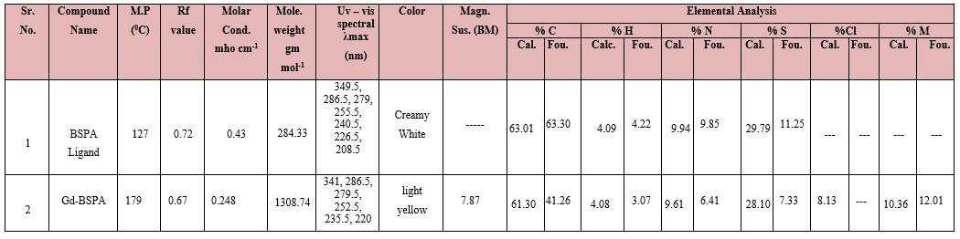
BSPA = 2-(1,3-Benzoxazole -2-yl - sulfanyl )-N-phenyl acetamide, Mole.=molecular,Mag.Sus.=Magnetic susceptibility, Cal. = calculated, Fou. = Found, Cond. = conductance, %M carried out by EDTA method, *Solvent system for TLC = 70% toluene + 30% methanol
The physical data of the complex and ligand are presented in the table no.2. The Gd(III) complex was colored while BSPA is colorless. The melting point of ligand is different than the complex. The TLC of solid was carried out using silica gel as the stationary phase and toluene : methanol (7:3) as the mobile phase. The Rf value of complex are different than ligand. Together, all these provide confirm formation of single complex in each case. The molar conductance of complex confirms that are quite low and these complex are nonionic.
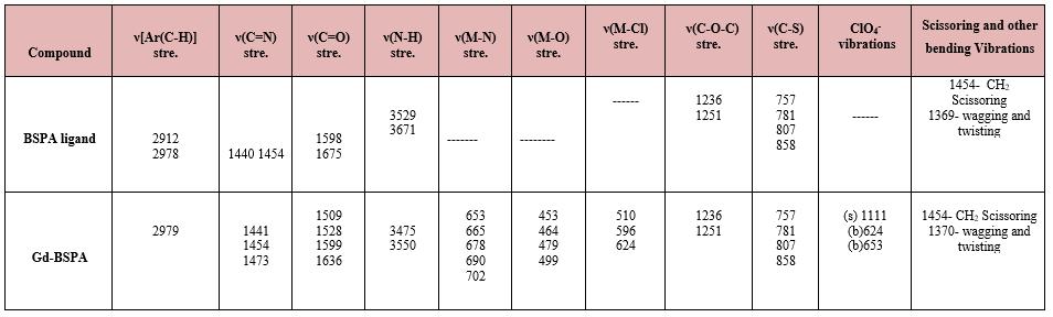
IR bands corresponding to BSPA and its complexes are shown in table no.3. The IR spectra of ligand and metal complexes were recorded inthe 4000-400cm-1 range usingKBr pellet. IR spectral[6,7] analysis confirms the presence ofcharacteristic groups in the compound. In the ligand bands at~3344 and 3333 cm-1is assigned to the ν(N-H)stretching of the amine group The sharp band at 1598cm-1 can be attributed to the ν(C=N) stretching mode in the ligand. This band shifted in the complexes.This indicates participationincoordinate bond. The band at 1675cm-1ofthe ligand is due to the C=O stretching. In the Gd-BSPA, it is shifted to 15-40 cm -1 either at lower energy or at higher energy indicating the coordination by the carbonyl-oxygen atom [6,7]
Further conclusive evidence of this ligand with metal ion was shown by the appearance of weak low frequencies new bands at 650-750cm-1 and 450-510cm-1 . These were assigned to the metal[8,9] nitrogen(M-N) and metal oxygen(M-O) vibration respectively, and were observed in the spectra of the metal complexes and not in the spectra of the ligand. Thus, this is the confirmation that the N and O atoms participated in the coordination[8,9]
ISSN 2348-313X (Print)
International Journal of Life Sciences Research ISSN 2348-3148 (online) Vol. 10, Issue 4, pp: (27-34), Month: October - December 2022, Available at: www.researchpublish.com
Table 3: IR spectra of BSPA ligand with Gd3+ complex
(1) *Metal –Sulphur stretching (below 300 cm-1 ) could not be measured by the instrument (2) All figures are in cm-1
The Gd-BSPAare analyzed for UV-Visible spectra and magnetic moments.These f-block metals have a usual characteristic of absence of d-d transition because no space for excited electron is present in the d orbital which is completely filled in these ions. The results indicate paramagnetic nature of the complexes along with metal to ligand charge transfer band.[10,11]
The room temperature magnetic moment of the solid Gd-BSPA was found to be 7.87 BM This indicates seven unpaired electrons per Gd (III) ion in tricapped distorted trigonal prism [12] environment
The group with longest wavelengths lies to near 3100 0A. The ground state of Gd3+ is 8S7/2, the only known level of any of the triply ionized rare earth ions which has primarily S character (where thus L= 0, with angular momentum entirely due to the spin) which in first approximation does not interact with the electric crystal field. The 8S level is, therefore, single, or at least[13,14] appears to be so unless very high resolution is employed, in which case a structure of the order of 0.1 cm-1 is revealed. [13,14] There are nominal S levels for the other rare earth ions, but their eigenvectors always have so much mixture with L ≠ 0 that they are split quite appreciably in a crystal field.[13,14] Table 4:
ISSN 2348-313X (Print)
International Journal of Life Sciences Research ISSN 2348-3148 (online) Vol. 10, Issue 4, pp: (27-34), Month: October - December 2022, Available at: www.researchpublish.com
Thermal analysis of metal complexes is carried out by thermo gravimetric analyzer. The heating was carried out until there was[17.18] no further loss in weight (up to ~1000 0C) Change in weight was recorded with time. A regular temperature change makes possible to plot a graph of weight as a function of temperature Thermo grams were analyzed by increasing the temperature at a uniform rate of 10 0C minute-1 . The temperature range wise % weight loss data of metal complexes are shown in table.6[17.18
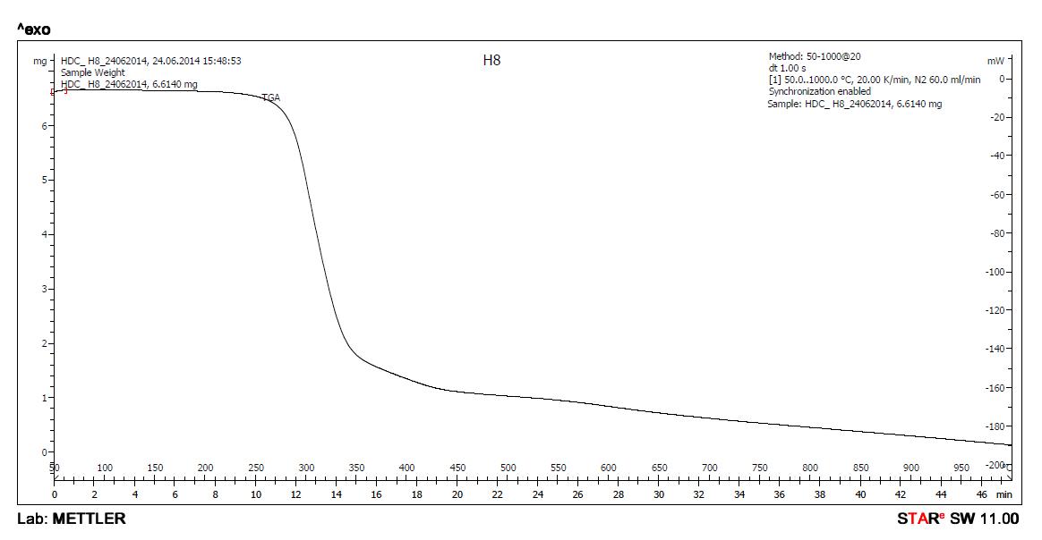
Loss in weight of the sample between room temperature and approximately, upto 150 0C – correspond to water of crystallization. Loss in weight of the sample between approximately 1500C to 2500C correspond to loss in weight due to water of coordination. After approximately 800 0C, almost constant weight is observed. [17.18 This is due to oxide of corresponding metal hence from this constant weight, percentage of metal can be obtained.
Table 6: TGA data of Complexes of BSPA
RT-150 0C
Compound
RT = Room temperature Table 7: Complexes and Coordination numbers
ISSN 2348-313X (Print)
International Journal of Life Sciences Research ISSN 2348-3148 (online) Vol. 10, Issue 4, pp: (27-34), Month: October - December 2022, Available at: www.researchpublish.com
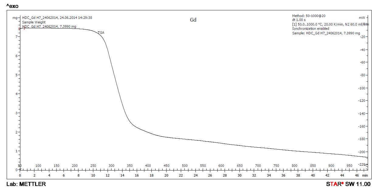
IthasbeenobservedthatGd-BSPAdoesnotshowanylossinweightintherange150 0 C -250 0 C therearenowater molecules that coordinate with gadolinium ion in the Gd-BSPA complex It has been also observed that Gd-BSPA lose weight corresponding to no water molecules up to 150 0C hence, no water molecules are there for water of crystallization.
The reported values for Gd-BSPA are based upon the results of complexes without water of crystallization When thermal analysis was carried out, the sample, because of its hygroscopic nature, absorbed water molecules from air and all the corresponding results were uniformly lower than anticipated. Therefore, the results have been expressed for the complex on dried basis. This hygroscopic complex was dried at 50 0C for 90 minutes in oven to remove absorbed water.
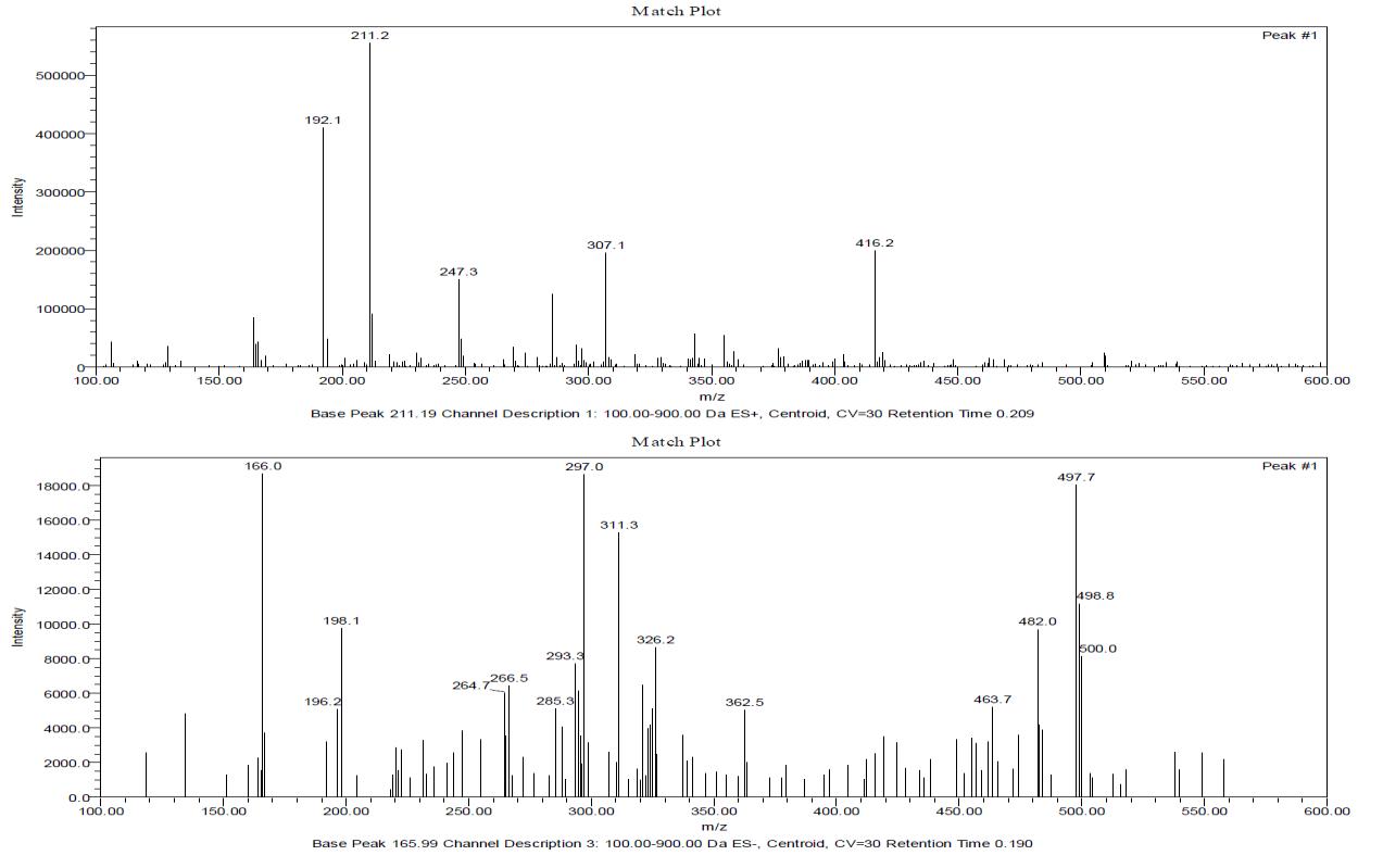
The most common use of mass spectrometry by the organic chemist is for the accurate determination of molecular weight and from that molecular formula so is the case for the inorganic chemist. Asecond important use is to provide information about the structure of compounds by an examination of the fragmentation pattern and also from the relative intensities of M+1, M+2, M+4 peaks.[19,20] The individual information and interpretation of mass spectra is shown below.
Probable peak – 441 amu (Metal + Ligand) (weak)
Base peak (B. P. ) - 211.2 amu (ES+) , 166.0 amu (ES-), 297 amu and 497.7 amu (ES-) (B. P. +1) is 16 % of B.P. therefore 15 carbon atoms present in fragment corresponding to base peak
Peak , peak +2, peak + 4 (ratio 3:3:1) in certain fragments therefore 3 chlorine atoms may be present in the fragment and hence in the molecule.
Ligand (- ) Ph- NH2 + 1 (ES+) peak = 192.1 amu
Lignad (–) Ph-NH-CO peak = 164.1 amu (ES+)
Ligand ( - )1,3-benzoxazole (118) peak = 166..0 amu
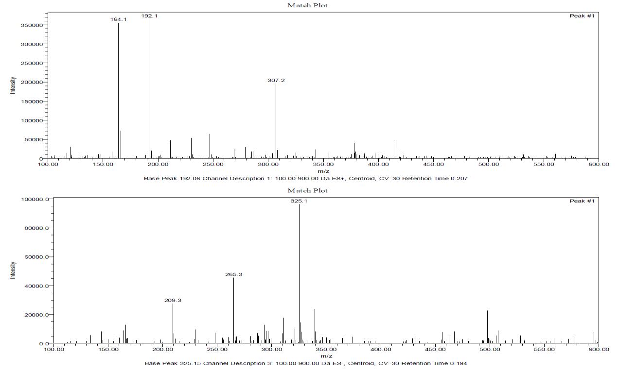
Metal + Ligand - 1,3-benzoxazole (118) peak = 323.0 amu
Metal + Ligand - 1,3-benzoxazole (118) – CH2 peak = 307.1 amu
ISSN 2348-313X (Print)
International Journal of Life Sciences Research ISSN 2348-3148 (online) Vol. 10, Issue 4, pp: (27-34), Month: October - December 2022, Available at: www.researchpublish.com
Metal + Ligand – Ph – (ES-) peak = 362.5 amu
Molecular weight of all complexes are higher than 1000 amu, the mass spectra are taken upto 600 amu only. The fragments present in the spectra largely confirm the probable structures proposed.
Figure 3: Mass Spectra Ligand BSPA
Figure 4: Mass Spectrum of Gd-BSPA
ISSN 2348-313X (Print)
International Journal of Life Sciences Research ISSN 2348-3148 (online)
Vol. 10, Issue 4, pp: (27-34), Month: October - December 2022, Available at: www.researchpublish.com
Based upon all the experimental data of physico chemical analyses, the structures of the Gd3+metal complex can be shown as below.
Figure 5: Gd-BSPA probable structure
This portion describes preparation of the ligand BSPA and its complexes with three lanthanide ions. Usual laboratory tests (M.P., U.V-visible spectra, TLC, colour etc.) confirmed formation [21,22,23] of coordination compounds which were finally characterized by (IR, Mass, TGA etc.). Electronic spectra and magnetic moment values gave information regarding number of unpaired electrons, spin –orbit coupling[21,22,23], charge transfer bands, probable geometry etc. Combining all these information, the tentative structures were assigned to the new complex [21,22,23]
[1] Vovna, V. I., Korochentsev, V. V., Cherednichenko, A. I., & Shurygin, A. V. Photoelectron spectroscopy and electronic structures of β-diketonate complexes of rare-earth elements. Russian Chemical Bulletin, 64(8), (2015).p1701-1712.
[2] Romero-Freire, A., Joonas, E., Muna, M., Cossu-Leguille, C., Vignati, D. A. L., & Giamberini, L. Assessment of the toxic effects of mixtures of three lanthanides (Ce, Gd, Lu) to aquatic biota. Science of the Total Environment, 661, (2019).p-276-284.
[3] Hernandez, A., Jenkins, J., Maslen, H., Zeller, M., Horner, G., Dempsey, C., ... & Zehnder, R. A. Stress compensation in an extended series of lanthanide 2-sulfonatoterephthalates [Ln (TPSO3)(H2O) 2] n (Ln= Ce− Lu, except Pm). Inorganica Chimica Acta, 471, (2018).p-104-112.
[4] Satyendra, R. V., Vishnumurthy, K. A., Vagdevi, H. M., Rajesh, K. P., Manjunatha, H., and Shruthi, A. “Synthesis, in vitro antioxidant, anthelmintic and molecular docking studies of novel dichloro substituted benzoxazole-triazolothione derivatives. European journal of medicinal chemistry, 46(7), (2011). p- 3078-3084.
[5] Paramashivappa, R., Kumar, P. P., Rao, P. S., and Rao, A. S. “Design, synthesis and biological evaluation of benzimidazole/benzothiazole and benzoxazole derivatives as cyclooxygenase inhibitors”. Bioorganic and medicinal chemistry letters, 13(4), (2003). p-657-660
[6] Rizwana, B., & Lakshmi, S. S. Synthesis, characterisation and antimicrobial studies of Zn (II), Ni (II) and Cu (II) complexes of a Schiff base derived from o-vanillin and N-allyl thiourea. International Journal of ChemTech Research, 4(1), (2012).p- 464-473.
ISSN 2348-313X (Print) International Journal of Life Sciences Research ISSN 2348-3148 (online) Vol. 10, Issue 4, pp: (27-34), Month: October - December 2022, Available at: www.researchpublish.com
[7] Zamfir, G., Stanica, N., Draghici, C., & Kriza, C. A. A. Some Transitional Metal Complexes of 4dimethylaminobenzilidene-2 mercaptoaniline. Rev. Chim, 63, (2012).p-1176-1180.
[8] Asakuma, N., Hirashima, H., Imai, H., Fukui, T., & Toki, M. Crystallization and reduction of sol-gel-derived zinc oxide films byirradiation with ultraviolet lamp. Journal of Sol-Gel Science and Technology, 26(1), (2003).p-181-184.
[9] Stone, G. W., Ellis, S. G., Cannon, L., Mann, J. T., Greenberg, J. D., Spriggs, D., ... & TAXUS V Investigators, F. T. Comparison of a polymer-based paclitaxel-eluting stent with a bare metal stent in patients with complex coronary artery disease: a randomized controlled trial. Jama, 294(10), (2005).p-1215-1223.
[10] Wang, X. Y., Avendaño, C., & Dunbar, K. R. Molecular magnetic materials based on 4d and 5d transition metals. Chemical Society Reviews, 40(6), (2011).p-3213-3238.
[11] Anisimov, V. I., Aryasetiawan, F., & Lichtenstein, A. I. First-principles calculations of the electronic structure and spectra of strongly correlated systems: the LDA+ U method. Journal of Physics: Condensed Matter, 9(4), (1997). p767.
[12] Nakamoto, Kazuo, Marvin Margoshes, and R. E. Rundle. "Stretching frequencies as a function of distances in hydrogen bonds." Journal of the American Chemical Society 77.24 (1955): p-6480-6486.
[13] Liu, G., & Jacquier, B. (Eds.). Spectroscopic properties of rare earths in optical materials (Vol. 83). Springer Science & Business Media. (2006).
[14] Benedek, N. A., & Birol, T. ‘Ferroelectric’metals reexamined: fundamental mechanisms and design considerations for new materials. Journal of Materials Chemistry C, 4(18), (2016).p-4000-4015.
[15] Jotham,R.W., S.F. A. Kettle, andJ.A.Marks."Antiferromagnetismintransition-metalcomplexes.PartIV.Lowlying excited states of binuclear copper (II) carboxylate complexes." Journal of the Chemical Society, Dalton Transactions 3 (1972): p-428-438.
[16] Osborn, J. A., Jardine, F. H., Young, J. F., and Wilkinson, G. “The preparation and properties of tris (triphenylphosphine) halogenorhodium(I)andsomereactionsthereofincludingcatalytic homogeneous hydrogenation of olefins and acetylenes and their derivatives.” Journal of the Chemical Society A: Inorganic, Physical, Theoretical,(1966) p-1711-1732.
[17] Quan, C., Li, A., & Gao, N. Thermogravimetric analysis and kinetic study on large particles of printed circuit board wastes. Waste Management, 29(8), (2009).p-2353-2360.
[18] López-González, D., Fernandez-Lopez, M., Valverde, J. L., & Sanchez-Silva, L. Kinetic analysis and thermal characterization of the microalgae combustion process by thermal analysis coupled to mass spectrometry. Applied Energy, 114, (2014).p-227-237.
[19] Buhr, K., van Ruth, S., & Delahunty, C. Analysis of volatile flavour compounds by Proton Transfer Reaction-Mass Spectrometry: fragmentation patterns and discrimination between isobaric and isomeric compounds. International Journal of Mass Spectrometry, 221(1), (2002).p-1-7.
[20] Schwarz, K., Filipiak, W., & Amann, A. Determining concentration patterns of volatile compounds in exhaled breath by PTR-MS. Journal of Breath Research, 3(2), (2009). P-027002.
[21] Gauthama, B. U., Narayana, B., Sarojini, B. K., Manjunatha, J. G., & Suresh, N. K. Colorimetric ‘naked eye’sensor for fluoride ion based on isatin hydrazones via hydrogen bond formation: design, synthesis and characterization ascertained by nuclear magnetic resonance, ultraviolet–visible, computational and electrochemical studies. Inorganic Chemistry Communications, 121, (2020). P-108216.
[22] Kumar, S. M., Hezam, A. O. F., Manjunath, B. C., Shamprasad, V. R., Mohammed, Y. H. E., Mahesh, N., & Byrappa, K. Crystal packing analysis of 1-(3, 4-dimethoxyphenyl)-3-(4-bromophenyl) prop-2-en-1-one exhibiting a putative halogen bond CBr⋯ O. Journal of Molecular Structure, 1156, (2018).p-216-223.
[23] Shah, M. P. Microbial Degradation of Acid Orange Dye by an application of Pseudomonas spp. ETL-1979 isolated from the textile dye effluent: An Environmental Bioremedial Approach. Biotechnology, 3(1), (2014).p-3.