
International Journal of Engineering Research and Reviews ISSN 2348-697X (Online) Vol. 10, Issue 2, pp: (43-47), Month: April - June 2022, Available at: www.researchpublish.com
AReview onAutomatic Detection and Recognition of Hard Exudates in Color Retinal Fundus Images
Rajesh I S1, Bharathi M A2, Bharathi M R3 , Nazneen Kiresur4
1*BMSIT & M, Bengaluru, Karnataka
2 BMSIT&M, Bengaluru, Karnataka 3BEC, Bagalkot, Karnataka
4Student REVA University, Karnataka
DOI: https://doi.org/10.5281/zenodo.6778796
Published Date: 29-June-2022
Abstract: Diabetes is a condition where glucose level in the body is high. It is one of the prominent causes of retinal diseases. The survey done by the International Diabetes Federation (IDF) in 2017 states that, as of now 425million people on the earth are experiencing diabetes and that India is the second biggest nation [1]. People with diabetes may experience retinal disease such as Diabetic Retinopathy (DR), Diabetic Maculopathy (DM), etc. Knowing at the right time about these diseases and keeping the glucose level in the body properly helps in avoiding retinal diseases. DR is one of the retinal diseases found in diabetic patients which may lead to painless vision loss. Hard Exudates (HEs) are the accumulated fat formation in the retina leaked from damaged blood vessels of the retina and it is an early clinical sign of DR and DM. Thus, detection of HE at an early stage helps in preventing vision loss in patients. This survey paper gives a short presentation about the human eye, retina, diabetes and its complications. This paper also talks about the various methods/techniques that are used in automatic detection of hard exudates and the various issues involved in it.
Keywords: Hard Exudates (HEs), Fovea, Macula, Optic Disk, Diabetic Retinopathy (DR), Diabetic Maculopathy (DM).

I. INTRODUCTION
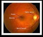
The human eye is a vital body part answerable for vision [2]. It is housed in the orbit, a bone socket that is protected from the outside world by the eyelids. The retina is a nerve layer that runs from the back of the eye to the front. It is made up of millions of photo receptors that translate light signals into electrical signals, which are then sent to the brain via the optic disc, where they are converted into images. These signals travel to the brain via optic disc where they turn into images [3]. Figure1 (a) shows the cross section of the eye, while that of the retina is shown in Figure1 (b).
Figure1: (a) Cross Section of the Human Eye, (b) Human Retina
Page | 43 Research Publish Journals
(a) (b)
International Journal of Engineering Research and Reviews ISSN 2348-697X (Online) Vol. 10, Issue 2, pp: (43-47), Month: April - June 2022, Available at: www.researchpublish.com
Diabetes is a condition where glucose level in the body is high due to the deficit of insulin production or the body is not responding to the insulin produced. Disease to the retina because of diabetes is called Diabetic Retinopathy (DR). It damages the small blood vessels present in the retina and also this might lead to vision loss. If diabetic retinopathy is diagnosed early enough, laser photo coagulation therapy can help delay the disease's progression. However, this is a difficult job because patients with DR have no early signs until their vision begins to deteriorate. To prevent vision loss and to ensure that the treatment is received at an early stage, the patients who are suffering from diabetes need to undergo a yearly retinal fundus examination [4].
Hard exudates are one of the early clinical signs of DR seen during a retinal fundus test. Figure 2 shows how HEs can range in size from a tiny dot to huge patches, all with straight edges. Since light is unable to penetrate the retina, this may impair vision.
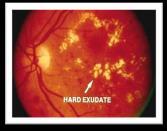
Figure 2: Hard Exudates
The remainder of the paper is structured as follows: Section 2 provides a brief overview of the literature on hard exudate detection. The issues involved in hard exudate detection are mentioned in section 3. The conclusion is given in section 4.
II. LITERATURE SURVEY
Hard exudates are one of the first clinical signs of DR on a retinal fundus test. Hard exudates can differ in size with clear edges from a tiny dot to large patches. This can affect vision by preventing light from reaching the retina. Several methods for detecting and recognising hard exudates have been suggested. Diabetic retinopathy and maculopathy are some of the most recent methods/techniques that are summarized in this section.
Author Chenxi Huang, et al. [5] (2020) proposed a method for detecting hard exudates in color retinal images. Super pixels considered as candidates are first created on each resized image using a super pixel segmentation algorithm called Simple Linear Iterative Clustering (SLIC). After that, each feature generates 25 features from resized images and patches.Patches are then used to train a deep convolutional neural network that distinguishes the hard exudates from the surrounding environment.The proposed method is evaluated on the E-Ophtha EX, DIARETDB1 and IDRiD database and achieved sensitivity, specificity and accuracy of 97.96%, 90.84%, 97.58% with e-ophtha EX,98.21%, 91.38%, 98.01% with DIARETDB1 and 98.40%, 90.67%, 98.19% with IDRiD.
Author Shengchum Long, et al. [6] (2019) proposed a method for recognising hard exudates in color retinal images. Preprocessing of images is initially performed followed by optical disc detection and masking. Then, in accordance with the global FCM-based threshold, the HE candidate is defined using the variable threshold. Finally, eight texture characteristics were extracted from the nominee HE area and fed into the SVM classifier for automatic HE classification.For the e-ophtha EX database, this approach yields 76.5 %, 82.7 %, and 76.7 % sensitivity, PPV, and Fscore, respectively and 97.5 %, 97.8 % and 97.7 % sensitivity, specificity and accuracy for the DIARETDB1 database. DiptoneelKayal, et.al, [7] (2018) presented a TABUsearch method based on dynamic thresholding to identify hard exudates in fundus images. In this technique grayscale image of the input image is used to eliminate the optic disc using SobelEdge Detection and Hough Transform. Later, additive noise is removed by using a median filter. Finally, image thresholding is applied to detect hard exudates. The method is verified using DIARETDB0 and DIARETDB1 database and achieved a sensitivity of 97.45% and specificity of 96.85%.
Author WorapanKusakunniran, et.al [8] (2017), proposed a hard exudates detection method in the color retinal image. Initially, color transfer is applied to increase the image quality. Then, using iterative blob detection hard exudates are
Page | 44 Research Publish Journals
International Journal of Engineering Research and Reviews ISSN 2348-697X (Online)
Vol. 10, Issue 2, pp: (43-47), Month: April - June 2022, Available at: www.researchpublish.com
detected by considering color, size and shape. Later for the final segmentation process, detected blobs are used as the initial foreground of hard exudates. The proposed method is tested on E-Ophtha, DIARETDB1 database and achieved sensitivity and specificity of 96%, 94% with e- ophtha and 96%, 98% with DIARETDB1.
Author Poonam M. Rokade, et al., (2015) proposed a method for identifying hard exudates in color retinal fundus photographs. In this process, the hard exudates are segmented using Haar wavelets transform, and then the hard exudates are classified using k nearest neighbor classification. This method is tested on four databases and obtained a sensitivity of 37.14%, 21.87%, 12.50%, and 25.47% for MISP, DIARETDB0, DIARETDB1 and STARE databases respectively.
AuthorDiptoneelKayal [10], (2014) et al. developed a new method for detecting hard exudates in color retinal fundus images. The input color image is converted to grayscale and the median filter is applied in this process. The grayscale image is then subtracted from the median filtered image. Later dynamic thresholding is used for image segmentation. Finally, thresholded and grayscale images are added for hard exudates. The proposed method was tested on 219 images from the DIARETDB0 and DIARETDB1 databases and found to have a sensitivity of 97.25 % and a specificity of 96.85 %.
H.B. Kekre et al. [11], (2013) presented a method for detection and recognition of hard exudate in fundus images. Initially detection using mathematical morphology is discussed followed by a hybrid approach. The input image is resized and Morphological Dilation is applied, followed by the Linde-BuzoGray clustering method. Finally, HEs are detected using the K-means algorithm.
KittipolWisaeng et.al [12], (2013) proposed a method for automated detection of exudates in poor quality color retinal images. The raw images were pre-processed and the optic disc is removed based on intensity property. In order to classify candidate regions as exudates and non-exudates, a set of extracted features from each region were used as inputs to the neural network.
The author Xiang Chen et al. [13] (2012) proposed a novel method for detecting hard exudates in color retinal pictures. In this method the exudate candidate regions are extracted by merging Histogram Segmentation and Morphological Reconstruction later 44 features are defined for each candidate region to extract true exudates. Finally, Supervised SVM is trained and tested to classify the HEs. DIARETDB1 is used to test the method, which yields a sensitivity of 94.7 % and a positive predictive value (PPV) of 90.0 %. Multi features of hard exudates help in distinguishing efficiently HEs from Cotton Wool Spot (CWS).
Hussain et.al [14], (2012) proposed a system using adaptive thresholding for hard exudate detection in retinal images. Using region-based segmentation technique, the image is decomposed into a number of homogeneous sub images. Later edges are detected using a morphological gradient technique. Finally, using rule-based classifier exudates are classified from non-exudates. To overcome the overhead of merging operations faced in the split-and-merge method, a puresplitting method is used.
Berrichi Fatima Zohra et al. [15] (2010) used color retinal fundus images to suggest a computer-based method for identifying Non-Proliferative Diabetic Retinopathy (NPDR) and Proliferative Diabetic Retinopathy (PDR). The SVM classifier is used to train and evaluate features extracted from segmented images using image processing techniques.
Automated detection of hard exudates in color retinal fundus images is presented in [16] (2010). In this method candidate regions are identified using Split-and-Merge technique and Adaptive Thresholding. Then hard exudates are extracted using a Local Variation Operator. The Split-And-Merge method is overhead because of merging operations.
AkaraSopharak et.al [17], (2007) presented an automated method for hard exudates detection incolor retinal fundus images having low-contrast. For segmentation, Morphological Reconstruction and the Fuzzy C-Means clustering process are used. K-means clustering is used to distinguish between Exudates and Non-Exudates.
III. ISSUES IN HARD EXUDATES DETECTION
The scale of hard exudates fluctuates, making identification difficult. ∙ Hard exudates detection may fail because of similarity in brightness of the optic disc and hard exudates.
Blood vessels in the retina affect the exudate segmentation. ∙ Poor illumination of the retinal image makes hard exudates detection difficult.
Page | 45 Research Publish
Journals
International Journal of Engineering Research and Reviews ISSN 2348-697X (Online) Vol. 10, Issue 2, pp: (43-47), Month: April - June 2022, Available at: www.researchpublish.com
The appearance of yellowish lesions on the retina, such as cotton wool spots, makes segmentation of hard exudates difficult.
It is very difficult to segment exudates which are formed near the OD region because of OD brightness.
It is very difficult to detect hard exudates if more drusens are present. ∙ The vessel reflection and optical artifacts complex the small exudate detection.
Presence of bright regions along the FOV border makes exudate detection difficult.
IV. CONCLUSION
In this survey paper, a wide variety of methods for detection of diabetic retinopathy and maculopathy has been presented. In the medical field, there are many challenges in hard exudate detection. Based on this survey, it is found that very little research has been done by using deep learning. To increase the accuracy of automated systems possible direction is to use deep learning. This survey paper can act as a resource for the upcoming scholars and students who are interested in retinal image analysis. In addition to this, it also helps them to get an overview about the structure of the human eye, retinal anatomical components, diabetes and diabetic retinopathy.
REFERENCES
[1] https://www.idf.org/our-activities/congress/hyderabad-2018.html
[2] http://www.sightwavers.or.uk/html/eyeconditions/huma_eye_detailed.html
[3] http://www.myeyeworld.com/files/eye_structure.html
[4] Iqbal, M.I, Aibinu, A.M, ''Automatic Diagnosis of Diabetic Retinopathy Using Fundus Images'‘, Master’s Thesis, Department of Signal Processing, Blekinge Institute of Technology, Year. Oct 2006.
[5] Huang, Chenxi, et al. "A new deep learning approach for the retinal hard exudates detection based on superpixel multi-feature extraction and patch-based CNN." Neurocomputing (2020). Huang, C., Zong, Y., Ding, Y., Luo, X., Clawson, K., & Peng, Y. (2020). A new deep learning approach for the retinal hard exudate’s detection based on superpixel multi-feature extraction and patch-based CNN. Neurocomputing.
[6] Huang, Chenxi, Yongshuo Zong, Yimin Ding, Xin Luo, Kathy Clawson, and Yonghong Peng. "A new deep learning approach for the retinal hard exudates detection based on superpixel multi feature extraction and patch-based CNN." Neurocomputing (2020).
[7] Huang, C., Zong, Y., Ding, Y., Luo, X., Clawson, K. and Peng, Y., 2020. A new deep learning approach for the retinal hard exudate’s detection based on superpixel multi-feature extraction and patch-based CNN. Neurocomputing
[8] Huang C, Zong Y, Ding Y, Luo X, Clawson K, Peng Y. A new deep learning approach for the retinal hard exudate’s detection based on superpixel multi-feature extraction and patch-based CNN. Neurocomputing. 2020 Dec 24.
[9] Shengchun Long, Xiaoxiao Huang, Zhiqing Chen, Shahina Pardhan, Dingchang Zheng, "Automatic Detection of Hard Exudates in Color Retinal Images Using Dynamic Threshold and SVM Classification: Algorithm Development and Evaluation", BioMed Research International, vol. 2019, Article ID 3926930, 13 pages, 2019. https://doi.org/ 10.1155/2019/3926930
[10] Kayal, Diptoneel, and Sreeparna Banerjee. "Dynamic Thresholding with Tabu Search for Detection of Hard Exudates in Retinal Image."In Industry Interactive Innovations in Science, Engineering and Technology, pp. 553560. Springer, Singapore, 2018.
[11] Kusakunniran, Worapan, QiangWul,et.al,. "Three-stages hard exudates segmentation in retinal images." In Information Technology and Electrical Engineering (ICITEE), 2017 9th International Conference on, pp. 1-6. IEEE, 2017.
[12] Rokade, Poonam M., and Ramesh R. Manza. "Automatic detection of hard exudates in retinal images using haar wavelet transform." eye 4, no. 5 (2015): 402-410.
Publish Journals
Page | 46
Research
International Journal of Engineering Research and Reviews ISSN 2348-697X (Online) Vol. 10, Issue 2, pp: (43-47), Month: April - June 2022, Available at: www.researchpublish.com
[13] Kayal, Diptoneel, and Sreeparna Banerjee. "A new dynamic thresholding-based technique for detection of hard exudates in digital retinal fundus image." In Signal Processing and Integrated Networks (SPIN), 2014 International Conference on, pp. 141-144. IEEE, 2014.
[14] H. B. Kekre,Tanuja K, et.al, “Hybrid Approach for Detection of Hard Exudates'', International Journal of Advanced Computer Science and Applications, Vol. 4, pp. 250-255, Year. Feb-2013. [12] KittipolWisaeng, Nualsawat Hiransakolwong, et.al, “Automatic Detection of Retinal Exudates Using a Support vector Machine”, Research on Applied Medical Informatics, Vol. 32, pp. 33-42, Jan-2013.
[15] Hussain F. Jaafar, et.al, “Detection of Exudates from Digital Fundus Images Using a Region based Segmentation Technique”, 19th European Signal Processing Conference, pp. 1020-24, Year. Aug 2011.
[16] Xiang Chen, Wei Bu, et.al, “A Novel Method for Automatic Hard Exudates Detection In Color Retinal Images”, Proceedings Of The 2012 International Conference On Machine Learning And Cybernetics, pp. 1175-1181, Year. July-2012.
[17] Berrichi Fatima Zohraet.al,”Automated diagnosis of retinal images using the Support Vector Machine (SVM).”, International Journal of Computer Applications, Vol. 4, pp. 176-183, Year. July 2010.
[18] Hussain F. Jaafar et.al, “Automated Detection of Exudates in Retinal Images Using a Split and Merge Algorithm”, 18th European Signal Processing Conference, pp. 23-27, Year. Aug-2010 [17] AkaraSopharak, Bunyarit Uyyanonvara, “Automatic Exudates Detection from Diabetic Retinopathy Retinal Image Using Fuzzy C-Means and Morphological Methods”, Proceeding of Third IASTED International Conference Advance In Computer Science And Technology, pp. 359-364, Year. April-2007.
Page | 47 Research Publish
Journals