HEMICHORDATA
BY SIDNEY F. HARMER, Sc.D., F.R.S. Fellow of King's College, Cambridge.
CHAPTER I
HEMICHORDATA
CHORDATA AND VERTEBRATA HEMICHORDATA ENTEROPNEUSTA EXTERNAL CHARACTERS AND HABITS—STRUCTURE—GENERA—DEVELOPMENT— PTEROBRANCHIA—CEPHALODISCUSAND RHABDOPLEURA—PHORONIDEA— PHORONISAND ACTINOTROCHA AFFINITIES OF THE HEMICHORDATA.
The Hemichordata, a marine group which includes the worm-like Balanoglossus, owe much of their interest to the fact that they are believed by many zoologists to be related to the lower Vertebrates. This view is one of a number of mutually exclusive hypotheses, which seek to derive Vertebrate animals from various Invertebrate ancestors. It is supported by many striking resemblances between Balanoglossus and the lowest forms which are by common consent regarded as belonging to the Vertebrate alliance; but it must be distinctly understood that Balanoglossus is at most the much modified modern representative of extinct forms which were also the ancestors of Vertebrates.
The axis of the backbone of all Vertebrates is formed by an elastic rod known as the "notochord" (Figs. 72, 115), which lasts
throughout life in some of the lowest forms, but in the higher forms appears only in the embryo. The universal occurrence of this structure has been regarded as the most important characteristic of the Vertebrata and their allies, which are accordingly grouped together in the Phylum CHORDATA. The members of this Phylum are further distinguished from other animals by several important features. Of these one of the most important appears to be the existence of lateral outgrowths of the pharynx, which unite with the skin of the neck and form a series of perforations leading to the exterior. These structures are the gill-slits, and in the Fishes their walls give rise to vascular folds or gills. With the assumption of a terrestrial life, the higher Vertebrates lost their gills as functional organs, respiration being then performed by entirely different organs, the lungs. But even in these cases, the gill-slits appear in the embryo; and remains of one pair can usually be recognised in the adult state of even the highest Vertebrates. Another fundamental characteristic of the Chordata is given by the central nervous system, which lies entirely above the alimentary canal, just dorsal to the notochord. Not only does this position of the nerve-centres distinguish the Chordata from Invertebrates, but a further point of difference is found in the development. While in Invertebrates the ventral nerve-cord is formed as a thickening of the ectoderm or outermost layer of the embryo, in the Chordata the nervous system is usually formed as a longitudinal groove running medianly along the back of the embryo. This groove closes to form a tube of nervous matter, the cavity of which always persists throughout life as the "central canal" of the spinal chord and its anterior prolongation which constitutes the "ventricles" of the brain.
Although the animals which are considered in this chapter are not admitted by all zoologists to be related to the Vertebrates, there can be no question that their respiratory organs closely resemble typical gill-slits. Since, moreover, they possess structures which can be regarded, with a fair amount of probability, as agreeing in essential respects with the notochord and the tubular dorsal nervous system
of Vertebrates, it appears justifiable to include them in the Chordata, which are then subdivided into (1) HEMICHORDATA, in which a "notochord" occurs in the anterior end of the body only; (2) UROCHORDATA (Tunicata or Ascidians), in which the notochord is restricted to the tail; (3) CEPHALOCHORDATA (Amphioxus), in which the notochord extends the entire length of the body andofthehead; (4) CRANIATA, in which a brain is developed as an enlargement of the central nervous system, the notochord does not extend farther forward than the middle of the brain, and a vertebral column is present. These last are thus usually known as Vertebrata, although in distinguishing an "Invertebrate" from a "Vertebrate" it is more logical to regard all Chordata as Vertebrates, since the Invertebrata are in no sense a natural group with common characteristics, their union under one name merely implying that they have no close affinity to the Vertebrates. It is often convenient in practice to divide animals into Vertebrates and Invertebrates, but from a zoological point of view a division of the animal kingdom into Molluscs and Non-Molluscs would have as much or as little significance.
The sub-phylum Hemichordata[1] consists of the Orders:—(I.)
ENTEROPNEUSTA, [2] including Balanoglossus (Fig. 1); (II.) PTEROBRANCHIA, [3] represented by the genera Cephalodiscus (Fig. 9) and Rhabdopleura (Fig. 12). To these should possibly be added (III.)
PHORONIDEA, for the reception of Phoronis(Fig. 13).
Order I. Enteropneusta.
Worm-like Hemichordata, with numerous gill-slits, a straight intestine,andaterminalanus.Proboscisseparatedbyanarrowstalk from the simple ring-shaped collar , which is succeeded by an elongatedtrunk.
The structure of Balanoglossus, formerly the sole genus belonging to this Order, but now divided[4] into the genera Ptychodera, Balanoglossus, Glossobalanus, Glandiceps, Spengelia, Schizocardium, Harrimania, Dolichoglossus, and Stereobalanus, has of recent years formed the subject of elaborate investigations by Spengel,[5] Bateson,[6] and Willey.[7] More than thirty species are known, ranging in length from 25 mm.[8] (Pt. bahamensis) to 2500 mm. (B. gigas), and for the most part inhabiting shallow water; Glossobalanus sarniensis occurring between tide-marks in the Channel Islands. Glandiceps talaboti has, however, been dredged near Marseilles from as much as 190 fathoms, while G. abyssicola was found by the "Challenger" at a depth of 2500 fathoms, off the West Coast of Africa.
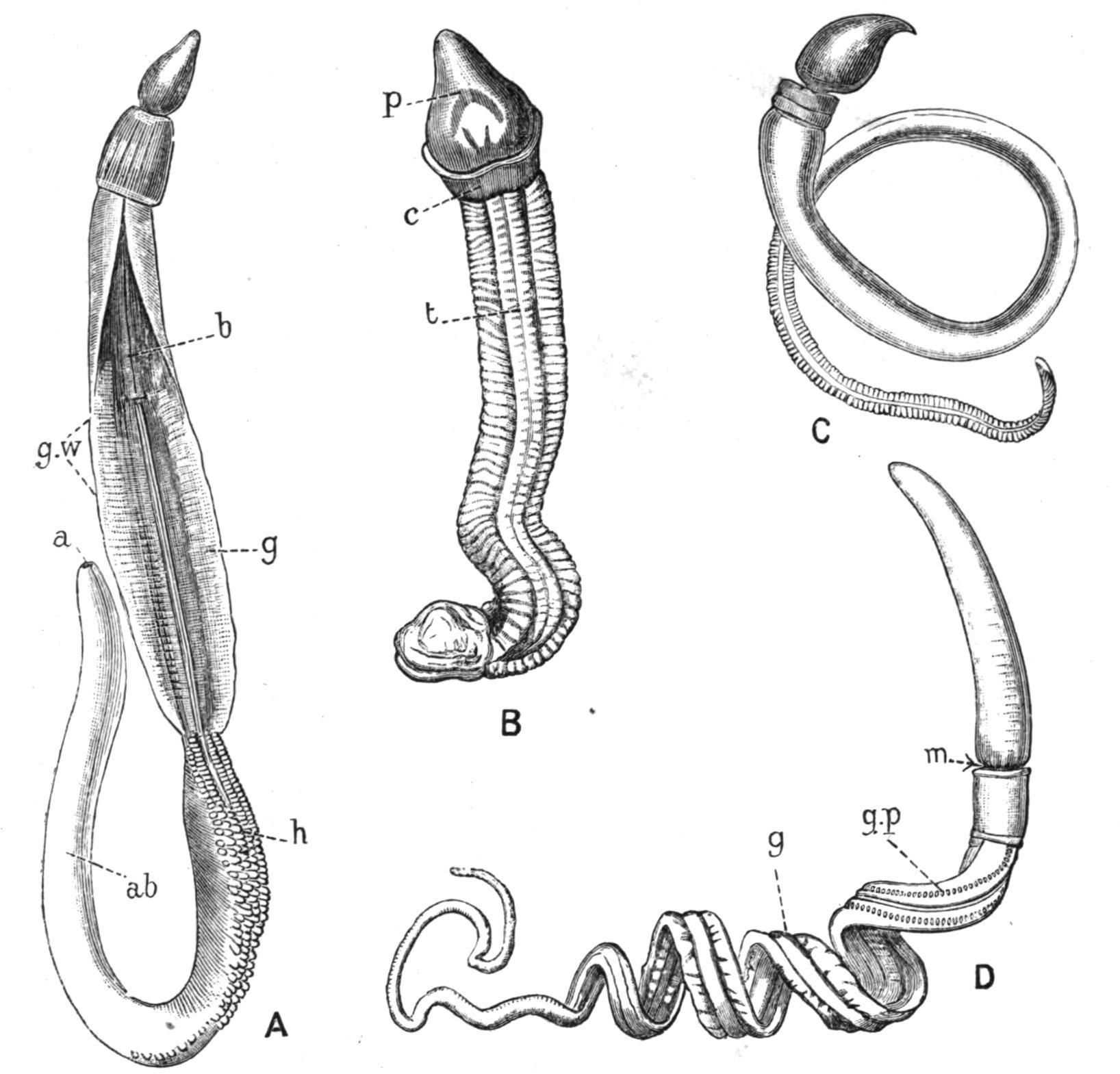
FIG. 1. Forms of Balanoglossus. A, Balanoglossus clavigerus, Eschsch., Naples, × ½; B, Glandiceps hacksi, Mar. (incomplete), Japan, × 1; C, Schizocardium brasiliense, Speng., Rio de Janeiro, × 1; D, Dolichoglossus kowalevskii, A.Ag., Chesapeake Bay, × 1. a, Anus; ab, abdominal and caudal regions; b, branchial region; c, collar; g, genital region; g.p, gill-pore; g.w, genital wing; h, hepatic region; m, position of mouth; p, proboscis; t, trunk. (A, B, and C from Spengel; D from Bateson.)
Balanoglossus, the largest genus now recognised by Spengel, appears to be practically world-wide in its distribution;
Schizocardiumis recorded from both sides of S. America; Glandiceps from the Atlantic, the Mediterranean, Japan, and the Indian Ocean; Spengelia from the South Pacific; and other species from the White Sea to New Zealand. The habitat is usually sand or gravelly sand, in which the animal forms a kind of tube by means of the abundant mucus secreted by its skin. Dolichoglossus kowalevskii (Fig. 1, D), according to Bateson,[9] lives between tide-marks at a depth of about eight inches. The greater part of the body is coiled in an even, cork-screw-like spiral, while the anterior end, including the front part of the branchial region, is maintained in a vertical position. The posterior end is also kept upright, and can be moved up and down in a vertical shaft opening on the surface, thus enabling the animal to eject the undigested sand from its anus.
The coloration of Balanoglossus is often brilliant. That of D. kowalevskii[10] is as follows:—The "proboscis" (cf. Fig. 1, B, p) is yellowish white; the "collar" (c) is brilliant red-orange (especially in males), with a white ring posteriorly; the "trunk" (t), the subdivision of which into "branchial," "genital," "hepatic," "abdominal," and "caudal" regions is better indicated in other species (Fig. 1, A, b, g, h, ab), is orange-yellow, shading to green-yellow in the semitransparent caudal region. The genital region is grey in females and yellow in males, a sexual difference in colour being common in Enteropneusta. The hepatic papillae of other species may be bright green.
The odour of D. kowalevskiiresembles that of "chloride of lime with a faecal admixture," while that of Balanoglossus aurantiacus suggests iodoform. All Enteropneusta are said to have a more or less offensive smell. A species of Balanoglossus is known to be intensely phosphorescent.[11]
The mouth (Fig. 7, m) is situated on the ventral side, at the base of the proboscis, and is concealed by the free anterior edge of the
collar, which encircles the thin "proboscis-stalk" (Fig. 3, p.s). The animal has the singular peculiarity of being unable to close its mouth;[12] and thus, as it burrows through the ground, the sand which passes into the alimentary canal leaves it in a continuous column through the terminal anus.[13] The large coiled "castings" formed in this way between tide-marks enable the experienced collector to infer the presence of Balanoglossus; and in a West Indian species described by Willey[14] they are so large as to form "an important feature in the landscape at low tide."
The principal agents in burrowing are the proboscis and collar. An animal observed by Spengel pushed the tip of its proboscis into the sand, waves of muscular contraction meanwhile passing over the surface of the proboscis. At first the animal made slow progress; but the collar, becoming surrounded by sand, soon became a point of resistance by means of which the proboscis could bury itself yet more deeply. The animal quickly disappeared as soon as the first two regions of its body were engaged in the task of burrowing[15]
This action is due partly to the muscles of the body-wall, but largely to the power possessed by the proboscis and collar of becoming swollen and turgid. Spengel has observed that these parts become flaccid when the animal is taken out of water, and can only swell again when it is replaced therein; and it may thus fairly be concluded that the enlargement is due to the taking in of water. This is probably in fact the most important function of the "proboscispore" and of the "collar-pores" which are described below.
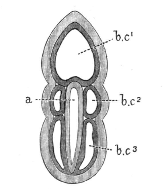
FIG. 2. Diagram of a dorsal view of a Balanoglossus-embryo, after the formation of the body-cavities, a, Alimentary canal; b.c1 , body-
cavity of the proboscis; b.c2 , of the collar; b.c3 , of the trunk. (From Bateson.)
Body-Cavities. The existence of five separate body-cavities (Fig. 2) is one of the most fundamental facts in the anatomy of Balanoglossus. The first body-cavity, or cavity of the proboscis (b.c1), is single and unpaired; the second body-cavities (b.c2) are paired spaces, one belonging to each side of the collar; the third body-cavities (b.c3) are similarly paired, and correspond with the trunk. While there is no connection between successive bodycavities, there are in certain regions communications between the two cavities of the same pair. Each of the paired cavities is at one time a closed lateral space between the skin and the alimentary canal. As the two spaces which constitute the pair grow towards one another, both above and below the alimentary canal, they come into such close apposition that they remain separated only by their conjoined walls. In this way are formed the dorsal and ventral mesenteries (Fig. 4, d.m, v), the former being the only one to persist in the higher Vertebrates. The body-cavities of the adult become to a large extent disguised by being traversed by connective tissue and muscles.
The hinder part of the proboscis-cavity is divided by the forward growth of the notochord (Fig. 3, n) into dorsal and ventral portions. The dorsal cavity in extending backwards becomes further subdivided into right and left halves, the latter typically opening dorsally to the exterior on the proboscis-stalk by an asymmetrical "proboscis-pore" (p.p.). Two symmetrical proboscis-pores may, however, occur, or a median pore connected with the left division of the proboscis-cavity. These may be individual variations within the limits of a single species, or may occur as a normal feature of a species.
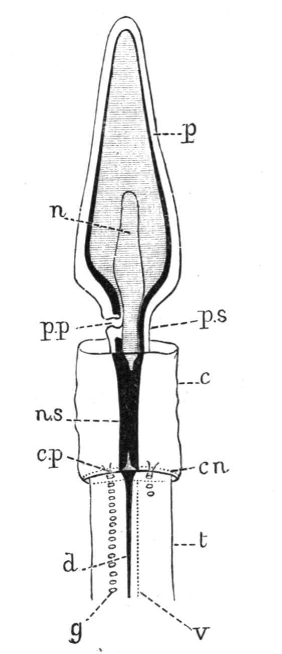
FIG. 3.—Dorsal view of the anterior end of the body of Dolichoglossus kowalevskii, × 3. c, Collar; c.n, circular nerve; c.p, collar-pore; d, dorsal nerve; g, gill-pore; n, notochord; n.s, central nervous system, showing the anterior and posterior neuropores; p, proboscis; p.p, proboscis-pore; p.s, proboscis-stalk; t, trunk; v, ventral nerve. The nerve-plexus of the proboscis is represented as a black line. (After Bateson.)
The collar-cavities open by two "collar-pores" (Fig. 3, c.p.), situated at the posterior end of the collar, into the first pair of gill-pouches, near their external opening. Willey has recently described[16] vestigial pores in relation with the "perihaemal spaces," a pair of dorsally situated outgrowths of the third body-cavities into the collarregion. Narrow "peripharyngeal spaces," also a forward growth of the third body-cavities, closely invest the pharynx in some species.
Body-Wall and Nervous System.—The body-wall (Fig. 4) consists externally of a thick ciliated epidermis (e), containing numerous gland-cells which secrete an abundant mucus. Beneath the epidermis is a basement-membrane, while more internally are layers of muscles, whose arrangement differs in different parts of the body and in different species.
The nervous system consists of a plexus of cells and fibres which lie in the basal part of the epidermis of all parts of the animal, outside the basement-membrane; the thicker portions of the plexus forming definite nerve-tracts. This intimate connexion between the epidermis
and the nervous system is usually restricted to embryonic life in other animals.
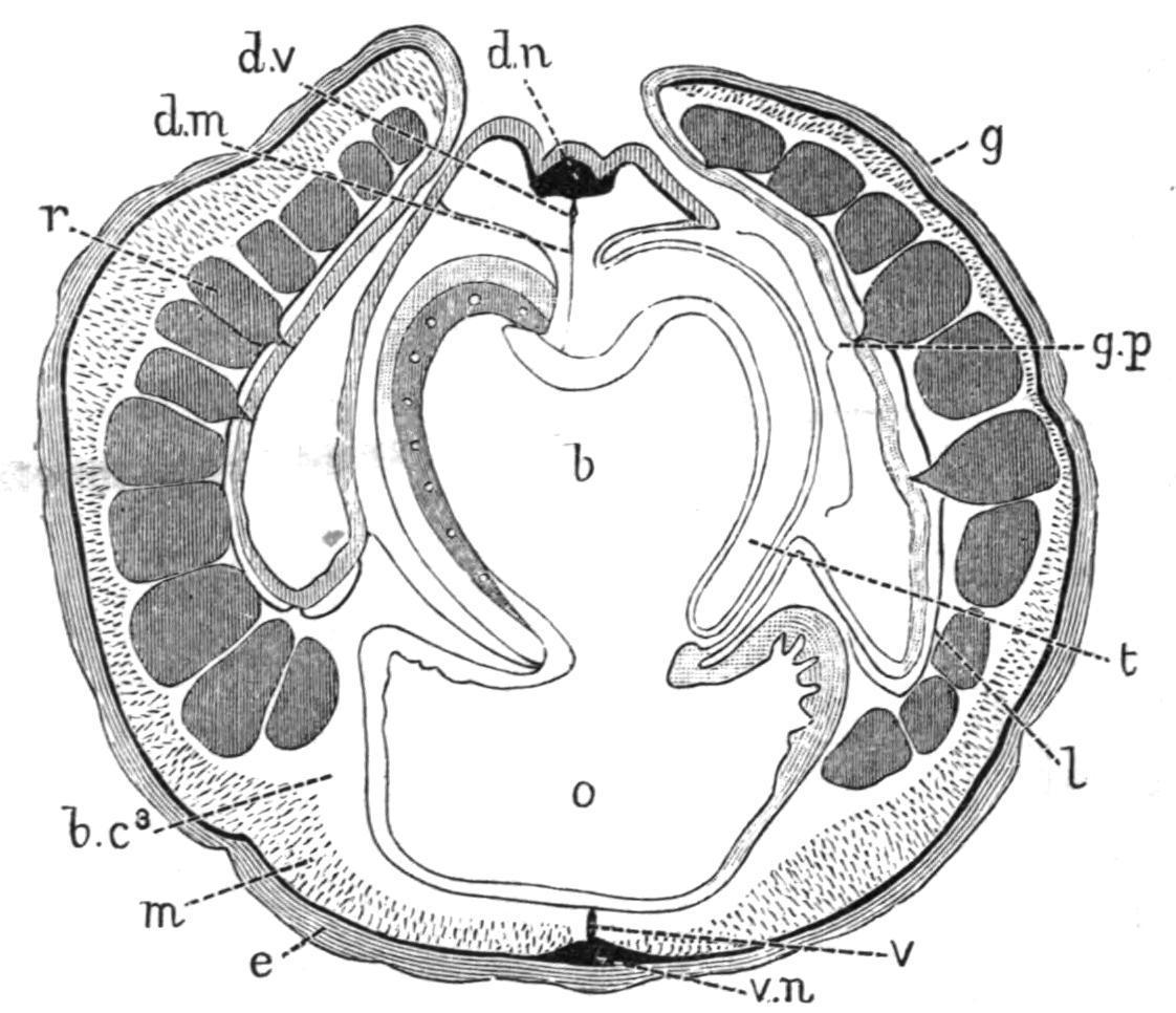
FIG. 4.—Ptychodera bahamensis, Bahama Is. Transverse section through the branchial region. b, Branchial part of pharynx; b.c3 , third body-cavity; d.m, dorsal mesentery; d.n, dorsal nerve; d.v, dorsal vessel; e, epidermis, with nerve-layer (black) at its base; g, genital wing; g.p, gill-pore, encroached on by the tongue-bar (t); l, lateral septum; m, longitudinal muscles; o, oesophageal or alimentary part of pharynx; r, reproductive organ; t, tongue-bar; v, ventral mesentery and ventral vessel; v.n, ventral nerve. (After Spengel.)
The main nerves of Balanoglossus are a dorsal and a ventral tract in the trunk region (Fig. 4, d.n, v.n), a circular tract (Fig. 3, c.n) connecting these two at the posterior edge of the collar, and a strong concentration of nerve-tissue round the whole of the proboscis-stalk, and of the posterior end of the proboscis (Fig. 3). In the region of the collar the nervous system attains its highest development, taking the form of a median cord passing above the alimentary canal. This cord, known as the central nervous system (Fig. 7, n.s), runs through the cavity of the collar, but is connected with the epidermis at each end. It thus becomes continuous in front with the nerve-layer on the proboscis-stalk, while posteriorly it passes into the dorsal and the circular nerve-tracts. In nearly all cases the epidermis is pushed into the cord at the points where it passes into the skin, in the form of an anterior and a posterior "neuropore" (Fig. 3). A transverse section through the extreme front or hind end of the collar accordingly shows a tubular nervous system. In certain species, as in Glossobalanus sarniensis and
Ptychoderaflava, a central canal, opening in front and behind, exists throughout the entire length of the central nervous system, while in G. minutus a canal of this kind occurs in the young animal, but not in the adult. The central nervous system is developed as a longitudinal dorsal groove in the larva,[17] and in a similar manner in the collar which is formed as the result of regeneration after injury. [18] Balanoglossus is thus typically provided with a dorsal, tubular, central nervous system, and although this arrangement does not extend beyond the limits of the collar, it shows a noteworthy resemblance to Vertebrate animals.
In some cases the central nervous system is connected with the dorsal epidermis by a varying number (1-17) of median "roots," which have been compared by Bateson with the dorsal roots of the spinal nerves of Amphioxus, and are probably remains of the embryonic connexion of the collar nervous system with the dorsal epidermis.
Alimentary Canal.—The mouth (Fig. 7, m) leads widely into the alimentary canal, which, passing through the collar, enters the branchial region, where it is characterised by the existence of communications with the exterior. These, the gill-slits, are developed, as in Vertebrates, as paired outgrowths of the alimentary canal, and new gill-slits are constantly being formed at the posterior end of the branchial region with advancing age. The maximum number of the gill-slits, and the extent of the branchial region, are by no means uniform throughout the Enteropneusta. Thus Dolichoglossus otagoensis is said to have no more than 12 pairs, Glossobalanus minutus only 40 pairs, while Balanoglossus aurantiacusmay have as many as 700 pairs. In Ptychoderaflavathe variation is so great that Willey distinguishes[19] two extreme conditions as "macrobranchiate" and "brachybranchiate" respectively, although intermediate conditions are also found. It should be noted
that Balanoglossus agrees with Amphioxus in the indefinite number of the gill-slits.
The gill-slits usually have the form of the so-called "branchial pouches" or "gill-sacs" (Figs. 5, 6, g.s). Each ordinarily opens to the exterior by a small pore (Fig. 1, D, 5, g.p) or slit, situated on the dorsal side, in a shallow longitudinal groove not far from the middle line. The gill-sac has a complete wall of its own, and lies between the alimentary canal and the body-wall, communicating with the former by a U-shaped slit. While a dorsal view of the animal thus shows a linear series of simple pores, a view of the pharynx from the inside appears as in Fig. 5.
At the hind end of the pharynx the inner opening of the developing gill-sac is circular. Slightly further forward the dorsal side of the pore is indented into a crescent, which grows longer in a dorso-ventral direction, and becomes a U, whose two limbs are nearly separated by a mass of tissue, the so-called "tongue-bar" (Fig. 5, t). The special interest of this mode of development is that it is identical with what occurs in Amphioxus (p. 120), which is universally admitted to belong to the Chordata.
The gill-sacs of Balanoglossus follow one another closely, the hind wall of one being in contact with the front wall of the next, and constituting a "branchial septum" (b.s). Both septa and tongue-bars are supported by chitinous rods, which are special thickenings of the membrane at the base of their epithelium. Two rods occur in each tongue-bar, separated by an interval of body-cavity (Figs. 5, 6), and only one rod in each septum. Originally of this form—∩∩ ∩∩—the rods have joined in pairs, the united limbs forming the single rod of each branchial septum. In this respect again we have a similarity between Balanoglossus and Amphioxus, except that in the latter the concrescence proceeds one step farther, and the two rods of the tongue-bar unite, like those of the branchial septum. The latter, the
so-called "primary" skeletal rods of Amphioxus, are forked ventrally as in Balanoglossus (Fig. 5).
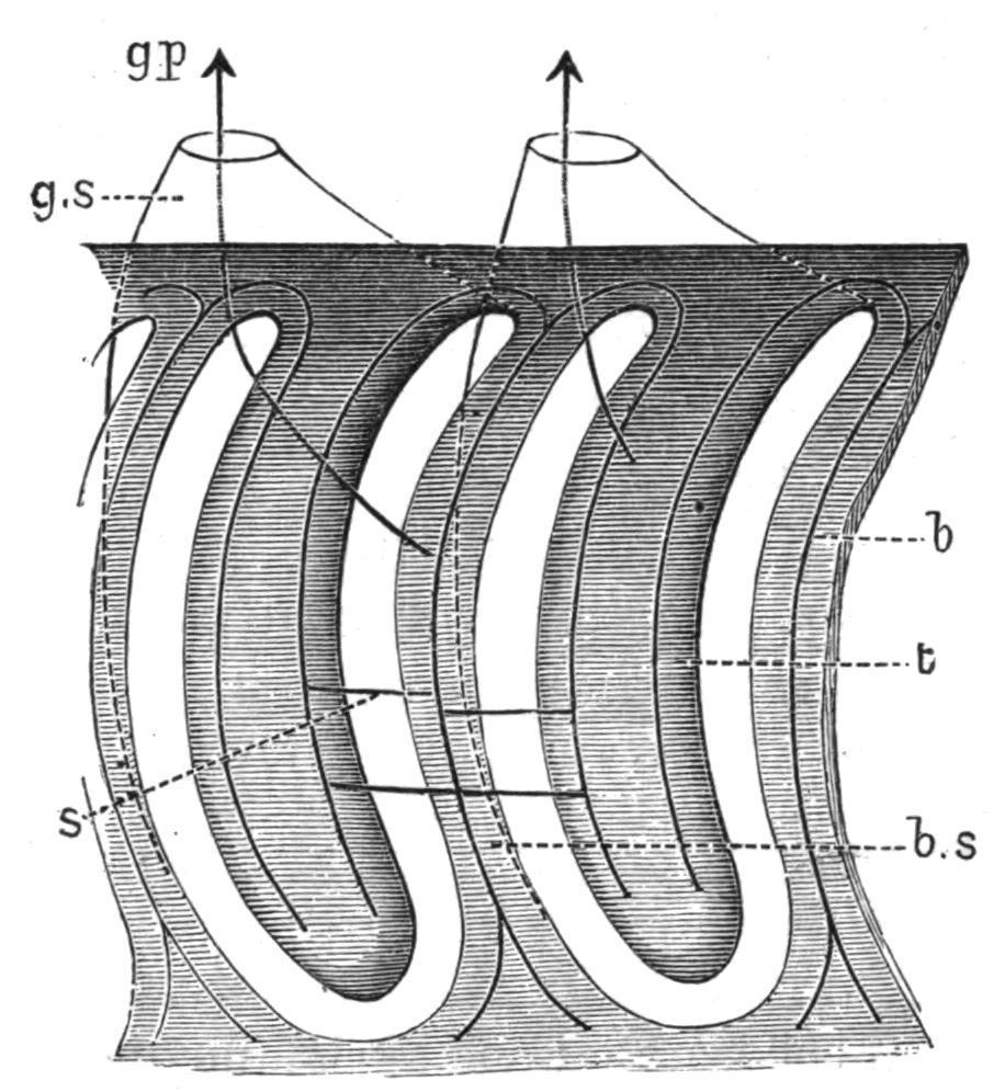
FIG. 5.—Diagram of two gill-sacs of Balanoglossus, seen from the inside of the pharynx. b, Branchial skeleton, consisting of a single forked bar in each branchial septum (b.s), and of two bars in each tongue-bar; g.p, gill-pore, opening on the dorsal surface of the trunk; g.s, gill-sac; s, synapticulum (only one or two shown); t, tongue-bar. The arrows indicate the communications of the gillsacs with the exterior and with the pharynx.
In Amphioxus, as in most Enteropneusta, adjacent rods are connected at intervals by chitinous "synapticula" (Fig. 5, s), which traverse one or the other of the halves of the gill-slit. In Dolichoglossus, where no synapticula occur, the tongue-bars may be turned inside out by slight pressure, and then project to the exterior through the gill-pores.
The subdivision of the branchial region of the alimentary canal into two parts, as shown in Fig. 4, is characteristic of Glossobalanusand its allies. In Dolichoglossus and Glandiceps there is no such constriction, the region occupied by the gill-slits being merely the dorsal half of a tube with a simple circular section. Schizocardium (Fig. 6) agrees with Amphioxus in the fact that the gill-slits occupy nearly the whole of the wall of the pharynx; the only parts not perforated by gill-slits being the small dorsal and ventral portions.
In Ptychodera (Fig. 4), the gill-sacs are practically absent. The Ushaped slits of the pharyngeal wall thus open directly to the exterior,
[20] and can be seen from the outside. In species which have this arrangement, the genital wings are greatly developed, so as to arch over the back of the branchial region. The gill-slits thus open into a kind of "atrium," resembling that of Amphioxus in its relation to the gill-slits, and in having the generative organs on its outer side, but differing from it in being dorsal to the pharynx.
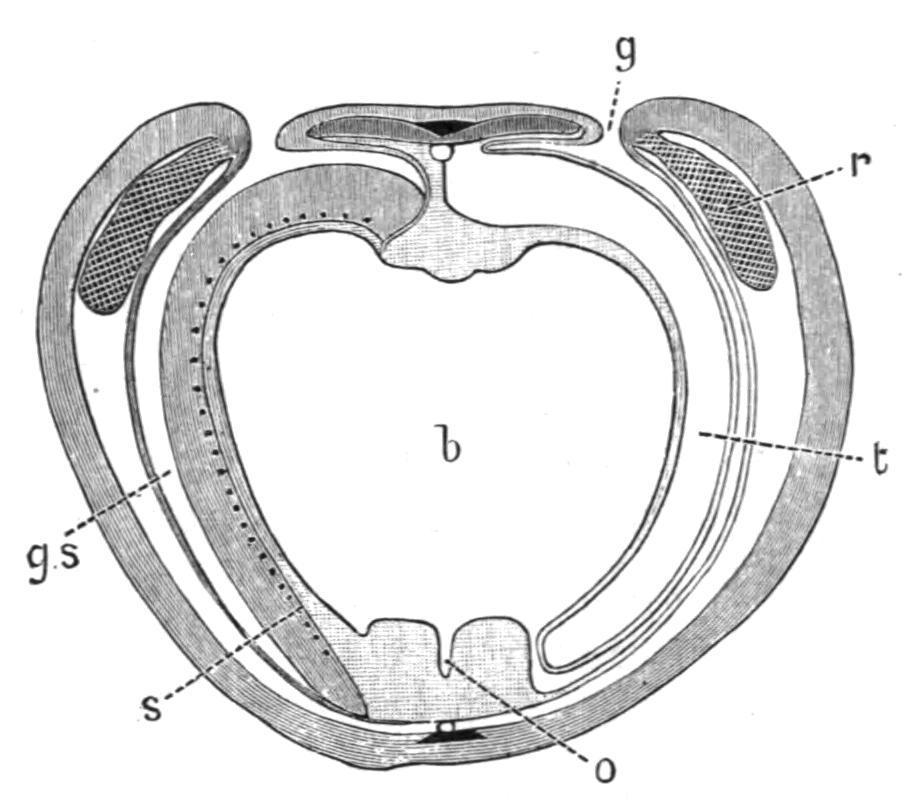
FIG. 6. Schizocardium brasiliense; transverse section through the branchial region, showing the great extent of the branchial part (b) of the pharynx; the oesophageal part (o) is reduced to a mere groove; g, gill-pore; g.s, gill-sac; r, reproductive organ; s, synapticula (cf. Fig. 5); t, tongue-bar. The muscles of the bodywall are not indicated: in other respects the figure corresponds with Fig. 4, except for the absence of genital wings in this region of the body. (After Spengel.)
At a certain distance behind the branchial region, the alimentary canal in Balanoglossus and Schizocardium is produced into a series of outgrowths, into which food does not pass. These "liver-sacs" give rise to corresponding folds (Fig. 1, A, h) of the dorsal body-wall, a conspicuous external feature of the species in which they are present. The most interesting peculiarity of the digestive tract in this region is the existence, in certain species, of pores, possibly vestigial gill-slits, leading from it to the exterior.
Notochord and Skeleton. The structure compared by Bateson with the Vertebrate notochord is a hollow dorsal outgrowth of the alimentary canal of the collar-region (Fig. 7, n). Near its origin it is slender, but in the proboscis it dilates into a comparatively large organ, which in most cases retains its cavity. Its cells have a
vacuolated appearance, which recalls the fine structure of the Vertebrate notochord. In Schizocardiumand Glandiceps, the organ is produced into a slender "vermiform process" (v), which extends nearly to the tip of the proboscis.
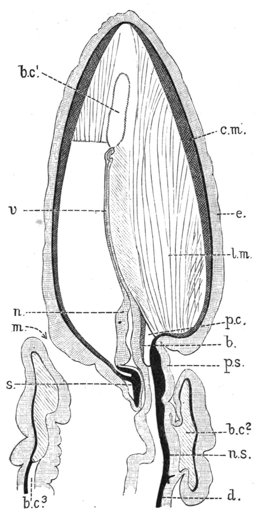
FIG. 7. Schizocardium brasiliense; longitudinal, median section through the proboscis, the collar, and the first part of the trunk; b, main blood-space of the proboscis; b.c1 , b.c2 , b.c3 , first, second and third body-cavities; c.m, circular muscles of proboscis; e, epidermis; l.m, longitudinal muscles of proboscis; m, mouth; n, notochord; n.s, central nervous system, continuous with the subepidermic nerve-plexus (black) of the proboscis, and with the dorsal nerve (d); p.c, pericardium; p.s, proboscis-stalk; s, proboscis-skeleton; v, vermiform process of notochord. (After Spengel.)
The main support of the proboscis-stalk is the "proboscis-skeleton" (s), a Y-shaped organ whose median part lies beneath the base of the notochord, its diverging legs extending backwards along the outer side of the alimentary canal of the collar. The proboscisskeleton, like the branchial skeleton, is a special development of the structureless membrane which is found at the base of the layers of cells of Balanoglossus, and in most species it grows merely by the deposition of laminae of chitin from the notochord, and from the ventral epidermis of the proboscis-stalk.
In some species, however, and particularly in Balanoglossus aurantiacus and Glandiceps, the primary skeleton becomes surrounded by an extensive development of a secondary cartilaginoid skeleton, consisting of a structureless substance into which the adjacent body-cavities of the proboscis and collar send cellular outgrowths. The possibility of a relation between this tissue, more or less surrounding a part of the notochord, and the cartilage of Vertebrates cannot be overlooked.
The caudal region may be stiffened (?) by a "pygochord"[21] which is a median derivative of the alimentary canal on its ventral side.
Vascular System and Proboscis-Gland.—The main vessels are a dorsal and a ventral vessel (Fig. 4, d.v, v), lying in their respective mesenteries. The details of the vascular system are complicated, and have not been thoroughly made out, the nearly colourless character of the blood making their investigation a difficult matter. The following points may, however, be noted. The blood is said to pass forwards in the dorsal vessel, which, like the ventral vessel and a pair of lateral vessels in the hepatic region, is contractile. In the collar the dorsal vessel lies between the two perihaemal spaces, on the dorsal side of the base of the notochord. The principal bloodspace in the proboscis (Fig. 7, b) lies between the notochord (n) and an organ known as the "heart-vesicle" or "pericardium" (p.c). The latter has muscular walls and it contracts rhythmically in the larva. Its behaviour in the adult is not so easily made out, but it is probable that, although it does not communicate with the vascular system, its contractions propel the blood contained in the space immediately beneath it. The blood, after passing to a glandular organ, the "proboscis-gland" or "glomerulus," which lies at the sides and in front of the notochord, appears to pass round the collar to the ventral vessel. Various systems of vessels are connected with the skin, the gills, the alimentary canal and the generative organs.
The function of the proboscis-gland is possibly excretory. In this case it is probable that the proboscis-pore eliminates the waste products discharged by the gland into the anterior body-cavity, though this view is not favoured by Willey.
Reproductive Organs. The sexes are separate, the reproductive organs consisting of a series of simple or branched glands which occur along the dorso-lateral lines of the anterior part of the body; being usually found throughout the branchial and generative regions and ending at the beginning of the hepatic region. The reproductive organs may pass into great extensions of the body-wall known as the "genital wings," specially developed in some species of Balanoglossusand Ptychodera(Figs. 1 A, 4).
Stereobalanus canadensis, a species with long slit-like external gillpores, is interesting in possessing a well-developed genital wing both dorsally and ventrally to the series of gill-pores of each side.
Each reproductive gland opens by its own pore or pores directly to the exterior. Several glands and pores may occur in the same transverse section.
According to Spengel there is no definite relation between the number of the reproductive organs and that of either the gill-sacs or the liver-outgrowths. The only definite segmentation exhibited by Balanoglossus is thus the division into three regions which is so distinctly shown by the arrangement of the body-cavities; though the gill-sacs may indicate an incipient further segmentation of the major part of the body. In this connexion it is interesting to notice MacBride's statement[22] that the body-cavity of Amphioxus develops in the embryo as five cavities, just as in Balanoglossus; the segmented part of the body being formed by a secondary segmentation of the third body-cavities.
Regeneration.
—Balanoglossus, like Phoronis (p. 30), possesses great powers of regenerating lost parts. The posterior part of the body is readily re-formed, while Spengel has shown[23] that even the proboscis, collar and branchial region can be regenerated, apparently from a fragment of the body.
Genera of Enteropneusta.
—Spengel, whose Monograph is indispensable to every student of the Enteropneusta, formerly proposed to divide the old genus Balanoglossus into four; but he now recognises no less than nine.[24] Some of the more important characters are given below, but for the arrangement of the muscles, important from a systematic point of view, reference must be made to the original sources.
A. Notochord with a vermiform process (Fig. 7, v); pericardium with anterior diverticula more or less developed. .......... GLANDICIPITIDAE
(a) Liver-sacs and synapticula present; gill-slits almost equalling the pharynx in depth, so that the ventral, non-branchial part of the pharynx is reduced to a mere groove (Fig. 6); nerve-roots absent; pericardial diverticula long. .......... Schizocardium, Speng.
(b) Liver-sacs absent;[25] ventral part of pharynx well developed; pericardial diverticula short.
(i.) Synapticula and nerve-roots absent. .......... Glandiceps, Speng.
(ii.) Synapticula present; nerve-roots present or absent; genital region with dermal pits. .......... Spengelia, Willey.
B. Notochord with no vermiform process; pericardium simple; ventral part of pharynx large, and sometimes more or less separated from the branchial part (Fig. 4).
(a) Liver-sacs,[26] synapticula and nerve-roots present. .......... PTYCHODERIDAE
(i.) Genital wings well developed.
(α) Gill-sacs opening by long slits. .......... Ptychodera, Eschsch.
(β) Gill-sacs opening by small pores. .......... Balanoglossus, Delle Chiaje.
(ii.) Genital wings hardly developed. .......... Glossobalanus, Speng.
(b) Liver-sacs, synapticula and nerve-roots absent. .......... HARRIMANIIDAE
(i.) Proboscis long; one proboscis-pore. .......... Dolichoglossus, Speng.
(ii.) Proboscis short; two proboscis-pores.
(α) Two pairs of genital wings. .......... Stereobalanus canadensis, Speng.
(β) No genital wings. .......... Harrimania, Ritter.
The name Balanoglossuswas introduced by Delle Chiaje in 1829 for B. clavigerus (Fig. 1, A), from the neighbourhood of Naples. As Spengel has shown, its etymology has been much misunderstood. The second half of the name refers to a fancied resemblance between the Balanoglossus, with its largely developed genital wings, and the tongue of an ox. Βάλανος means "acorn," and it has usually been supposed that this name was suggested by the resemblance of the proboscis, projecting from the collar, to an acorn in its cup, a view which finds its expression in the name "Eichelwurm" used by German zoologists. But the idea expressed by Delle Chiaje was really a similarity between the collar of Balanoglossus and the outer shell of Balanus, the barnacle or "acorn-shell" found everywhere on rocks between tide-marks.
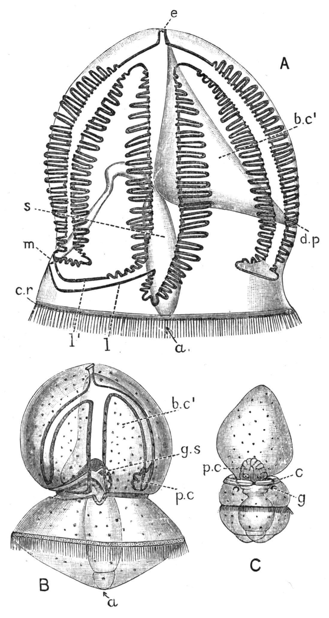
FIG. 8. Metamorphosis of Balanoglossus, probably of Balanoglossus biminiensis Willey, Bahama Islands. All the figures are magnified to the same scale (× 14). A, fully developed free-swimming larva, or Tornaria, side view; B, commencement of metamorphosis, side view; C, later stage, dorsal view. Increase in size takes place after this stage; a, anus; b.c1 , body-cavity of proboscis; c, collar; c.r, transverse ciliated ring; d.p (in A), dorsal pore (= proboscispore), seen also in C on the left side, just behind the reference line p.c; e, eyes and sensory thickening of skin (in A); g, gill-pore; g.s, gill-sacs, developing as outgrowths of the alimentary canal; three are already present in B, but are better seen in C, in which they are still without openings to the exterior; l, postoral part of the longitudinal band of cilia; l′, its praeoral part; both land l′ are produced (in A) into tentacles, over which the band of cilia is looped; the groove in the middle of the figure, between l and l′, conducts the food by the transverse groove to the mouth (m); p.c, blood-space of proboscis and pericardium ("heart" of larva); s, stomach. (After Morgan).
Development.—The free-swimming, larval stage of Balanoglossus is known as Tornaria (Fig. 8, A). Several distinct forms of the larva are known,[27] although it is not yet possible to refer them with certainty to their respective adults.
Tornaria was described and named by Johannes Müller, who regarded it as the larva of a Starfish,[28] in spite of his intimate knowledge of the development of these animals. Its correct systematic position was first demonstrated by Metschnikoff in 1869.
The larva agrees with many other pelagic forms in being excessively transparent. The form described by Spengel as T . grenacheriattains the remarkable length of 9 mm. (nearly ⅖th inch).
The full-grown larva is usually ovoid, and a complicated "longitudinal" band of cilia runs in several loops over its anterior twothirds. In side view, part of the surface limited by the ciliated band appears like a T with a double outline, the cross piece being bent downwards on each side, so as to form an anchor-like curve, the middle of which is at the anterior pole of the larva. In T . krohni, which occurs on our south coast,[29] the ciliated band has a wavy course. In the West Indian larva[30] shown in Fig. 8 A, the ciliated band is produced into numerous tentacles, which fringe the sides of the T-shaped areas or grooves of the surface. These grooves and the cilia which border them are used for conveying food to the mouth.[31] At the apex of the larva is a thickening (e) of the ectoderm, bearing two eye-spots. The main locomotor organ is a simple transverse band (c.r) of "membranellae," vibratile structures composed of fused cilia. The mouth (m), on the ventral side, leads into the oesophagus, and this into the stomach (s). The latter is separated by a marked constriction from the intestine, which opens by the anus (a) at the posterior pole.
On the dorsal side is a pore, the "dorsal pore" (d.p.), which leads into a thin-walled sac (b.c1) destined to become the proboscis-cavity of the adult. To the right of the dorsal pore lies the pulsating "heart," which apparently becomes the pericardium of the adult. Bourne and Spengel regard it as a right proboscis-cavity. In the older larvae, the
second and third body-cavities appear as paired thin-walled sacs in close contact with the hinder part of the stomach. The skin is very thin, and the five body-cavities do not nearly fill the space between it and the alimentary canal. This space becomes obliterated for the most part by the enlargement of the body-cavities, and its last remains persist, as in many other animals,[32] as the vascular spaces of the adult.
In Dolichoglossus kowalevskii, and probably in other species with large eggs,[33] development proceeds by gradual stages to the adult form, and no Tornaria-stage is passed through. The opaque young animal, on being hatched, creeps about in the muddy sand in which the adult is found, later moving in a leech-like manner, by alternately attaching itself by its two ends. The young stages were ingeniously obtained by Bateson, to whom our knowledge of the development of this species is due,[34] by allowing a large quantity of the mud to settle after being stirred up, the layer of the specific gravity corresponding with that of the young Balanoglossus being then separated by means of a siphon. The young stages previously contained in several hundredweight of mud were thus easily collected into a pint of water. Morgan recommends treating the layer obtained by a similar process with picric acid, which stains the young Balanoglossus yellow.
The embryo early becomes a "blastosphere" or hollow vesicle formed of a single layer of cells. One half of this is invaginated, or pushed into the other half, and a "gastrula" is thus developed, the cavity of which is the "archenteron," and the two cell-layers respectively "ectoderm" and "endoderm." The "blastopore," or orifice of invagination, is at the posterior pole of the larva, where it narrows and closes, the locomotor, transverse band of cilia developing round it. No other bands of cilia appear in this form of development. The proboscis becomes marked out externally by the appearance of a circular groove, near the middle; and behind this groove a second
one appears, which forms the posterior boundary of the collar. The larva, which now resembles Fig. 8 C, is usually hatched at this stage. Two gill-slits make their appearance, and the mouth and anus are perforated; the anus being in the position of the blastopore.
The body-cavities are formed as five derivatives of the archenteron. One of these is unpaired, and becomes the proboscis-cavity; while the others are the paired cavities of the collar and trunk (cf. Fig. 2). There is some uncertainty about the origin of the body-cavities of the free-swimming Tornaria, although it seems most probable that they are developed either from the wall of the stomach or intestine, [35] or from scattered mesoderm cells[36] which lie in the segmentation-cavity.
The metamorphosis of Tornaria is accompanied by a great diminution in size,[37] probably due to the loss of water; by this cause and by the simultaneous thickening of the skin, the larva loses its transparency.
The external features of the metamorphosis are sufficiently indicated by Fig. 8, the ciliated bands finally disappearing. The dorsal pore persists as the proboscis-pore; the notochord and numerous gill-slits are developed as outgrowths of the alimentary canal, the reproductive organs make their appearance, probably from the mesoderm,[38] the trunk meanwhile elongating so that the proportions of the adult are acquired.
TubicolousHemichordata,withonepairofgill-slitsornone,a Ushaped alimentary canal, and a dorsal anus situated near the mouth.Proboscisflattenedventrallyintoalarge"buccaldisc,"its
Order II. Pterobranchia.
base covered dorsally by the collar , which isproduced into two or more tentaculiferous arms. Trunk short, prolonged into a stalk.Reproductionbybuddingoccurs.
This group consists of the two genera Cephalodiscus (Fig. 9) and Rhabdopleura (Fig 12). The latter, first dredged by G.O. Sars, in 1866, from 120 fathoms off the Lofoten Islands, was included in a catalogue of deep-sea animals published by his father, M. Sars, in 1868 as Halilophusmirabilis, a name which has been superseded by Rhabdopleura normani, Allman,[39] based on specimens dredged by Canon Norman in 90 fathoms, off the Shetland Islands.
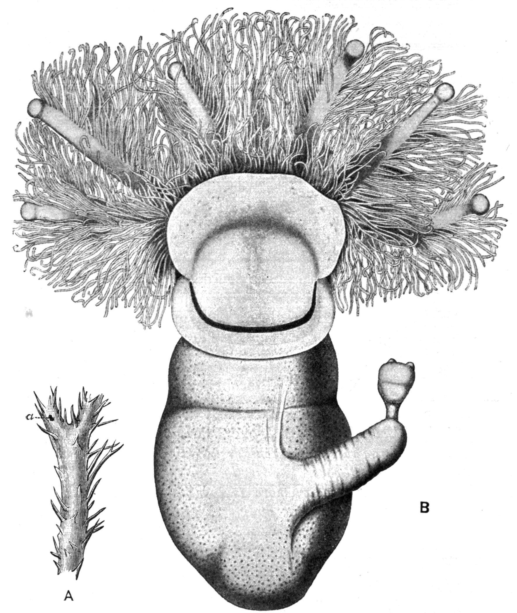
FIG. 9. Cephalodiscus dodecalophus, M‘Intosh, Straits of Magellan; A, small portion of the common "house," × 1; a, a single individual, shown also as B, × 65; six of the tentacular arms, belonging to the collar, are seen springing from behind the proboscis or "buccal disc." This has a crescentic band of pigment parallel with its posterior border, which conceals the mouth. The stalk, bearing a bud, which already shows the beginning of two tentacular arms, is seen to the right. (After M‘Intosh, B from Parker and Haswell.)
The structure of Rhabdopleura has been described by Sars,[40] Lankester,[41] and Fowler.[42] R. normani is common in certain Norwegian Fjords, at depths of 40 fathoms or more, and has been recorded by Fowler from the Tristan d'Acunha group in the S. Atlantic; R. compacta has been found off the N.E. coast of Ireland[43] and near Roscoff, on the N. coast of Brittany; while forms described by Jullien[44] as R.grimaldiiand R.manubialishave been dredged off the Azores. I have recently found a fragment of Rhabdopleura from South Australia. It is doubtful how far these species are distinct.
Cephalodiscus dodecalophus[45] was found in the Straits of Magellan, during the "Challenger" voyage, at a depth of 245 fathoms, and has recently been rediscovered in shallower water in the same neighbourhood by the Swedish Antarctic Expedition. Another Cephalodiscus, at present undescribed, has been obtained by Dr. Levinsen from 100 fathoms off the coast of Japan; while the Dutch expedition carried out by the "Siboga" has resulted in the discovery of two other specimens, one from a reef close to low-tide mark on the coast of Borneo, the other from 41-52 fathoms off Celebes. These three specimens differ markedly from one another and from the "Challenger" specimen of C. dodecalophus, and it is probable that they all belong to new species. The occurrence of a deep-sea animal at a great distance from the locality at which it was first found is not in itself a matter for great surprise; but in the present instance two of the newly discovered forms are from shallow water, and one of them is actually littoral. The occurrence of so many species of Cephalodiscus in Oriental waters suggests that the Pacific or the Indian Ocean may be the headquarters of the genus, which may prove to be far less of a rarity than has hitherto been supposed. There is evidence derived from the results of the "Siboga" expedition that abyssal animals may migrate into comparatively shallow water in the Malay Archipelago.
Cephalodiscus and Rhabdopleura are remarkable for their power of producing buds. In the former these arise from the apex of a stalk which is given off on the ventral side of the body, and they break off when they reach a certain age; in the latter they do not become free, and a colony results, which consists of a creeping "stolon" from which vertical branches are given off at intervals, each ending in an individual of the colony. Cephalodiscus forms a gelatinous "house" (Fig. 9, A), in the passages of which are found large numbers of the free individuals, together with their eggs and embryos. Rhabdopleura (Fig. 12) is protected by cylindrical tubes, one of which corresponds with each individual.
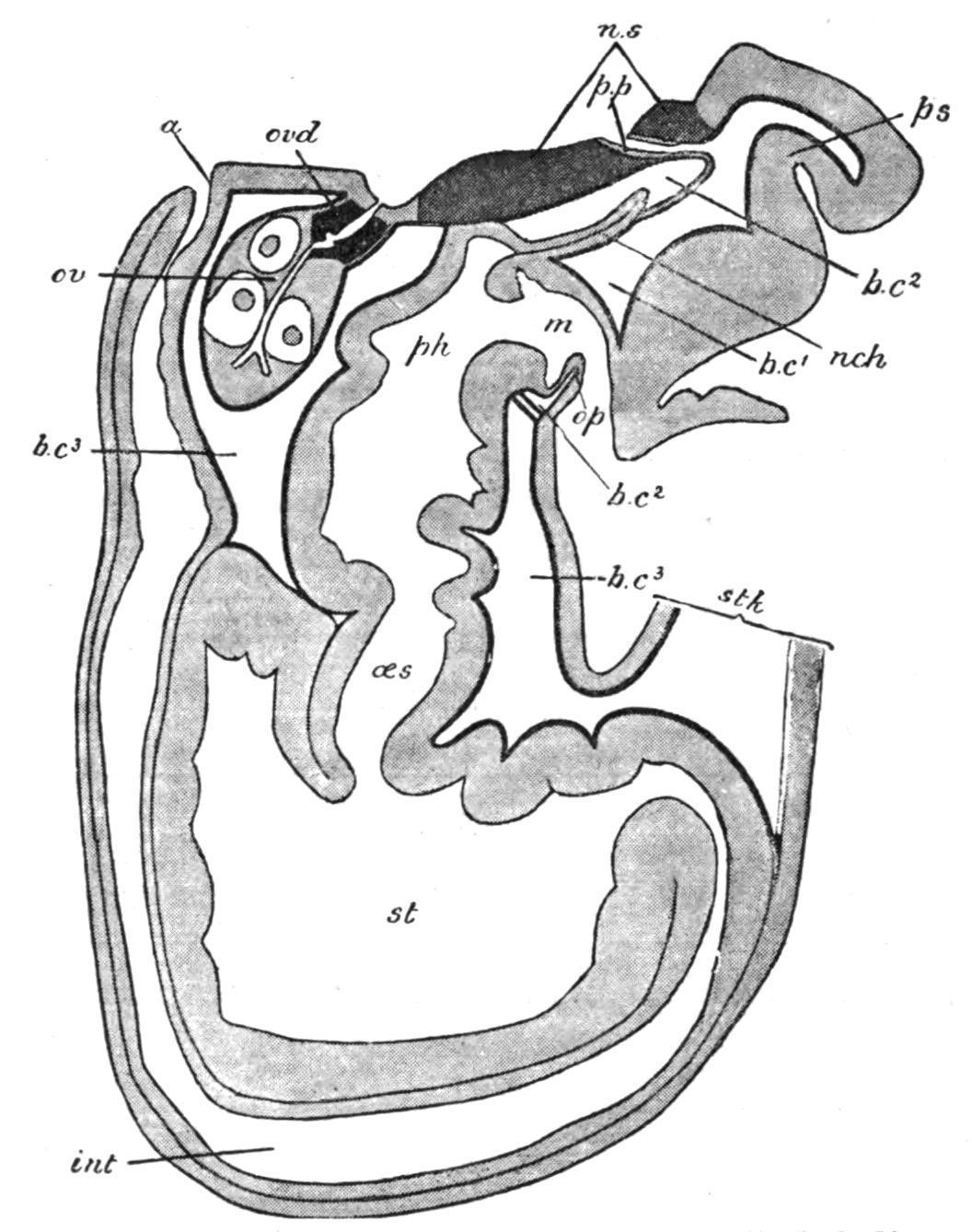
FIG. 10. Longitudinal median section of Cephalodiscus dodecalophus. a, Anus; b.c1 , b.c2 , b.c3 , first, second, and third body-cavities; int, intestine; m, mouth; nch, notochord; n.s, central nervous system; oes, oesophagus; op, operculum, the ventro-lateral part of the collar; ov, ovary; ovd, pigmented oviduct; ph, pharynx; p.p, proboscis-pore; ps, proboscis; st, stomach; stk, stalk.
Cephalodiscus, though no more than two or three millimetres in length, is provided with practically all the important organs possessed by Balanoglossus. Its proboscis or "buccal shield" (Fig. 10, ps) is a large flattened structure, which overhangs and entirely conceals the mouth. The anterior body-cavity opens to the exterior by two symmetrically placed proboscis-pores (p.p), just in front of the tip of the notochord (nch). The collar, which has paired bodycavities, is produced dorsally into 4-6 pairs of plume-like arms, which