Citation: Auger F, Comtois AS, Roy R (2015) Intra and Inter Examiner Reliability Study for the Characteristics of Evaluation of the SA201. J Spine 4: 245.doi:10.4172/21657939.1000245 Page 3 of 6
• •
One exercise feeling the instrument displacing the instrument and maintaining the approximate 45 degree angle One exercise feeling the instrument displacing the instrument and maintaining the approximate 45 degree angle and evaluating the cervical, thoracic and sacrolumbar areas on each other.
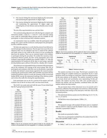
The site of the experimentation was a private clinic. Two communicating adjacent room with sharing one computer and instrument namely « room #1 » « room #2 » were used; the waiting room could sit comfortably fifteen persons and was available for the participants, to relax in between their evaluation sessions. An examination room was available, where the participants could change into a gown and the technician could prepare the participants, for their evaluation session. We had a site supervisor, to verify that the protocol was followed as erected and supervise the technician. The site supervisor was in the room whenever there was an evaluation and was in control of the software and made certain no one could see the screen as it was recording the data from the analysis. There was no comment made to the evaluator or participant during the procedure. He verified the exactness of the anatomical reference points, the flow of the participants and of the evaluator, respecting the establish time schedule (Tables 1-3). After the experimentation he transferred all the data to excel using a specially develop database query software (Chirosoft Inc., Quebec, QC, Canada). The technician was responsible for the recruitment of the participants and of the evaluators, the training of the novice group, take care of participants and evaluators while they were on the premise, responsible to obtain the declared consent forms signed for the research project, evaluating the questionnaires (ODI, NDI and BMI) associated with the inclusion/exclusions criteria, to verify the symmetry of the cervical and lumbar ROM, execute the anatomical marking of the reference points and to direct the traffic flow during the experimentation. Each participant was on site for a period of 90 minutes and each evaluator was on site for six hours with a one hour lunch break. The SA201 was onsite and used with the same 30 mm forked head that the N group had practiced with. Time
Room #1
Room #2
T1(0-5 min)
A6
B7
T2(5-10 min)
A8
B9
T3(10-15 min)
A10
T4(15-20 min)
A7
T5(20-25 min)
A9
T6(25-30 min)
B6 B8 B10
T7(30-35 min)
C6
D7
T8(35-40 min)
C8
D9
T9(40-45 min)
C10
T10 (45-50 min)
C7
T11 (50-55 min)
C9
T12 (55-60 min) T13 (60-65 min)
D6 D8 D10
E6
F7 F9
T14 (65-70 min)
E8
T15 (70-75 min)
E10
T16 (75-80 min)
E7
T17 (80-85 min)
E9
T19 (85-90 min)
F6 F8 F10
Table 2: Time for the procedures with the patient ID letter for the evaluator and number for the participant and room assignation, A1= evaluator A and patient 1.
J Spine ISSN: 2165-7939 JSP, an open access journal
Time
Room #1
Room #2
T1(0-5 min)
A11
B12
T2(5-10 min)
A13
B14
T3(10-15 min)
A15
T4(15-20 min)
A12
B11
T5(20-25 min)
A14
B13
T7(30-35 min)
C11
D12
T8(35-40 min)
C13
D14
T9(40-45 min)
C15
T10 (45-50 min)
C12
D11
T11 (50-55 min)
C14
D13
T6(25-30 min)
B15
T12 (55-60 min)
D15
T13 (60-65 min)
E11
F12
T14 (65-70 min)
E13
F14
T15 (70-75 min)
E15
T16 (75-80 min)
E12
F11
T17 (80-85 min)
E14
F13
T19 (85-90 min)
F15
Table 3: Time for the procedures with the patient ID letter for the evaluator and number for the participant and room assignation.,A1 = evaluator A and patient 1. Evaluators
Experienced
Novice
1-2–3
1-2–3
Measures
1st set
2nd set
1st set
2nd set
Sites
S1 … S30
S1 … S30
S1 … S30
S1 … S30
Participant 1 … Participant 15 Table 4: Statistical schematic.
The analysis was done in two steps. The first part consisted in the evaluator doing two complete analyses on a durometer 40 of a ¼ inch thickness which was resting on a metal base. Then he would proceed to do the same analysis on the patient. The analysis consisted on testing the cervical, dorsal and sacrolumbar areas representing 30 data per analysis. The analysis was done twice with a few seconds pause in between the analysis, and was therefore considered before and after analysis without any intervention. The pre-tension force of the instrument was at six pounds and it was factory calibrated. The participants were seated on a sitting massage chair with their head resting on the head piece with their neck in the horizontal position. The anatomical reference points were the following: C2, C7, T1, T6, T12, L1, L5 and S2. We had three waves of participants, (Tables 1-3).
Statistics We measured averages with standard deviation and we completed with an ANOVA, Cohen’s D and the Student T test [17]. Our analysis schematic is presented in Table 4.
Results Our results are presented as tables. There were no participants excluded from the analysis based on the NDI, ODI and BMI (Tables 5 and 6).
Discussion In Tables 7 and 8, we can visualize a great variation for both
Volume 4 • Issue 4 • 1000245
4
