
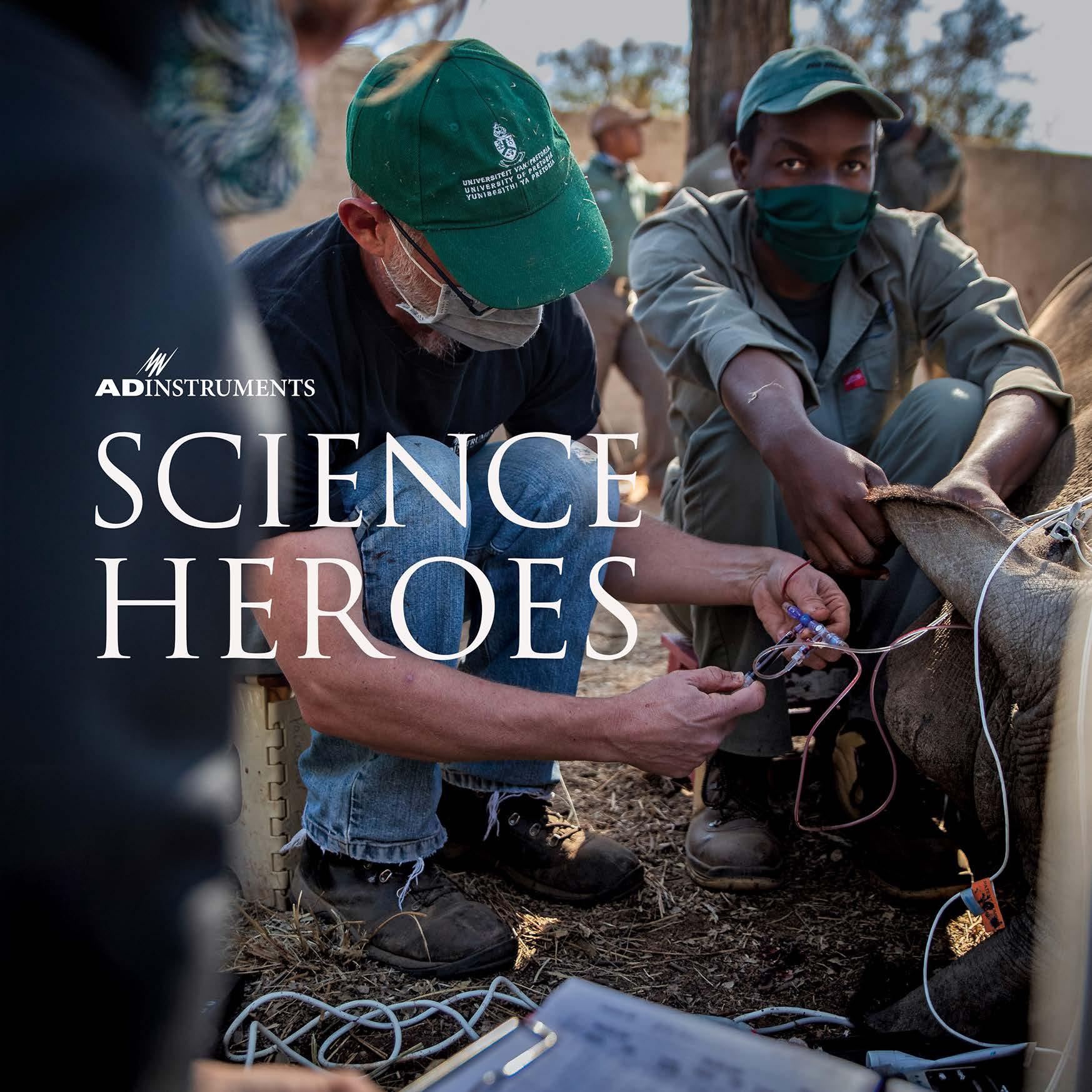
CONTENTS
Michael Macknight, ADInstruments Founder 03 Keeping anesthetized rhinos safe Professor Leith Meyer 11 Reining chaos to save lives Dr Manasi Nandi 19 Mapping a path to healthy development Associate Professor Emily Zimmerman 27 Lighting the way for Parkinson’s research Associate Professor Louise Parr-Brownlie 35 Lessons from the wild Associate Professor Tony Hickey 43 Extreme science in search of answers Professor Damian Bailey 51 Taking physiology to new heights Professor Trevor Day
01 Introduction

Heroes come in many forms
We are all inspired by people doing amazing things. Some people are incredible in the physical things they do, through talent and practice. Others create works of art or music that move us. Through their intelligence, compassion, talent, diversity, and drive, we all have heroes that make the world a better place.
Here at ADInstruments, we love science. We believe science can help us all live more worthwhile and healthy lives. The scientific method of repeated hypothesis and testing help us understand and control the world we live in.
Science has many heroes. Some of them are household names, but many of them work behind the scenes, following their interest and passion. Nevertheless, they have had a profound impact on our world.
We present to you a very small subset of the scientist heroes that we get to interact with every day. People like them are the reason we do what we do. To know that we have helped, even in a small way, heroes like these is why we come to work each day. What they all have in common are the passion and energy they bring to their field and the inspiration they give their colleagues and students.
We want to thank all our scientific heroes. It is a great pleasure and privilege to know you and work with you. We are honored whenever you use our products. ADInstruments Founder


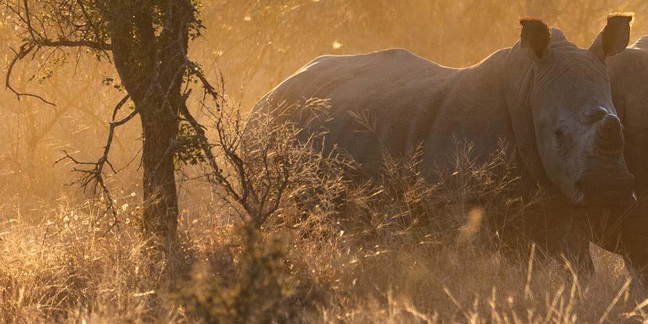
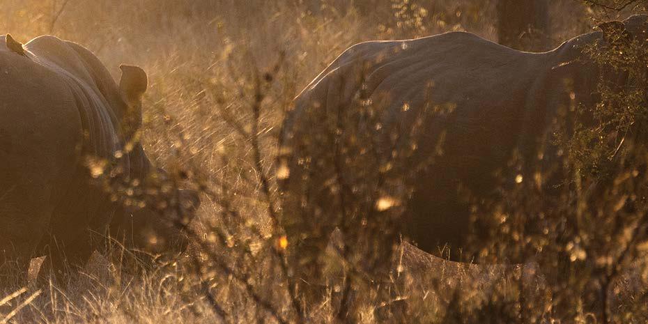
Keeping anesthetized rhinos safe
Developing safer capture and immobilization techniques for one of the world’s most endangered animals
Professor
LeithMeyer
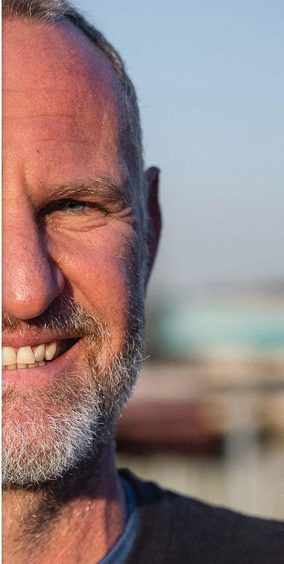 Director:
Centre for Veterinary Wildlife Research Professor in the Department of Paraclinical Sciences, University of Pretoria South Africa
exactly do
collect physiological data from rhino?
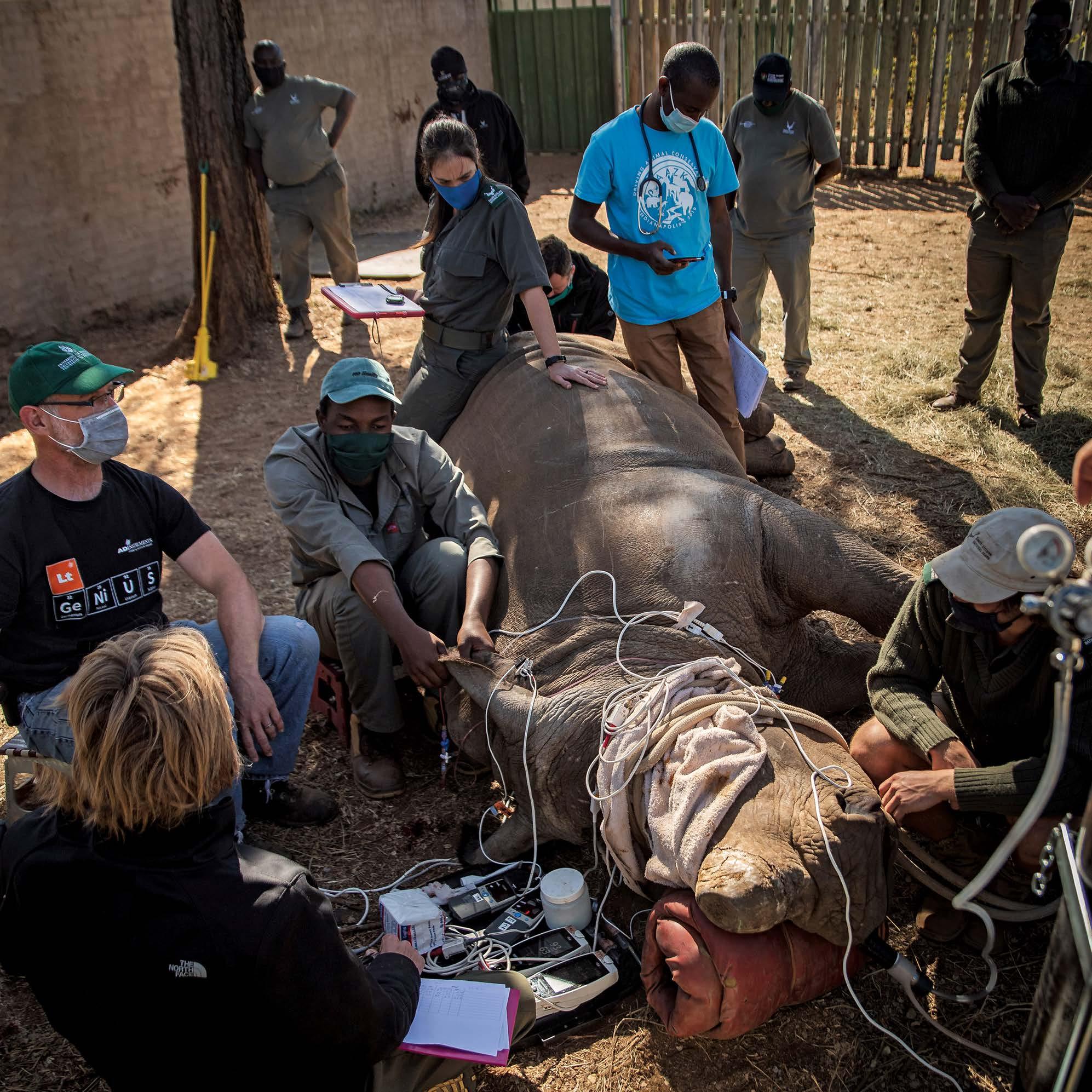
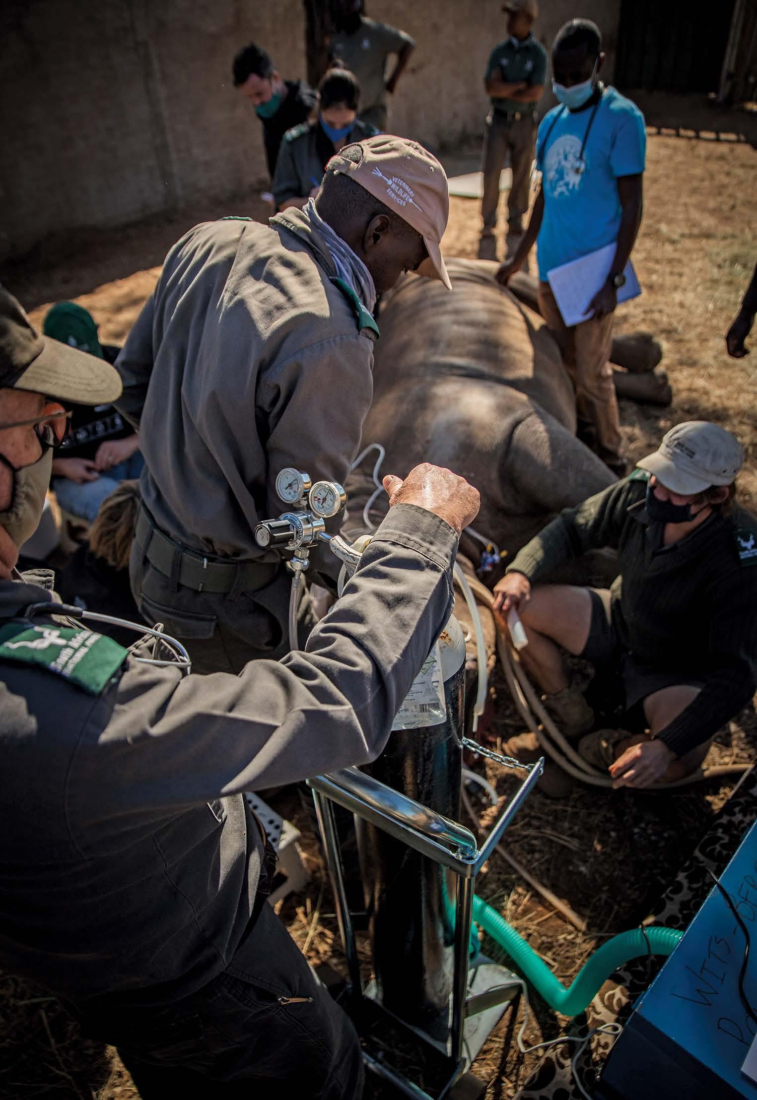
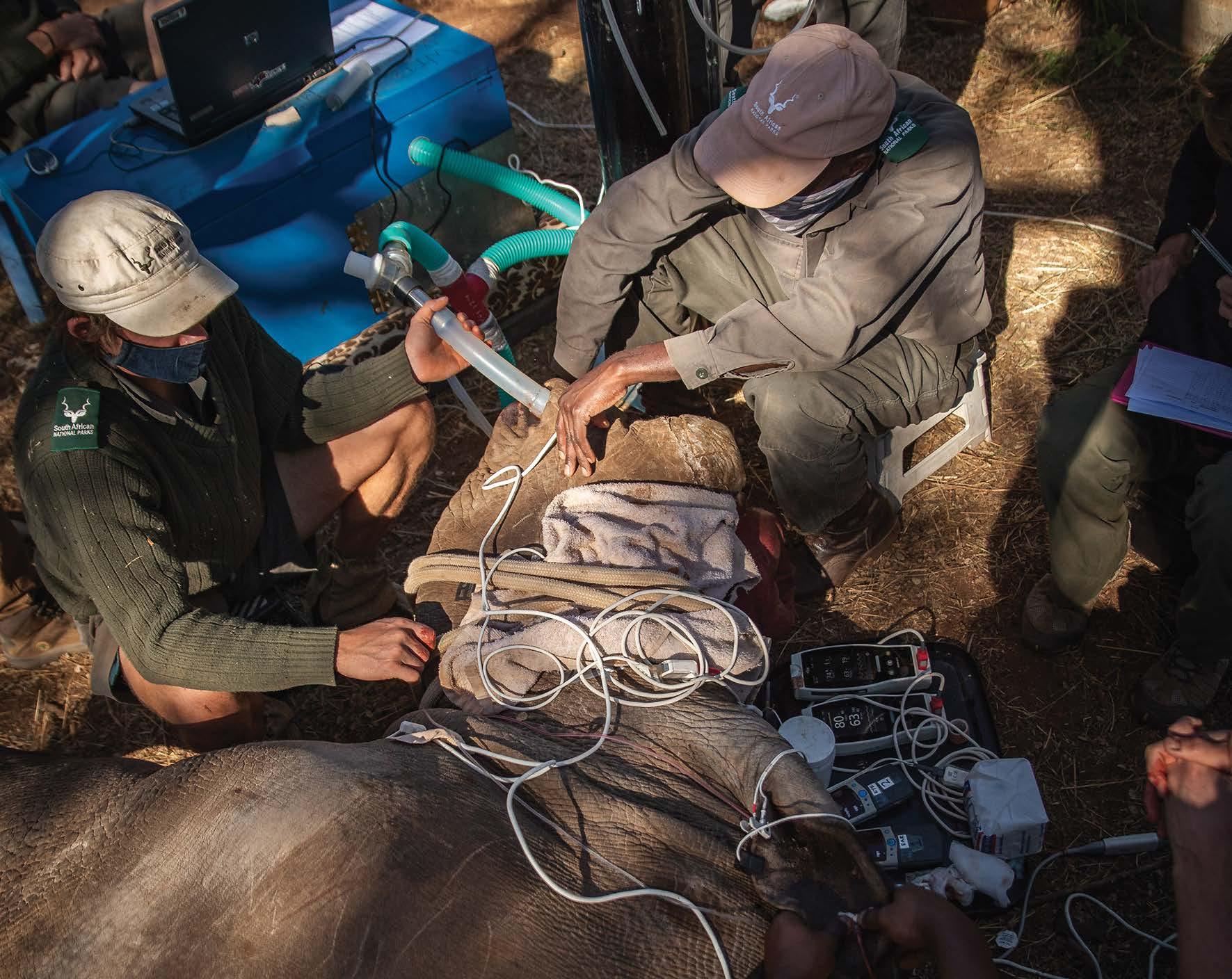
Poaching is driving the African rhino toward extinction, but the techniques used to protect them can be just as dangerous. Rhinos are particularly sensitive to anesthesia, which is needed to dehorn or relocate at-risk animals. They can suffer from severe cardiorespiratory depression, with respiratory rates dropping from a typical 19 breaths per minute down to as low as 3 or 4 breaths per minute. “When you look at the data of their physiology, these animals are actually sitting on a knife’s edge.” Leith says, “It’s really important for us to improve how we do things during rhino anesthesia.”
So, how exactly do you collect physiological data from rhino? Once the rhino is immobilized, Leith’s team use a PowerLab system with transducers adapted from human exercise physiology gear – mainly a gas analyzer and spirometer – to monitor the rhino’s ventilation, tidal volume, respiratory rates, and metabolism during anesthesia. Rhinos are obligate nasal breathers so all expired gases can be collected from the nostrils.
6
So, how
you

Signals recorded Blood Oxygenation Blood Pressure CO2 Oxygen pH VCO2 VO2
In place of a face mask, Leith uses adapted equine endotracheal tubes in their nostrils to collect the expired gases, measuring airflow and also the CO2 and O2 levels each time the rhino breathes out. Calculating VO2 and VCO2 gives an indication of how much O2 the rhino is burning up, and how much CO2 they’re producing. Capture drugs often make the animals hypermetabolic – Leith’s team are aiming to develop ways to reduce this elevated metabolism. To monitor blood pressure, and other variables such as blood oxygenation and pH, a catheter is inserted into an artery in the rhino’s ear.
8
The findings allow the capture process to be fine-tuned for the rhino, to keep them as healthy as possible. A better understanding is already paying dividends, with changes in how drugs are used during de-horning operations, where people spend up to an hour removing the horn that puts the rhino at risk from poachers. Alongside colleagues from South Africa National Parks, Leith now helps run an advanced capture course for experienced wildlife vets from around the world, where delegates learn about the latest developments and research in the field of wildlife medicine.
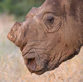
DE-HORNING
With rhino horn worth more than its weight in gold, de-horning is used to remove the incentive for poachers. Horns are made from keratin, similar to fingernails, and removing them doesn’t hurt. The horn takes around 2 years to grow back.
9
Drop in Kruger’s rhino population since 2013 due to poaching 1 Rhino were poached for their horn in South Africa in 2020 2 Increase in rhino poaching between 2007 - 2014 in South Africa 1 60% 394 9000% “There’s always something that we can do to help,” he says, “and there’s always so much that we can learn.” 1. https://www.savetherhino.org/rhino-info/poaching-stats 2. https://www.environment.gov.za/mediarelease/rhinopoaching_sa 3. https://ielc.libguides.com/sdzg/factsheets/whiterhino/population sa 150 20,000 RHINO POPULATION GROWTH 1929 3 2015 3 6 years CONTINUED DECREASE IN POACHING marked in 2020 3
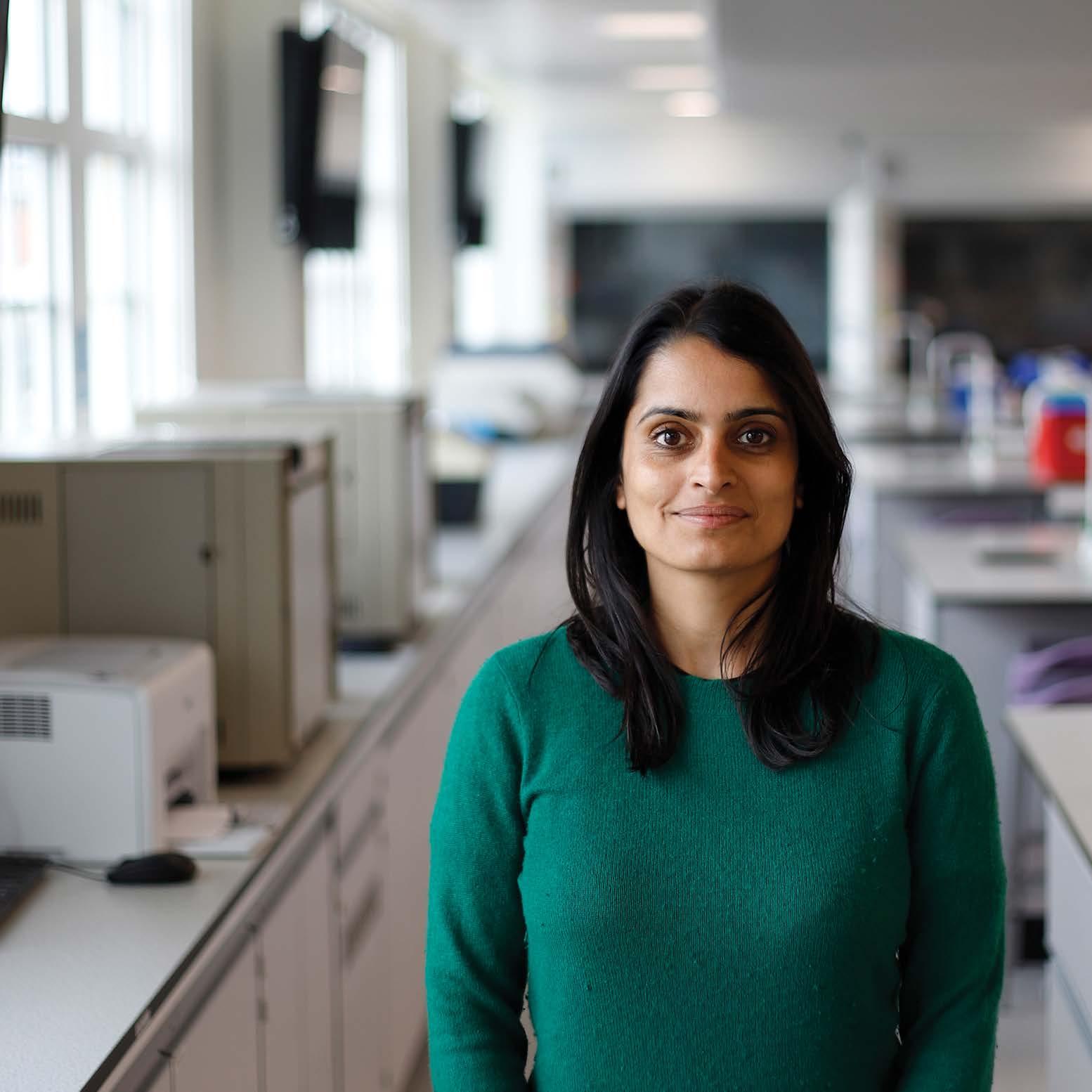
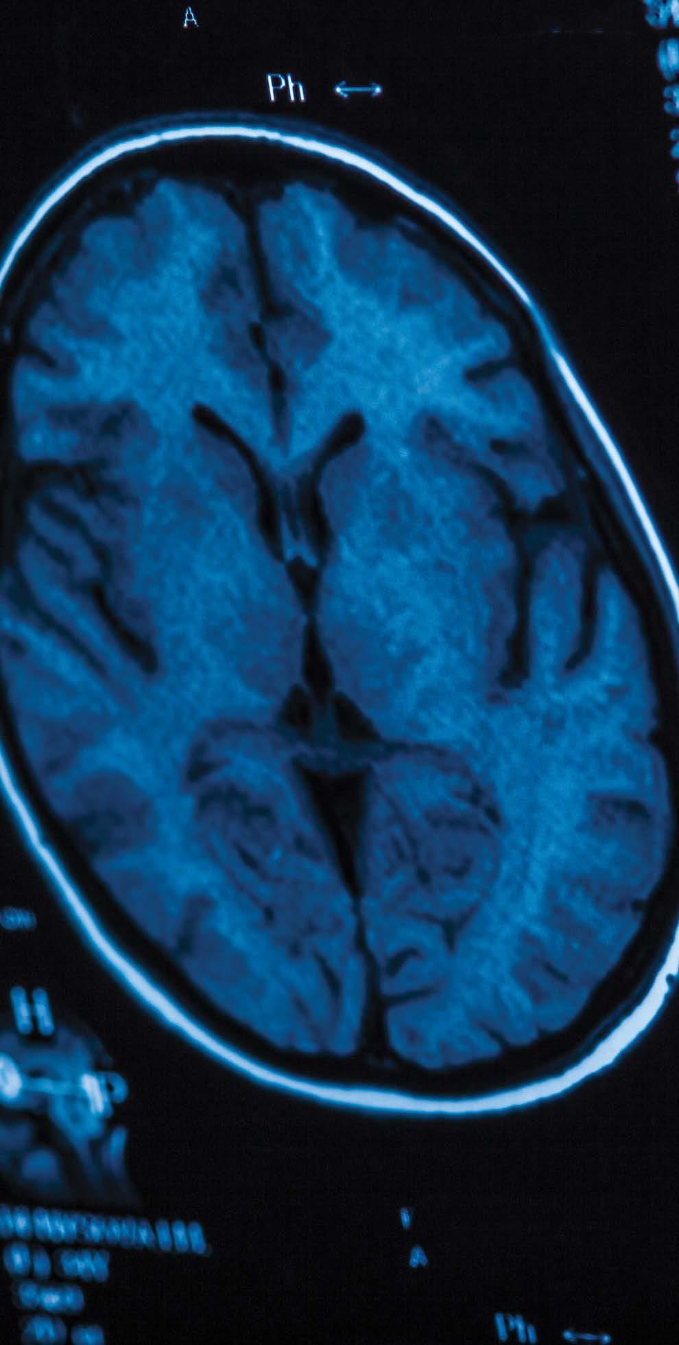
Reining chaos to save lives
Using maths and medicine to see when organs start to struggle
Dr Manasi Nandi Department of Integrative Pharmacology King’s College London United Kingdom
13
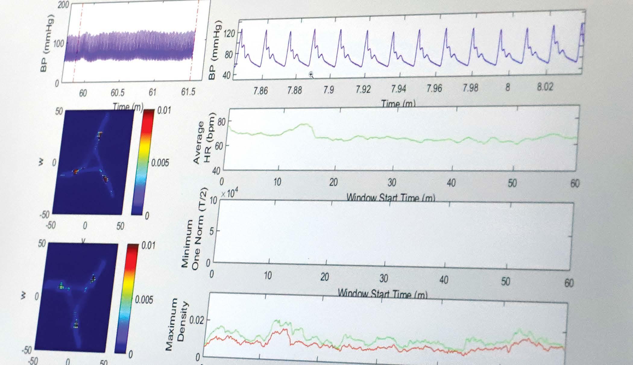
Modern hospital monitoring devices record a
huge amount of data about a patient’s body; sometimes thousands of readings a second.
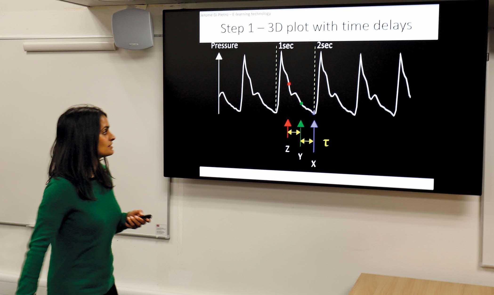
Dr Manasi Nandi has been working on a groundbreaking interdisciplinary project to get more out of that data. “You’ve got a waveform for, say, blood pressure, and in the background you’ve got all of these numbers that make up the shape of that waveform,” Manasi explains, “but we don’t really use it all, although we know that it contains important information.”
To glean more information from the raw data in the waveform, Manasi partnered with mathematician Professor Philip Aston from the University of Surrey. Their aim was to use mathematics to look at the waveform data differently and find new, creative ways of processing the data as a way to extract more information.
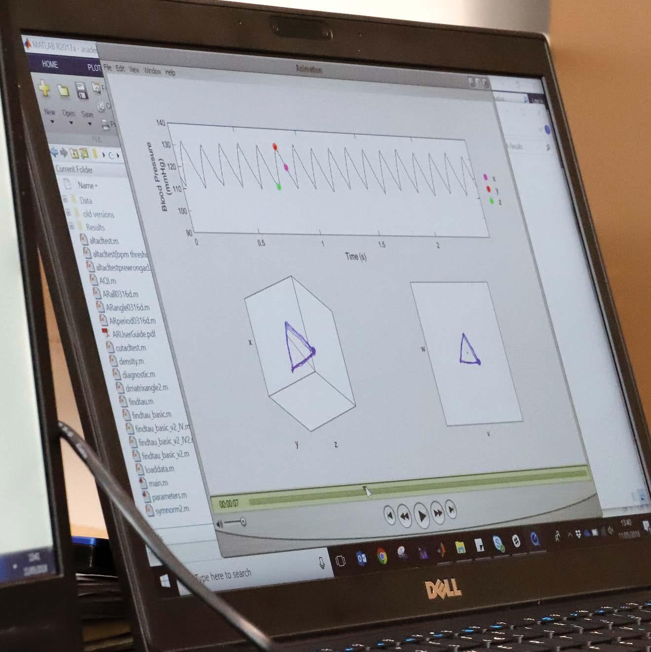
“What this project allows us to do is use every single piece of numerical data that has been captured by the monitoring devices and plot it in a particular way using some clever mathematics. We can handle the big data in a way that is manageable, and it gives us quantitative information from the signal that we previously couldn’t measure. Importantly, we don’t change the raw data, we just visualize it in a different manner.”
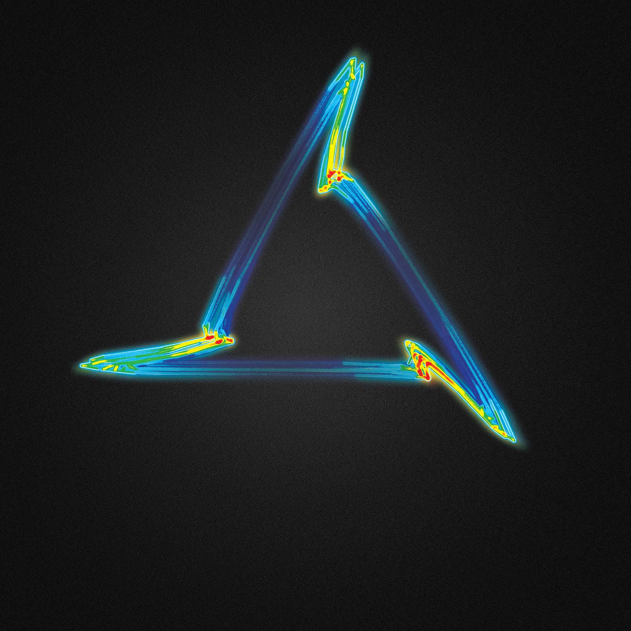
They call this new visualization an Attractor.
At first, the attractor was used to detect inflammation from the cardiovascular recordings of patients after surgery. However, since those early studies, Manasi and her team have examined how it works in other animals and organ systems.
14
“Tiny changes in the shape of the waveform appear as much bigger changes on the cardiomorph,” Manasi explains, “what we have shown so far is that those subtle changes in the shape of the wave give you clues about early stage cardiovascular failure, before obvious changes occur, like your maximum and minimum blood pressure dropping.”
15
Cardiomorph Attractor
Along with her PhD student, Miquel Serna Pascual, her team focused on the respiratory system. They were able to demonstrate that the attractor looks very different when the lungs are functioning properly vs when they’re under stress. “It’s very visually distinct,” Manasi says, “the attractor changes shape from a
tight triangle toward a more open circular shape.”
By examining the shape of attractor a patient had when they were discharged from the hospital, Miquel and Manasi were able to predict with 100% accuracy which patients would be readmitted within 30 days.
16
The attractor uses all of the data from a recording device and plots it in 3D.
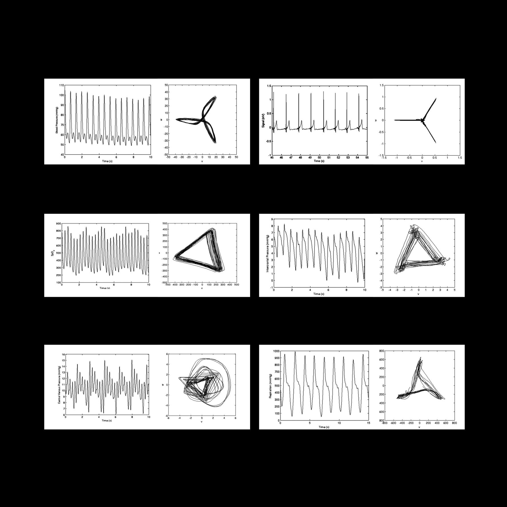
Blood Pressure Electrocardiogram
Central Venous Pressure Respiratory
Pulse Oximetry Intra Cranial Pressure
17
The crucial point to remember is that the data used by the attractor is the same as the information scrolling past on a typical hospital monitoring device, it’s just being presented differently. The highly visual nature of this presentation is not only more sensitive to changes in organ function but is much easier to understand.
At a glance researchers, clinicians, and patients can see when a change occurs. It’s a time and energy saver designed for an environment where both are under constant strain.
18
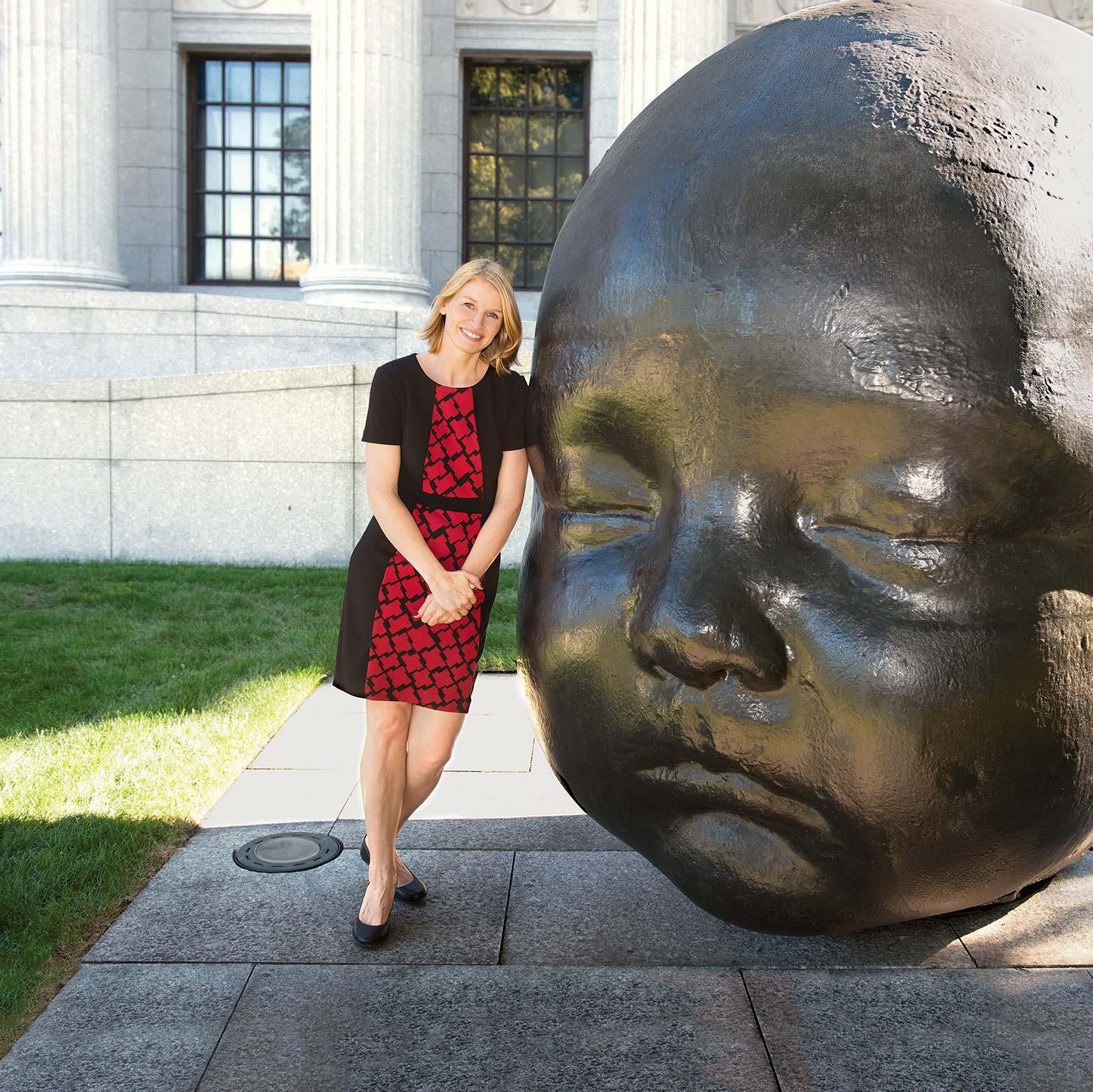
Mapping a path to healthy development
A novel approach to understanding preterm infant development and how to give all children a better start in life
 Associate Professor Emily Zimmerman Director: Speech and Neurodevelopment Lab
Bouvé College of Health Sciences
Northeastern University United States of America
The ability of the human body to grow and develop throughout life is both incredibly complex and beautifully precise.
And nowhere is the precision of biology more obvious than in a neonatal intensive care unit (NICU).
21
Here, the lives of infants born before 37 weeks depend on advanced technology - incubators, respirators, and feeding lines - alongside the cumulative knowledge and care of a team of nurses, pediatricians, and developmental biologists. Thanks to this technology and specialized treatment, the prognosis for preterm infants is continually improving. However, as the focus moves from simple survival to longterm, healthy development, the care has become more complex.
“When babies are born early, they are taken from a very rich and complex environment – the ideal recipe for development,” explains Associate Professor Emily Zimmerman, “part of our research is looking at ways to mimic the womb environment with the purpose of helping these preterm infants get back on track with their development.”

23
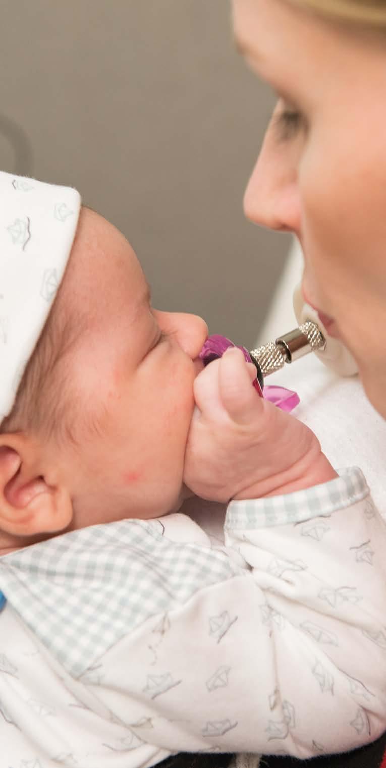
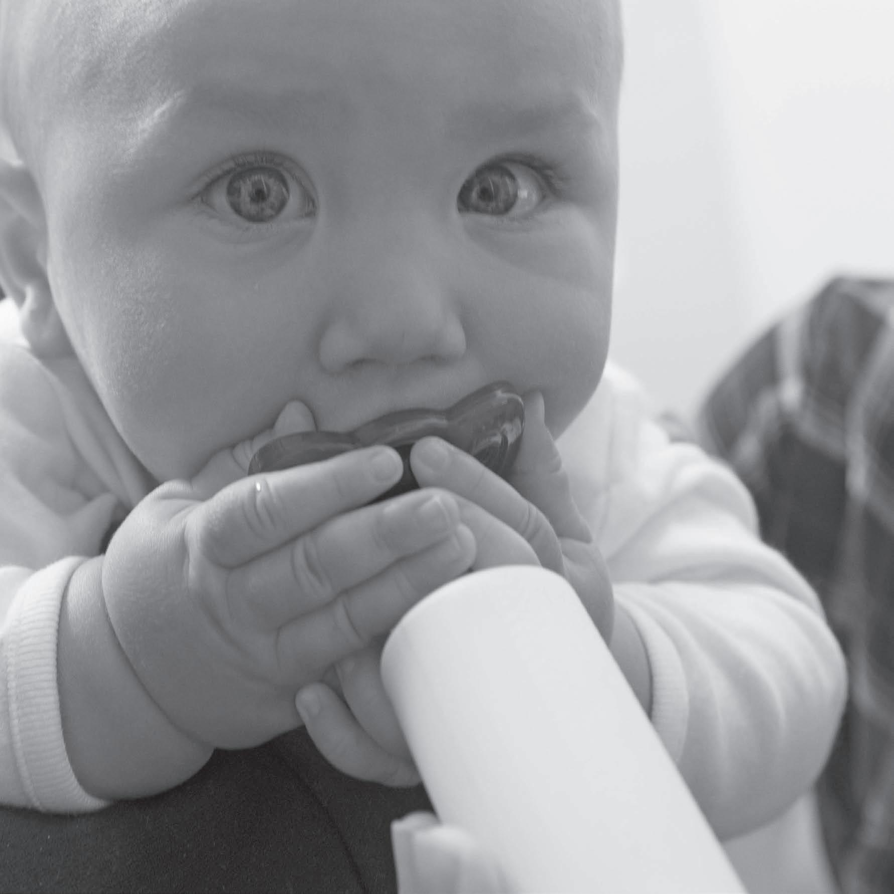
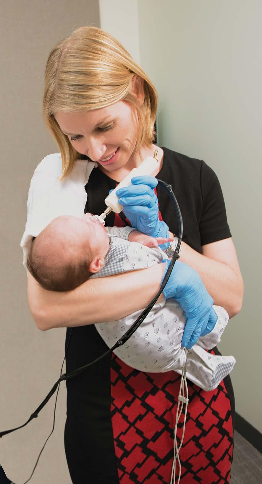
However, the kinds of tests you can run on fragile, infection-prone infants are very limited. In Emily’s lab, one of the key tests is suck patterning, also known as nonnutritive suck as it involves sucking without nutrients. This behavior is centered in the brainstem, providing an early indication of the integrity of the infant’s central nervous system.

“This knowledge can be useful for both preterm and healthy infant populations,” Emily says, “to not only give us early insights into the CNS but also to predict subsequent neurodevelopment.”
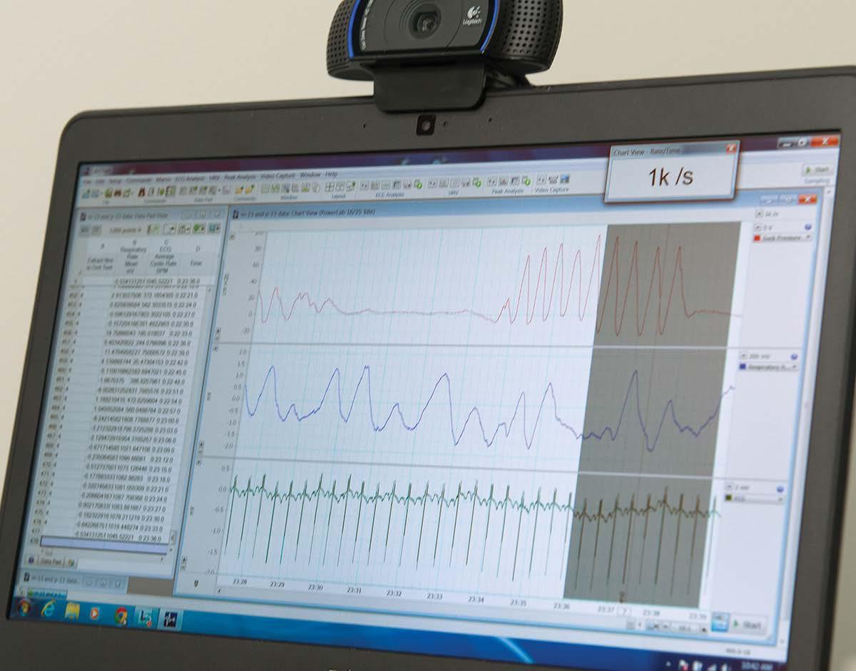
Emily worked with the Mechanical Engineering Department at Northeastern to design a suck device - a pressure transducer that attaches to a pacifier at one end and a PowerLab at the other, recording minute changes in patterns within each burst of sucking. “We look at suck amplitude, how many cycles occur within a burst, how many bursts occur within a minute, how many cycles occur within a minute, and how many cycles per burst. There are many fine spatio-temporal differences found in those measures that help us understand CNS activity.”
24
“To know you are contributing to your field and helping real people is an amazing feeling.”
Not satisfied with trying to fix the problem after the fact, Emily has been working with Dr. Jill Maron at Tufts Medical Center to find early, non-invasive biomarkers. “We are investigating expression levels of a speech and language gene,” Emily says, “to understand how it correlates with oral feeding ability and subsequently with speech and language development.”
25
She’s also taking on the broader causes of preterm birth with the Centre for Research on Early Childhood Exposure and Development in Puerto Rico (CRECE), investigating the role of environmental exposures in the high rate of preterm birth in Puerto Rico.

Preterm births in Puerto Rico occur at a higher rate than that of the continental USA
PUERTO RICO
According to the WHO, an estimated 15 million babies are born preterm. Preterm birth complications are the leading cause of death among children under 5 years of age, responsible for approximately 1 million deaths in 2015.
“We’re so excited to look for these outcomes,” Emily says, “we really hope that through this research we can influence maternal behaviors or even guide environmental regulations to result in healthy children who are free of delays.”
26
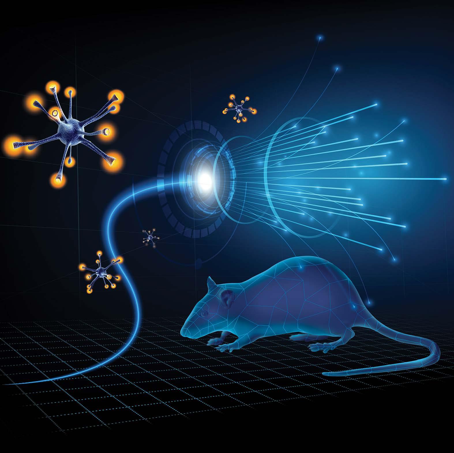
Lighting the way for Parkinson’s research
How optogenetic stimulation could be the next step in the treatment of Parkinson’s Disease
Associate Professor Louise Parr-BrownlieNgāti Maniapoto me Te
ArawaDirector: New Zealand National Science Challenge for Ageing Well Department of Anatomy University of Otago New Zealand
29
Parkinson’s disease is the second most common neurodegenerative condition, with more than six million people diagnosed worldwide.
The disease is caused by rapid death of dopamine cells in the brain, leading to slowness of movement, rigidity of the body, and muscle tremors.
Dr. Louise Parr-Brownlie (Ngāti Maniapoto me Te Arawa) is a neurophysiologist passionate about Parkinson’s disease research and outreach. “The key thing to be aware of,” Louise says, “is that, in most Western societies, we have an aging population. So for at least the next 50 years, there’ll be more people over 60 years of age.” The highest risk factor for Parkinson’s disease is being older than 60 years of age. With an aging population, the need for Parkinson’s research and awareness continues to grow.
Louise and her team are focused on improving the quality of life for those with the diagnosis. Her goal is to assist people with Parkinson’s disease to live independently, move more easily, and maintain their standard of living.
30
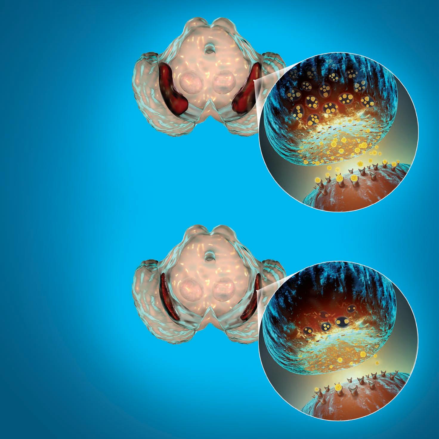
of dopamine neurons in the substantia nigra have already been lost by the time motor symptoms appear. NORMAL PARKINSON’S DISEASE SYMPTOMS Slow movement • Tremor Rigidity • Imbalance • Pain Sensory disturbances • Loss of smell Cognitive impairment • Sleep dysfunction 60-80% Substantia nigra Transmitting neuron Dopamine Dopamine receptor Receiving neuron 31
Opposite: The substantia nigra of the midbrain and its dopaminergic neurons. The substantia nigra regulates movement and reward, its degeneration is a key step in development of Parkinson’s disease.
Electrical brain stimulation can improve symptoms of Parkinson’s disease, leading to an extra 4-5 hours a day without dyskinesia. However, off-target stimulation is common with this treatment, and there is some concern over not knowing “which bits of the brain are being activated.” The solution appears to be in an emerging field of cell targeting and stimulation: optogenetics.
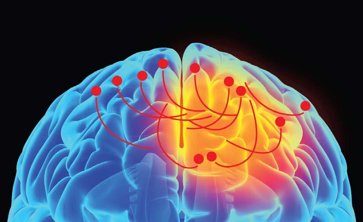
In the context of the brain, optogenetics involves using viral vectors to implant the genes for light-sensitive proteins, called opsins, into specific neurons. When a fibre optic cable shines light into the brain, only those neurons with active opsin proteins will be activated. It’s a whole new world for researchers like Louise, enabling specificity of not only the region but the individual neurons being activated.
ELECTRICAL STIMULATION OPTOGENETIC STIMULATION
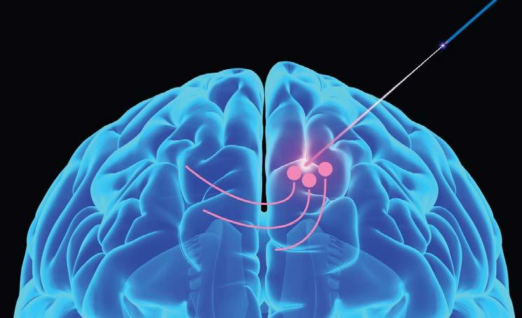
Neurons impacted by electrical stimulation Stimulated axons Neurons impacted by optogenetic stimulation Stimulated axons 32
“Ideally, I’d like to stop people from ever getting Parkinson’s disease,” Louise says, “but if we can slow down the progress of the disease so that instead of taking 2 years, it takes 10 or 15 years to become symptomatic; then that would be an amazing outcome.”
The Parr-Brownlie lab theorizes that optogenetic stimulation of the motor thalamus may reduce the abnormal activity associated with altered muscle control in Parkinson’s. Her current project aims to correct the abnormal pattern of cell activity in the motor thalamus of Parkinsonian rats by stimulating them to fire in a healthy pattern.
The key thing for these experiments was to validate a brain stimulation method that could be viable for months without interfering with the day-to-day activity of their animal models. When technology began to limit how long they could run their experiments, the Parr-Brownlie research group began testing implantable Kaha optogenetics telemeters.

“…If this is ever going to be a treatment for people, then we’ve got to have a device that’s probably implanted, can be turned on… and keeps going probably for the rest of the life of the device.”
Right: The Kaha optogenetics and biopotential telemeter, the first fully implantable, controllable, optogenetics stimulator for use with rats that can also record biopotential signals.
33
The prevalence of Parkinson’s disease has doubled in the past 25 years
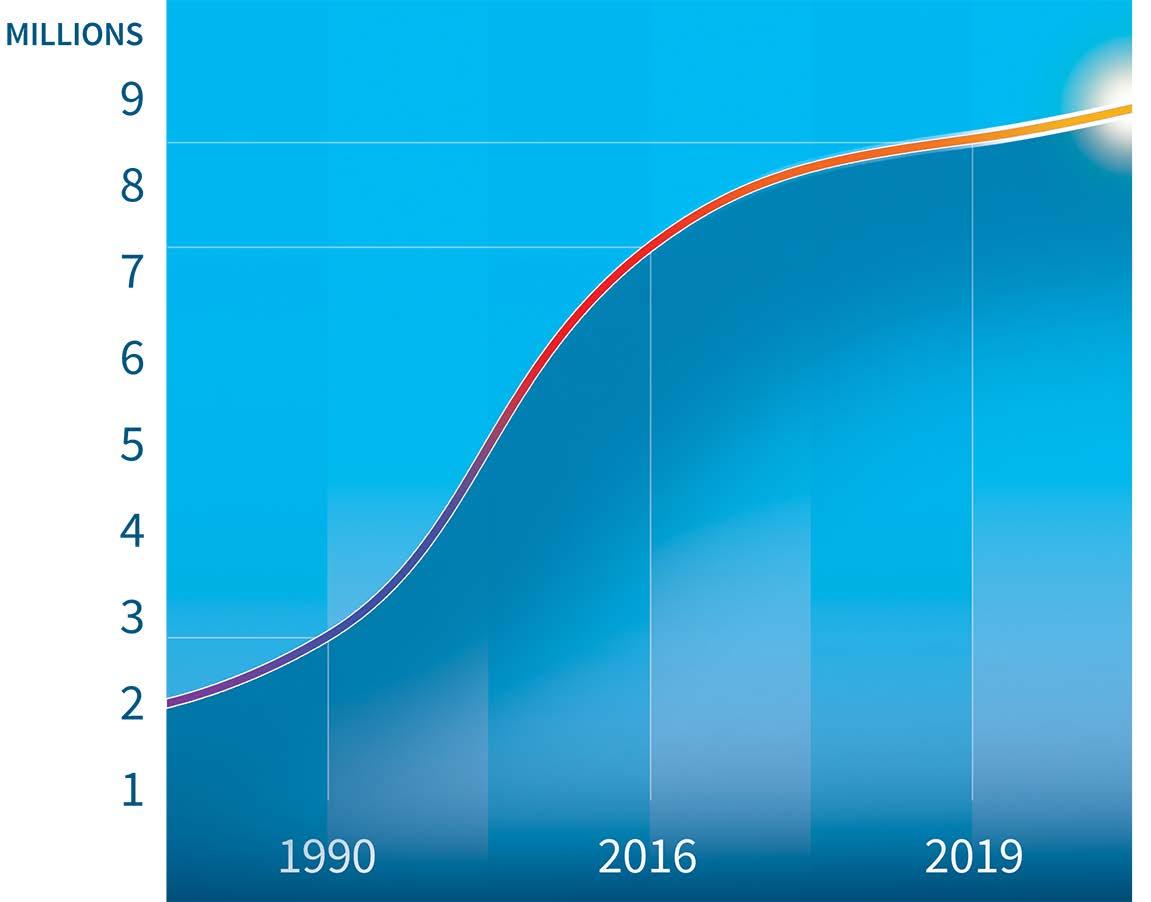
Above: Worldwide incidence of Parkinson’s disease from 1990 to 2019.
1% of people over 60 are affected by Parkinson’s disease. As our population ages we will see the number of cases continue to increase.
34
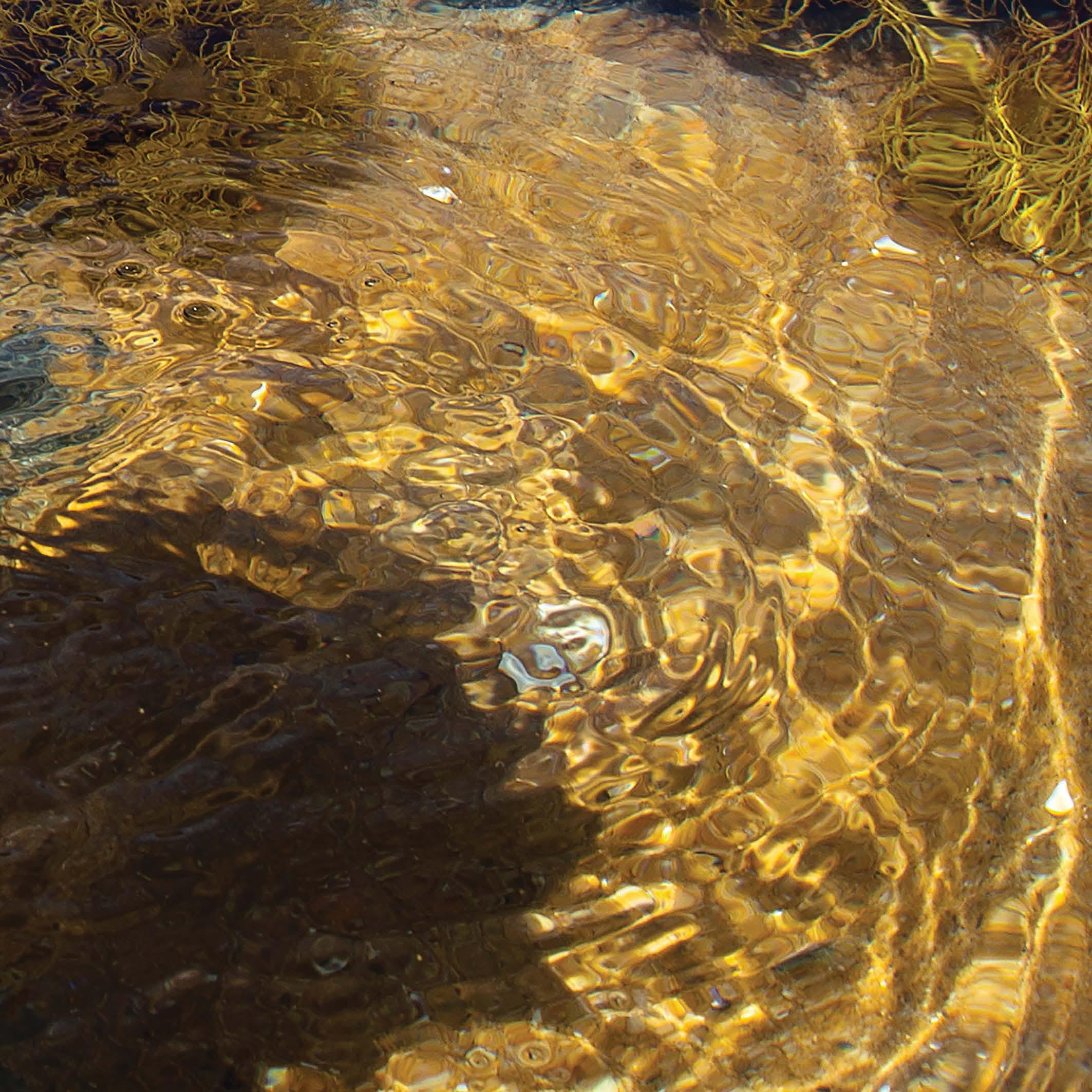
have two fish that are very closely related, almost identical, and one lives in rock pools that can tolerate hypoxia and one in deeper ocean that can’t, then we have a really good chance of identifying the factors that allow the fish to protect itself from its environment.”

“If we
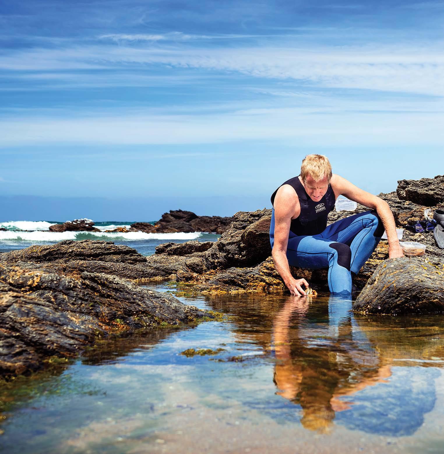
Lessons from the wild
How comparative physiology is putting our world into perspective
Associate Professor Tony Hickey Faculty of Science University of Auckland New Zealand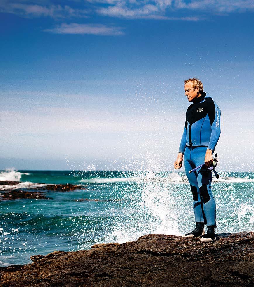
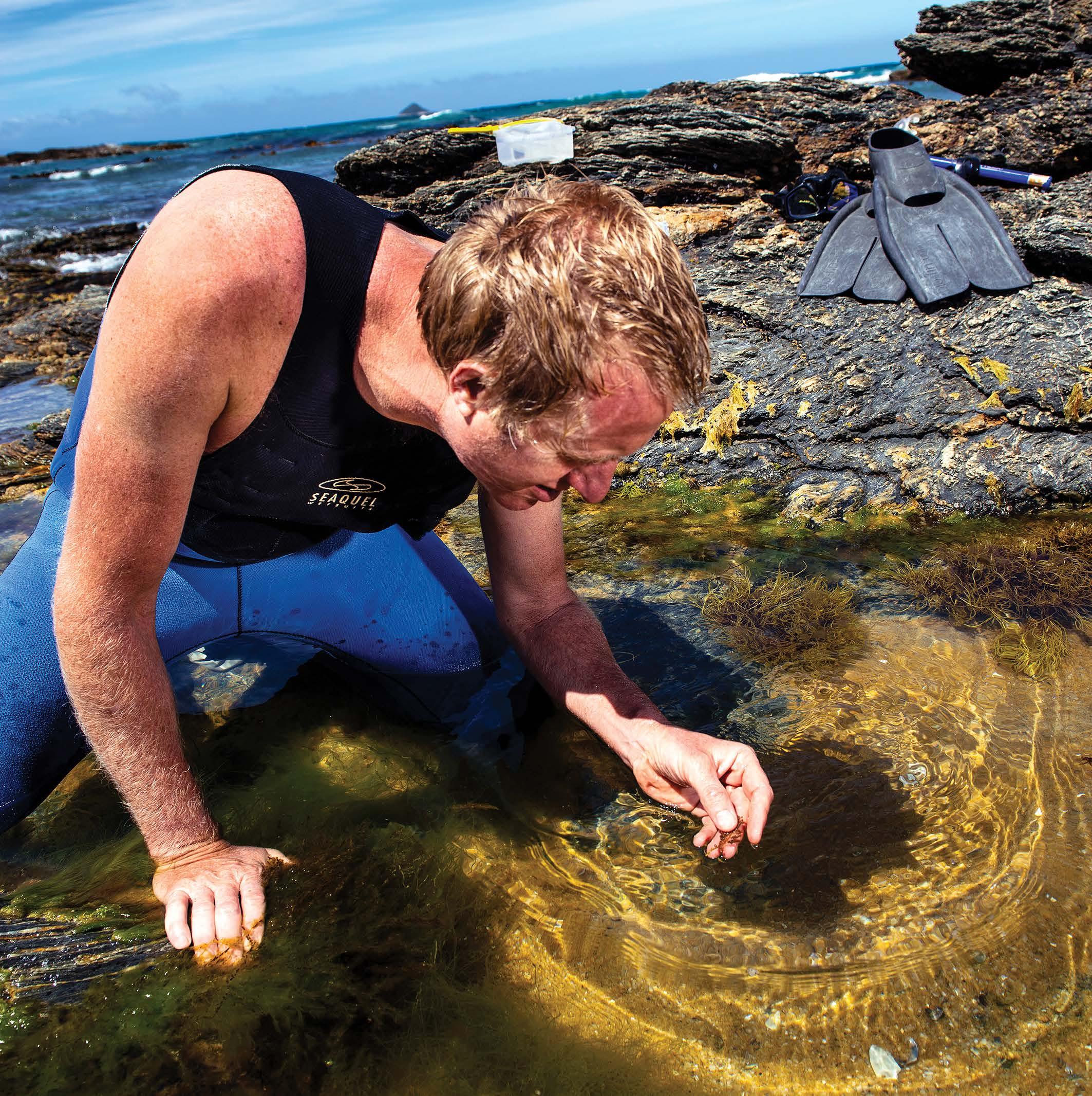
37
Human problems are rarely human-specific. We may have large brains and globe-spanning civilizations; but inside our cells, we’re not all that different from a rat, a fish, or a bee.
Associate Professor Tony Hickey takes advantage of these similarities, finding animal solutions to human problems.
When we first interviewed Tony he and a student had been exploring the impact of diabetes on heart failure. Using an ADInstruments Isolated Working Heart System, Tony examined how tissue from the heart behaves once oxygen runs out. “We thought we’d made a mistake with the experiment,” Tony explained, “once the oxygen was gone they were consuming ATP almost as fast as they could make it.” When the same experiment was run with a diabetic heart the increased ATP consumption was the same, but the production couldn’t keep up. This could be why a diabetic heart gives out faster than a non-diabetic heart.
The Isolated Working Heart System for Rats


38
Bellapiscis medius, a triplefin fish that makes its home in tidal pools, has evolved resilience against prolonged oxygen deprivation. However, like the combination of diabetes and heart attack, when the triplefin’s system has already been stressed by heat its ability to deal with oxygen deprivation begins to fall apart.
This kind of heat sensitivity isn’t specific to fish; it’s everywhere. “Honey bees are known to kill wasps by overheating them,” Tony says. “At first we thought it was just a Japanese honey bee that did this, but it’s a common behaviour.” The bees will swarm an invading wasp, vibrating to heat the surrounding air to 55°C. When he examined the flight muscles of wasps he found that they stopped functioning
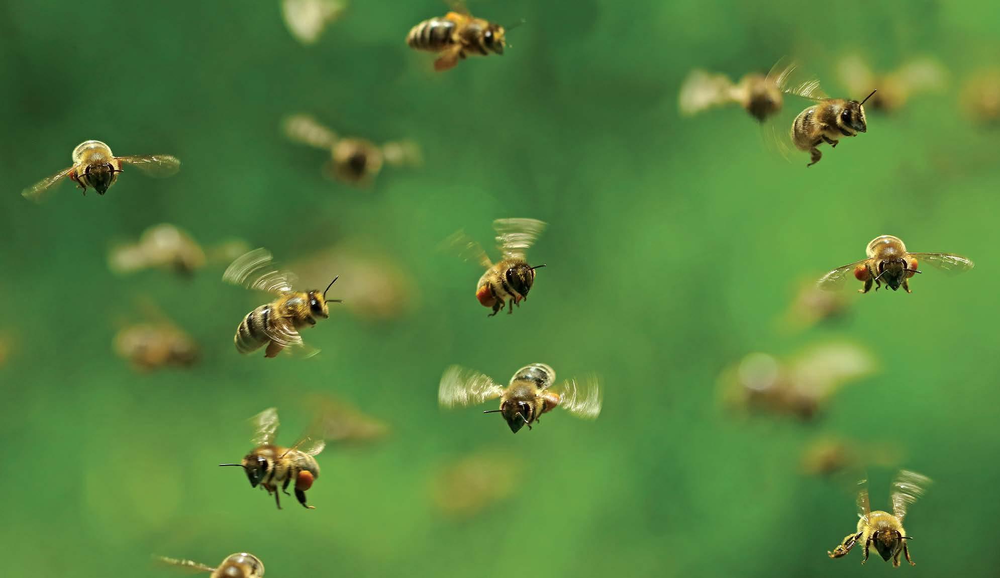
39
at 45°C, whereas the bees could keep going until they hit 55°C. These natural temperature limits change depending on the habitat of the species, but the effects of climate change are pushing all life closer to their limits.
“This is a wrasse heart,” Tony says, as a recording of the tiny heart pumps away on his phone screen. “It works perfectly until you get up to around 24-26°C, and then the mitochondria will start to give out and the heart fails.” Around Auckland, these fish are used to living in temperatures of 20-21°C; the sea surface temperatures in Auckland hit 28°C this summer.
Below: Mitochondria use oxygen to produce the vast majority of the body’s ATP, the cellular energy currency that keeps our proteins, cells, and organ systems running.
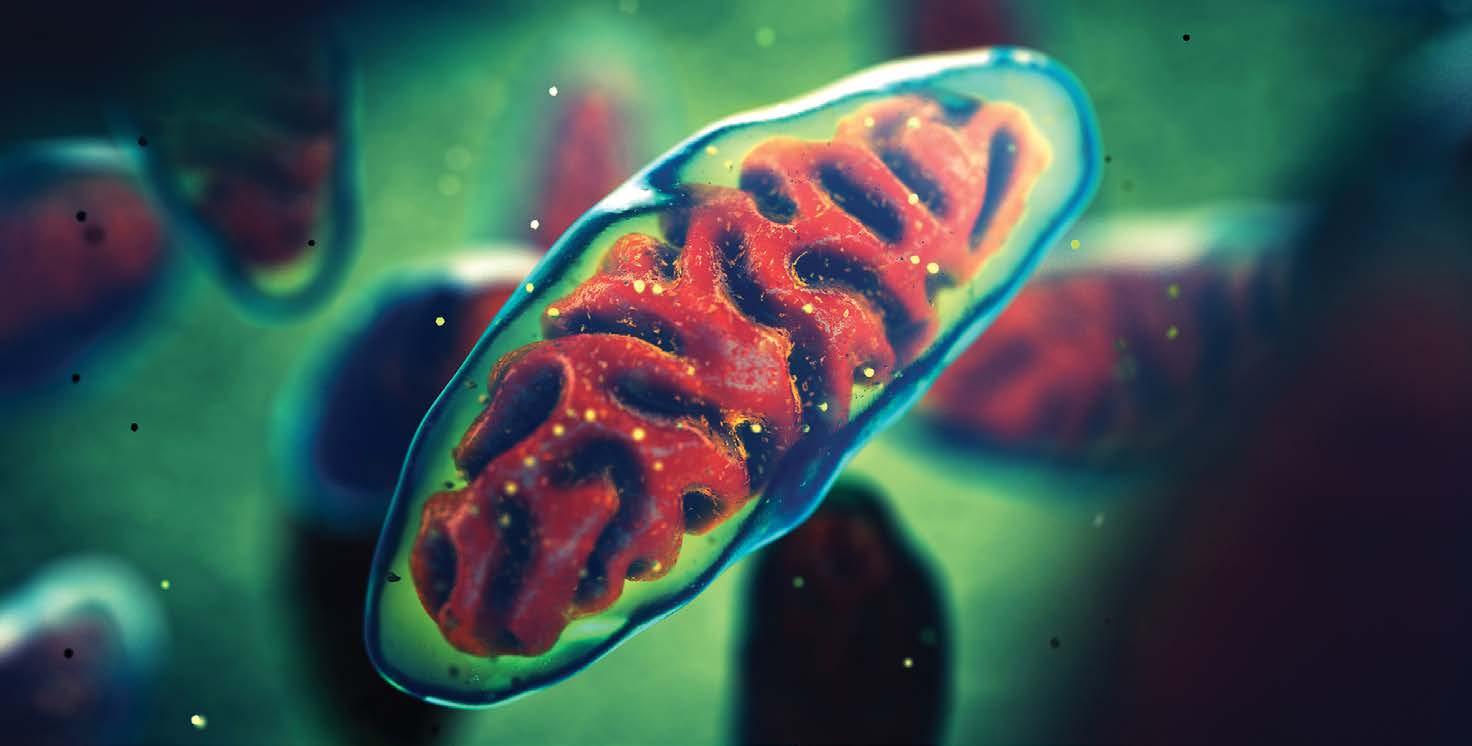
40
For Tony, heart failure, oxygen deprivation, and climate change are all peas in a pod. Human problems highlighted by the animals around us. These comparisons serve double duty, providing insights into the metabolic function of our own bodies, and warning us about the biological limitations of the global biota we rely on to survive.
“The organ that kills us as we get too hot tends to be the heart. As it gets hotter and hotter it suddenly fails.”
42
“A fundamental belief of my colleagues and I is that often big problems can be solved by looking at small solutions that animals have adapted.”
Extreme science in search of answers
Learning about age-related disease from the edges of the world
Professor Damian BaileyEditor-in-Chief of Experimental Physiology Royal Society Wolfson Research Fellow
Director of the Neurovascular Research Laboratory
Chair of the Life Sciences Working Group
European Space Agency
Department of Physiology and Biochemistry
University of South Wales United Kingdom
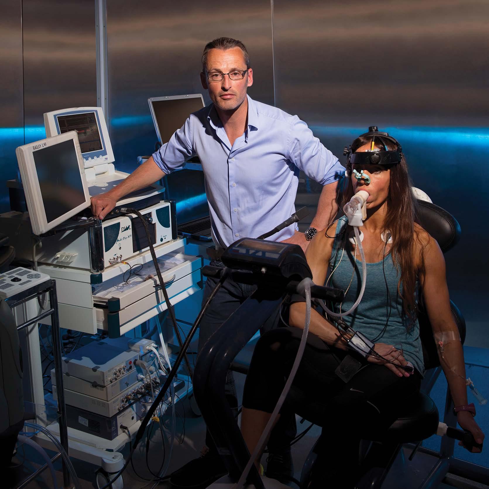
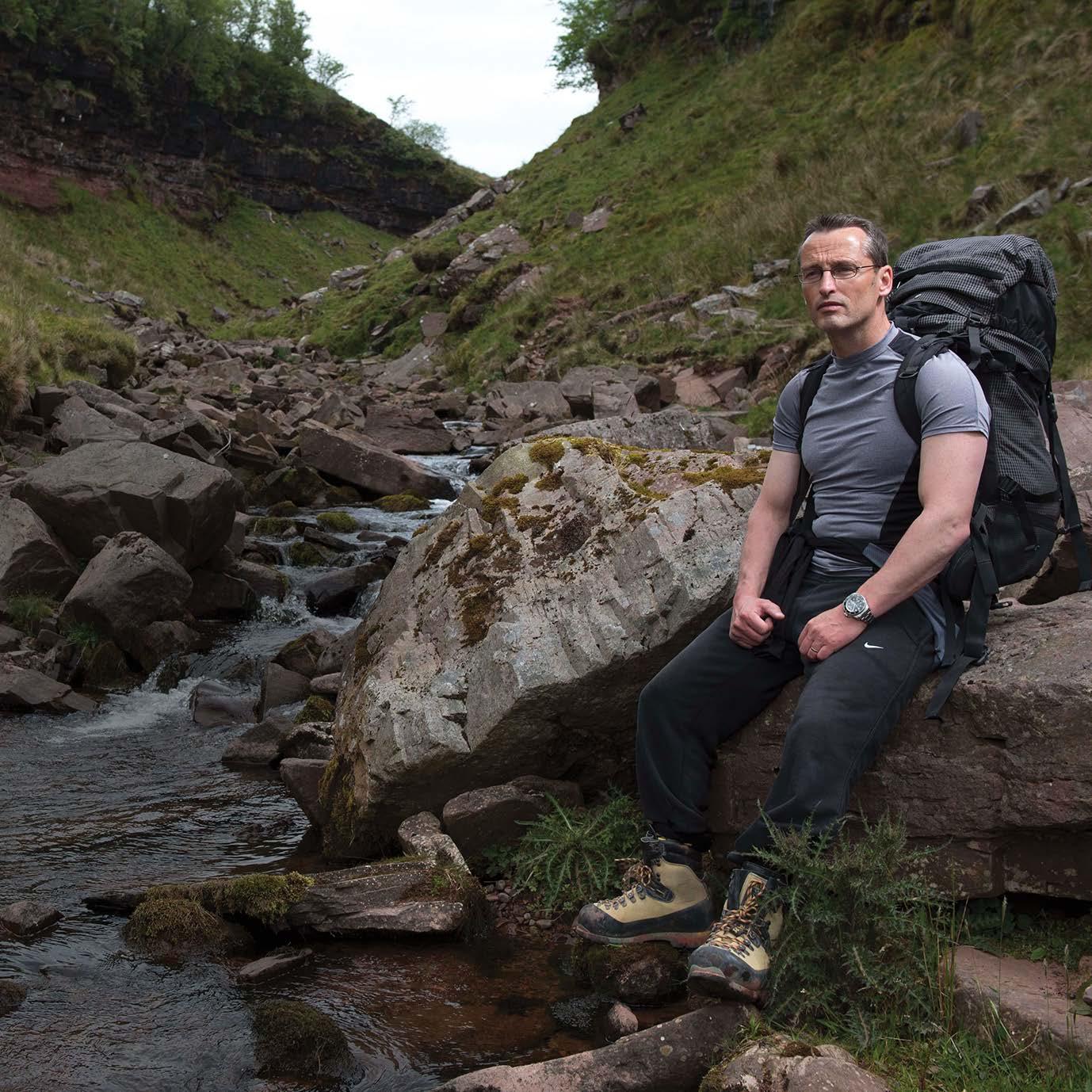
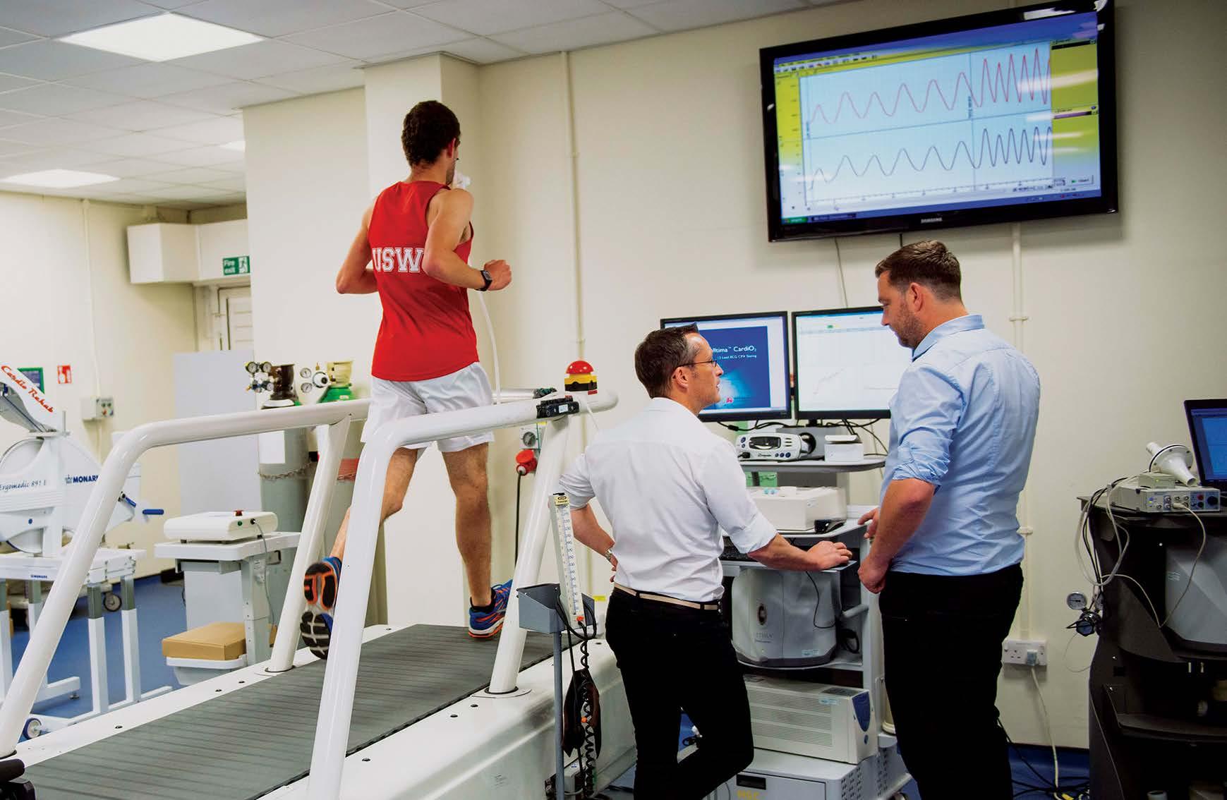
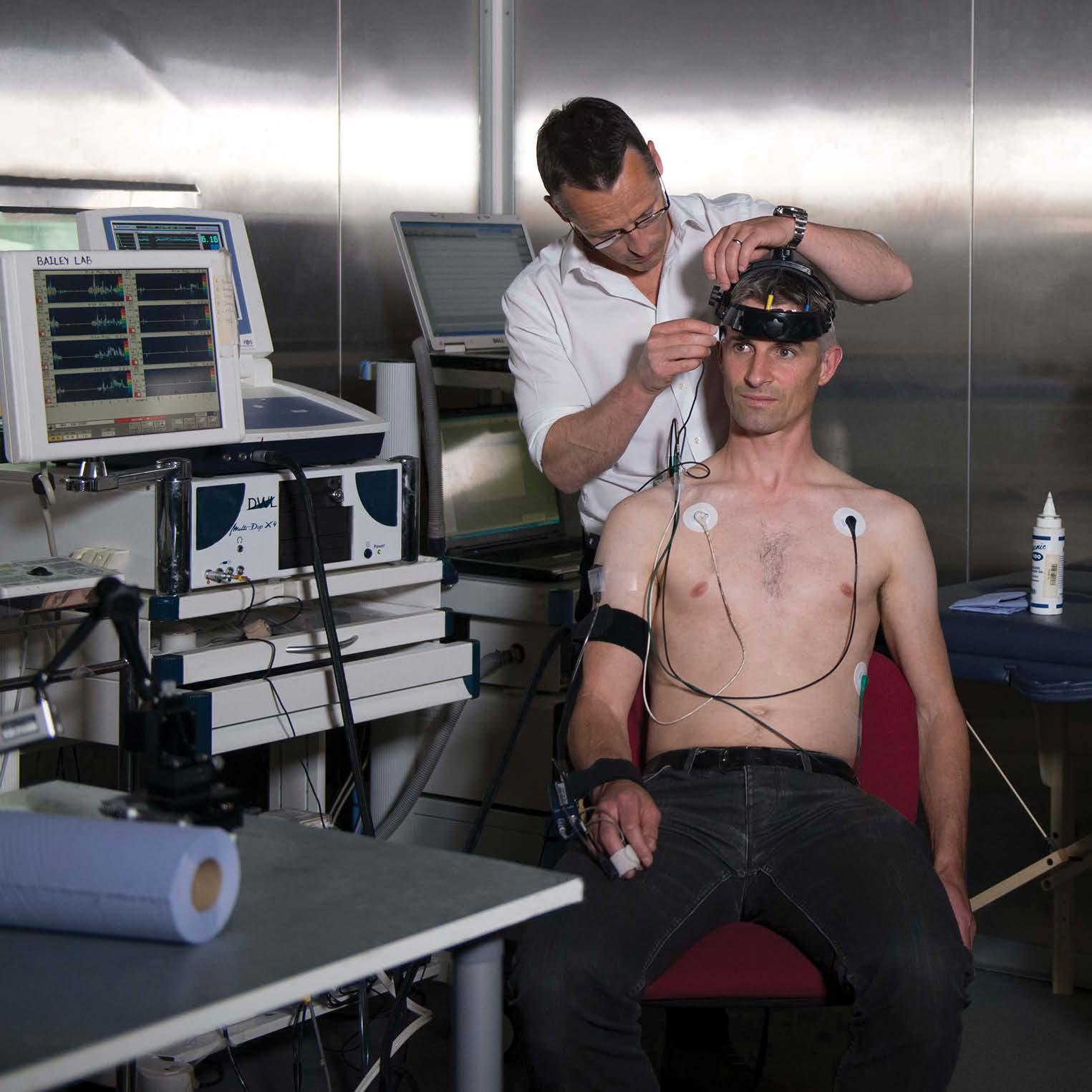
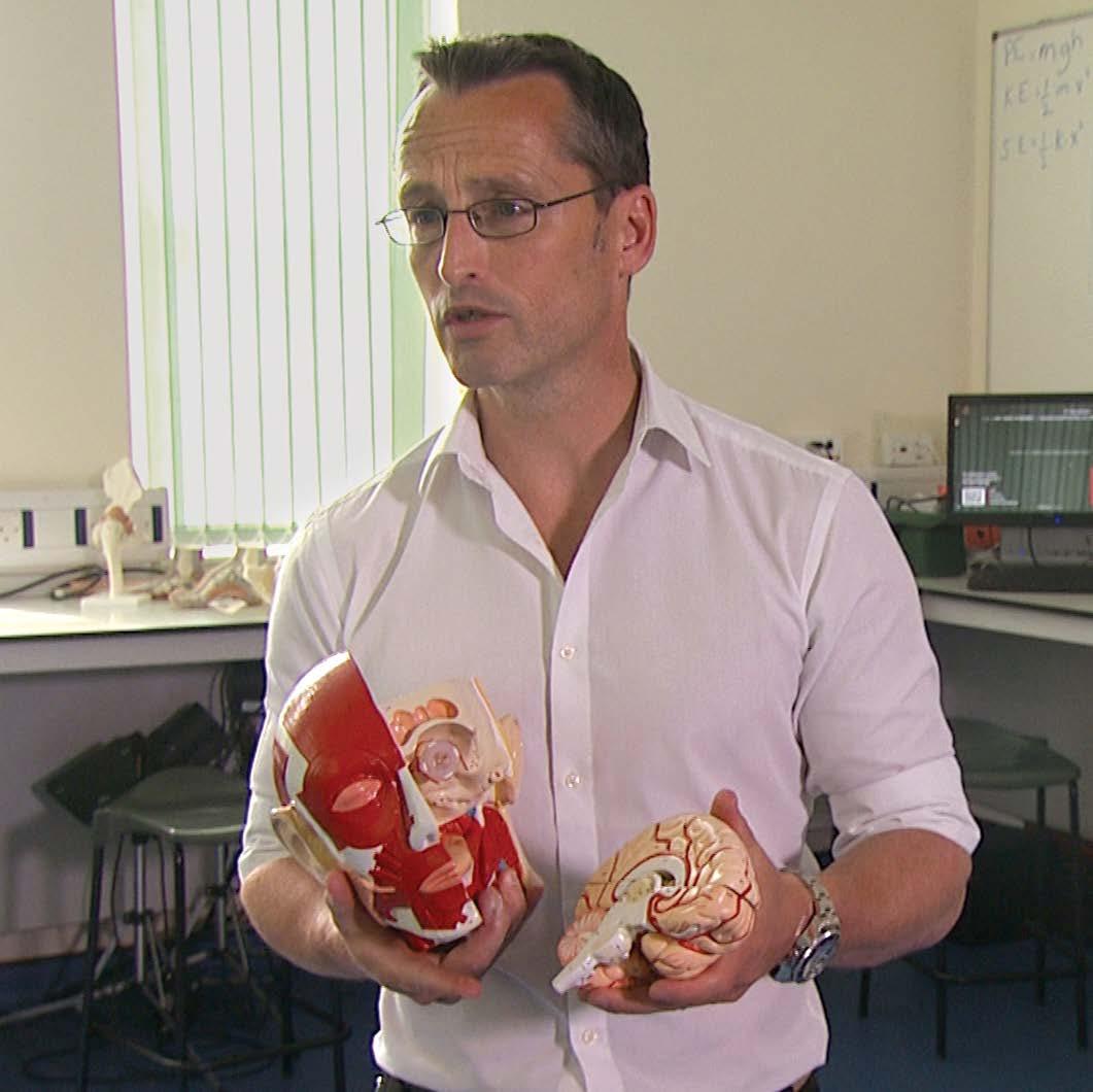
Professor Damian Bailey has been to the edges of the world exploring the human body’s relationship with oxygen; from the short-term oxygen starvation of freediving to the chronic mountain sickness that sets in at high altitudes. By studying how individuals cope with environmental extremes Damian opens a window into the regulation of oxygen transport in everyday life.
“Why have we evolved a brain that is so reliant on oxygen? And to what extent does oxygen availability in the brain influence our trajectory towards disease?”
His work with surgical patients, athletes, freedivers, and mountaineers, has provided a new perspective and greater understanding of ordinary people and the diseases they face as they age.
By examining free radical formation in the skeletal muscle of exercising humans, he was able to show that exercise can work as a countermeasure to aging and age-related neurodegeneration. Free radicals act as adaptive signaling molecules, responding to the metabolic needs of the muscles in use. By building up in exercising muscles they contribute toward improving vascular health within not only the muscle but also in the brain.
These adaptations allow the exercising brain to function as if it were over a decade younger, which can reduce the incidence of stroke and dementia. It is a dream result.
46
The power to fight age-related neurodegeneration could be available to everyone, at no cost and without the adverse side effects that can come with medication.
Since that finding, the extremes of neurodegeneration have continued to push Damian toward physical extremes. He and his lab have even taken up skydiving as a way of investigating dementia. By recording the neural adaptation of world-class skydivers to the cold and hypoxic conditions of high-altitude jumps, they hope to find a way of protecting the brain against age-related degeneration.
47
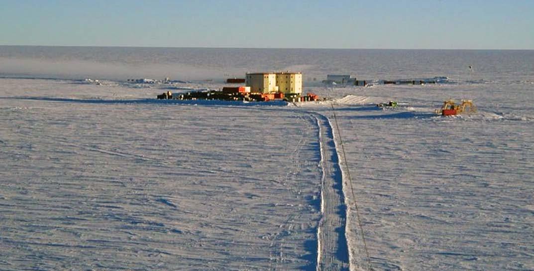
CONCORDIA BASE
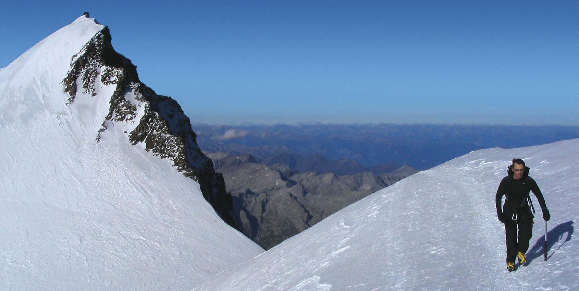
In 2024, with support from the European Space Agency, Damian will set out on a 10-month study at the Concordia base in Antarctica. Also known as ‘White Mars’, the base is a perfect location to examine the potential physiological effects of deep space missions, and at 4000ft above sea level, it also serves to show the body’s ability, or lack of ability, to adapt to high altitude.

As with the revelation that exercise can protect the brain against the effects of aging, these extreme leaps toward the edges of the human experience hold a tremendous amount of information. What can our oxygen-greedy brains adapt to? How can we train them to perform better, for longer, or with the least harm possible? If we don’t reach out and push as far as we can go, we might never know what our bodies are capable of.
49
“For me, the expeditions are about providing opportunities and inspiring the next generation of researchers to address some of the really complex questions that we are still wrestling with,” he says,
“And it’s a great way to show them that there is an opportunity to be creative, to combine excellent science with a sense of adventure and in so doing, have a lot of fun.”
50
Taking physiology to new heights
Creative approaches in high-altitude research
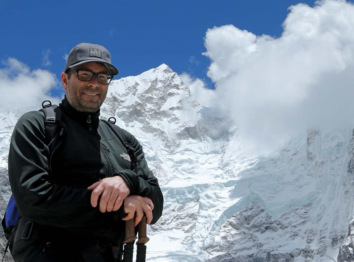
 Professor Trevor Day
Department of Physiology Mount Royal University Canada
Ordinarily, you’d think this predicament would provide some cause for concern, but at an altitude of over 17,000 feet (approximately 5,000 metres) I’ve been reassured this ‘periodic breathing’ is a normal physiological response for someone like me who usually lives at sea level. So a little unnerving, yes, but typical nonetheless. After a few moments and a deep, forced inhale, I’m breathing quietly again and able to drift back to sleep.”

“It’s three in the morning and I’ve woken up, unable to breathe.
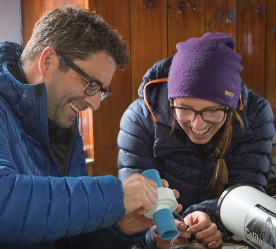
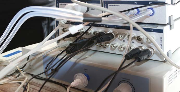
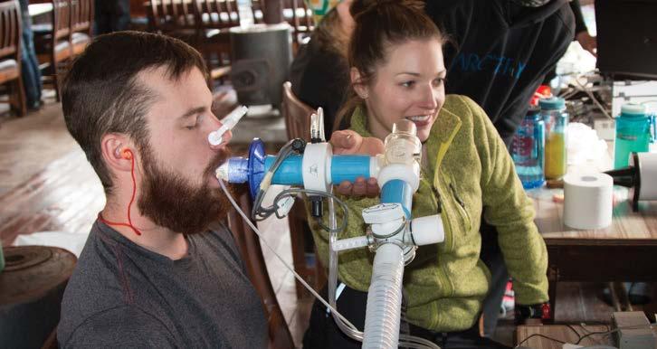
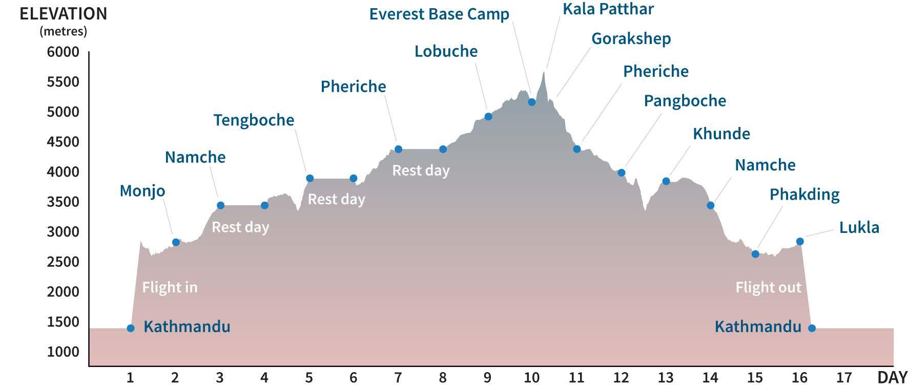
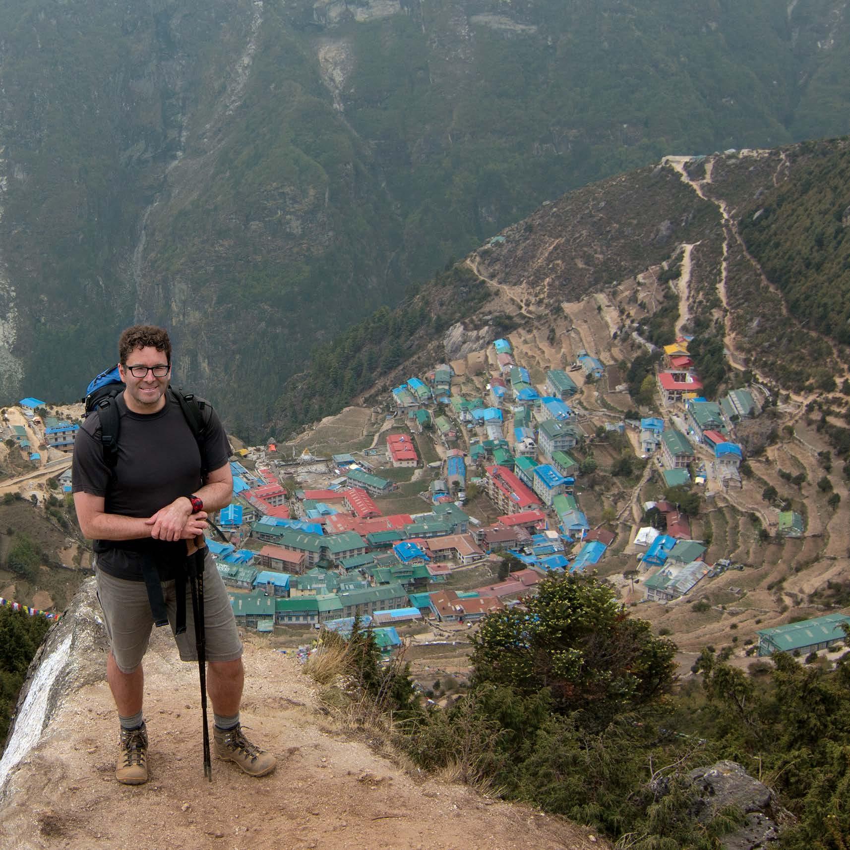
Professor Trevor Day has organized three research expeditions to Everest Base Camp in as many years, in search of a predictor for Acute Mountain Sickness. These expeditions, which include collaborators, participants, and students, comprise a 120-kilometer round trip from Lukla airport at 9,200 ft (2,800 m) above sea level in the base of the valley, to Everest Base Camp and back, over two weeks. It’s a significant altitude challenge, around 18,000 ft (5,300 m) in the end, but long and slow enough to track each trekker’s acclimatization to the increasingly oxygen-thin atmosphere. The goal was to develop a battery of simple metrics to assess acclimatization, with the aim to be able to predict who will get sick at high altitude before the sickness sets in. This unique “incremental ascent” research and teaching model allows for large groups of trekkers to ascend slowly, minimizing the chances of severe altitude sickness. “With ascent,
we sensitize our receptors that detect low oxygen, so we breathe more when we are exposed to a low oxygen stimulus,” Trevor says. “We also make more red blood cells, which improves our oxygen carrying capacity.”
The higher they ascend, the greater the chance that some trekkers will develop Acute Mountain Sickness - a combination of headache, dizziness, nausea, and fatigue that can ultimately deteriorate into life-threatening cerebral or pulmonary edema. “As you ascend, everyone will get some of these
53
symptoms,” Trevor says, “but what we’re looking for is a battery of tests you can do at sea-level and with ascent that will help to predict who is going to do well and who is going to be more affected.”
Predictive factors have been ruled in and out over the years. Exertion, hydration, food intake, rate of ascent; these all seem to play a part. Whereas age, sex, and even fitness level don’t appear to have an impact. Trevor’s focus, however, is on that core difference between sea-level and high altitude: oxygen. Before leaving Lukla at the
54
start of the trip they record weight, blood pressure, oxygen saturation, carbon dioxide and breathing. Each morning of the trek is a similar battery of tests with the added bonus of hematocrit and hemoglobin concentration. These tests measure how much of their total blood volume is taken up by red blood cells desperately trying to take in oxygen. On one expedition, they obtained arterial blood draws with ascent to better assess oxygenation and acid-base status.
Trevor has since expanded the types of expeditions he organizes, adding rapid ascent

and residence models to study new and related questions at high-altitude labs.
Ultimately, the Himalaya mountains and the Sherpa people are drawing him back. “We are set to return next year to compare exercise capacity in low-landers vs native highlanders (Sherpa) with the support of federal grants from both Canada and the US.” Trevor says, “I can’t wait to try and figure out some of the factors that make the Sherpa such incredible athletes on the mountain!”

•
•
•
•
•
•
•
•
• Food
•
•
•
• Fitness • Age • Gender •
•
• HRV • Breath
•
•
• Blood
• Resting
• Oxygen
•
•
•
•
• Oxygen
•
•
•
•
•
Acute Mountain Sickness (AMS):
Headache
Dizziness
Nausea
Fatigue
If not addressed by reducing altitude it can deteriorate into life-threatening cerebral or pulmonary oedema.
Possible factors:
Exertion
Hydration
intake
Genetics
Rate of ascent
Not Factors:
Rest day measures:
Hypoxic Ventilatory Response - breathing more as oxygen saturation in the air gets lower
hold duration Measuring the physiological effects of altitude
Before departure:
Weight
pressure
heart rate
saturation
Respiratory rate
End tidal CO2
Every morning of the ascent and descent:
CO2
saturation
Respiratory rate
Heart rate
Blood pressure
Hemoglobin
Hematocrit (ratio of red blood cells to total blood volume)
you ascend, everyone will get some of these symptoms, but what we’re looking for is a battery of tests you can do at sea-level that will help to predict who is going to do well and who is going to be more affected.”

“As
PHOTOGRAPHER CREDITS
James Stringer 1
Naudé Heunis 3-5, 7-8
Shutterstock, Inc. 9-10, 28, 31, 39, 40
Marcus Hughes 11, 13, 16, 18
Manasi Nandi 17
Frank Monkiewicz 19-23, 25
Joe Gallagher 27
Sharron Bennett 30
iStockphoto 31, 32
Alex Lovell-Smith 35-37, 41-45, 47-48
Damian Bailey 46, 50
James Elworthy 51-58
All images ©ADInstruments
SCIENCE HEROES
Copyright © 2022 ADInstruments.
All rights reserved. No part of this publication may be reproduced in any form (by any means, electronic or mechanical, including photocopying or recording) without written permission from ADInstruments or individual copyright holders.
Writers: Blaise Cahill-Lane, Alex Sides, James Elworthy, Sina Walker, Ella Williams
Design: Matthew TrbuhoviĆ
Cover image: Naudé Heunis
Read the full stories and watch our hero videos at www.adinstruments.com/heroes

www.adinstruments.com/lightning

www.adinstruments.com/powerlabc
www.adinstruments.com