

INSIDE
Optimal Management for Patients with Barrett’s Oesophagus and Dysplasia
Gastric Peroral Endoscopic Myotomy (G-POEM)
Faecal Microbiota Transplant
OLYMPUS CORNER
WELCOME TO THE OLYMPUS CORNER
A Future-Focused Olympus Shines at the 2024 NZSG & NZgNC Annual Scientific Meeting
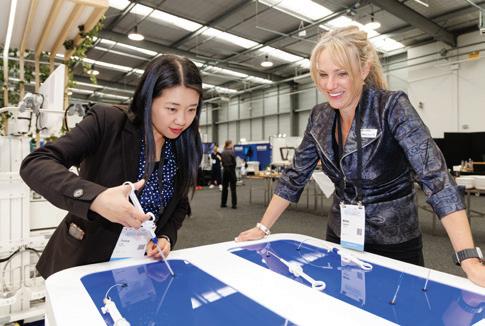
As a proud Platinum sponsor of the 2024 New Zealand Society of Gastroenterology (NZSG) and New Zealand Gastroenterology Nurses College (NZgNC) Annual Scientific Meeting, Olympus was at the forefront of innovation and education during the three-day event held at Claudelands, Hamilton. Bringing together local and international delegates, keynote speakers and industry leaders, the conference offered a platform for advancing gastroenterological science, sharing expertise, and exploring cutting-edge technology.
At the Olympus exhibitor stand, delegates had the opportunity to explore a host of industry-leading technologies, including the EVIS X1 platform, interventional EUS and ERCP equipment, and the revolutionary ENDO-AID, Olympus’ latest Artificial Intelligence technology for endoscopy. Attendees engaged in hands-on experiences with the interactive Endonix endoscopy simulator and a simulated colon model, tools that sparked conversation and provided a deeper understanding of the evolving possibilities in endoscopy training and practice.
One of the standout products on display was Olympus’ EndoCuff Vision, a mechanically enhanced colonoscopy device designed to improve visualization of mucosa during procedures. This device generated widespread interest and positive feedback from delegates, who were eager to incorporate it into their clinical practice.
Olympus also demonstrated its commitment to supporting gastroenterology nursing professionals by sponsoring the Nurses Hands-On Workshop. Attendees gained invaluable practical experience with activities including polypectomy in-service training, detailed scope structure education, and foreign body retrieval procedures. The workshop featured a variety of Olympus devices, such as grasping forceps, snares, and stone extraction baskets, enabling participants to enhance their technical skills in a collaborative environment.
The 2024 NZSG & NZgNC Annual Scientific Meeting was a true celebration of progress, education, and innovation. Olympus is honoured to have contributed to its success and extends its gratitude to all who participated. We’re already looking forward to reconnecting with this passionate community at the 2025 Annual Scientific Meeting in Palmerston North, where we will continue to inspire and advance the field of gastroenterology together.


The Olympus Continuum team is dedicated to educating the specialists of tomorrow. Olympus Continuum offers courses and webinars for reprocessing and endoscope training, specific topics include infection control, reducing the risk of cross contamination, and other adverse events related to the reprocessing of flexible endoscopes and surgical instruments. Getting started is easy—simply visit the website, locate your regional LMS site, and register for Olympus Continuum.
For Upcoming Olympus Continuum Courses: To book, visit: https://learn-nz.olympus.co.nz and register.
COMMITTEE
Chairperson
Karen Kempin
Secretary
Nicola Caine
secretaryofnzgnc@gmail.com
Treasurer
Fiona Williams
Hepatology Group
Nicola Caine
Nurse Endoscopist Group
Karen Kempin
Inflammatory Bowel Disease Group
Nideen Visesio
Committee Members
Emma Deere (also Co-chairperson)
Kate Lodge
Caroline McClutchie
Life Members
• Sandra Burton
• Karen Gower
• Sherry Sharp


NZNO NZGNC Report from the new chairperson – Karen Kempin
Hi everyone,
Welcome to 2025.
Since the very successful Gastroenterology ASM in Hamilton at the end of last year we have had some big changes on the gastroenterology nurses committee.
Firstly I would like to thank the Waikato nurse team who organised a quality conference program including the very memorable Hoe Down in H Town conference dinner. An incredible night of team building, dancing and the usual amazement from our international speakers about how much effort goes in to the theme and how much fun we have at our dinner. Congratulations to all the teams who planned their creative group costumes.
I would also like to congratulate the winners of our annual nurse awards. Makayla H for NZGNC Best Poster, Holly W and Andrea D for Boston Scientific Best Tube article, Gini B for the Insight Medical Best First Time Presenter and Caro S and Jay H for the Obex Best Presentation. All of these people received a cash prize and I would encourage you to consider submitting articles to the Tube or poster/talk abstracts for this year’s ASM so you can be in the mix for one of these prizes.
I would like to give grateful thanks to the NZGNC committee members who stepped off at the 2024 AGM. Merrilee W, Kirsten A, Jess S and Justin A all made huge contributions to the college, bringing some big projects to fruition and setting new goals for the next few years.
As we have had some members step off we have welcomed new volunteers to the team. Emma D, Kate L, Nideen V and Caroline M were all voted onto the committee. We have already had two meetings and have set some interesting new goals within our colleges annual plan.
The final part of my report for this edition is to introduce the new committee executive group.
I have stepped in as chairperson and representative for the Nurse Endoscopist sub group, as a committee we have selected Emma D as co-chair with a view to her taking the chair position when she is ready. Nicola C has kindly volunteered to become secretary and is also the representative for the Hepatology sub group. Fiona W has taken on the role of treasurer for which I am grateful and she has been reassured the role has been made easier through changes the NZNO has made to their accounting systems. Nideen V will be the representative for the IBD Nurse subgroup.
Gino B has been doing a sterling job as our IT and Comms person, making the Facebook account much more interesting and interactive and has been working with Justin A on a new NZGNC webpage. However a bit of late breaking news is that he will be moving to Melbourne to take up an endoscopy role, so is also stepping away from the committee. While we are sad to be losing another awesome nurse and committee member to Australia, we send him the best wishes for this big career step. I am hoping that 2025 will see some of our pending projects like updating the PEG First Assist learning package, the webpage going live and the release of on-line learning packages will be completed. As we have a committee vacancy I am also calling for volunteers to join us. Please email secretaryofnzgnc@gmail. com if you want to ask a question or volunteer. Keep in mind we will be running the gastroenterology leaders meeting later in the year in Wellington and or course you need to be thinking about the ASM and what abstract you are going to submit.
Until the next edition.
Karen Kempin
Your Chairperson NZGNC Secretary secretaryofnzgnc@gmail.com

Education 2025

GENCA National Conference
Adelaide Convention Centre: 16-18 May
https://www.genca.org/education/2025-nationalconference/#:~:text=GENCA%20is%20delighted%20to%20 announce%20that%20the%202025,of%20cooperation%20 in%20driving%20innovation%2C%20progress%20and%20 excellence
GENCA IBDNA ‘Ignite’ IBD Nurses Foundation School 2025
Registrations will open in March 2025- course starts Monday 28 April 2025.
Easy-access online learning opportunity for nurses who are either new to IBD nursing or interested in becoming an IBD nurse, to develop their techniques, skills and knowledge, and to prepare them for practice as an IBD Specialist Nurse.
https://www.genca.org/education/find-an-event/
GENCA Fundamentals of Endoscope Reprocessing
Workshop:
Auckland 25 July 2025
Wellington 5 September 2025
https://www.genca.org/education/find-an-event/
Olympus ERST (Endoscopic Reprocessing Specialist Training)
Auckland 10th March 2025.
https://learn-nz.olympus.co.nz/learn/courses/2981/ endoscopic-reprocessing-specialist-training-erst-aucklandclinical-expert-training
Olympus ERCP Course
Wellington 12 March 2025.
https://learn-nz.olympus.co.nz/learn/courses/2965/ endoscopic-retrograde-cholangiopancreatography-courseercp-wellington-hutt-valley-hospital
Olympus Anatomy through a Bronchoscope video – online module
https://learn-nz.olympus.co.nz/learn/courses/2185/elearning-module-anatomy-through-a-bronchoscope
Annual Scientific Meeting – Gastro 2025
Palmerston North, 26-28 November 2025- Save the Date and get your funding application in12
NZgNC Education Fund applications:
https://www.nzno.org.nz/groups/colleges_sections/colleges/ nzno_gastroenterology_nurses_college/education_fund
Next funding round closes 1st September.

Nurse Endoscopists News
Welcome to 2025! I cannot believe it is March already. Our small group of Nurse Endoscopists in New Zealand continue to find enormous support in the Nurse Endoscopist (NE) subgroup. Our quarterly zoom meetings provide a forum for discussing many topics including training and recruitment of NE’s, encouraging and supporting one another, as well as education.
Our yearly face to face study day in November 2024 was again very successful. We were privileged to have Dr Nick Smith as a guest speaker presenting on “the rectal cancer patients’ journey”, which was extremely informative. We also had reports from Karen Kempin & myself on our trip to Birmingham for BSG Live, as well as a few other educational talks. Besides the education, it was an invaluable experience for the NE subgroup members to be able to connect in person and share experiences.

We continue to have many enquiries from nurses throughout NZ regarding what we do as a NE, and how you train to become a NE, which is very exciting as we hope our group will gradually grow. Via our subgroup email, we have also had enquiries from NE’s in the UK looking to move to New Zealand and work as NE’s here. We have been able to offer advice and support, as well as put their names out into our gastroenterology community in the hope they may find employment here.
Our next zoom meeting is planned for 27 May at 7.30pm. If you are interested in joining NZNEA, attending our meeting, or connecting with us in any way, please see our page on the NZGNC website, or email us at nzneassoc@gmail.com
Tania Waylen Co-chair NZNEA.
Hepatology Nurses Update February 2025
As we move into 2025 and reflect on the year that has passed, we also look forward to the opportunities the next twelve months may bring for our dedicated community of nurses. Working in an environment that is constantly changing presents us with numerous challenges. However, as nurses, we often serve as a source of stability for those who matter most in our work: our patients and whānau. By prioritizing connection and maintaining our professionalism, we can effectively navigate these challenging times and continue to be a vital support for our patients. The Hepatology Nurses Committee aims to highlight the important and diverse work our group does throughout the region.
To help foster important connections, the Hepatology Committee is working on projects aimed at engaging our networks in a meaningful and constructive way. We look forward to sharing our ideas with you soon, so please keep an eye on your email inboxes.
In committee news, we have had a few changes. Jacqui Stone has stepped back from her role, and we wholeheartedly thank her for the invaluable work and expert guidance she has provided over the years. In her place, Nicola Caine has joined
the committee. Nicola works as a hepatology nurse practitioner at Bay of Plenty Hospital and also serves as a nurse practitioner at the Hepatitis Foundation NZ. She is the current secretary of NZgNC and a board member of the Australasian Hepatology Association (AHA). Nicola joins existing committee members, Chair Judith McLaughlin; Secretary Bridget Faire, Member Jessica Southall; and Member Susan Gale.
Recently, we completed and released the Hepatology Nurses Knowledge and Skills Framework at the AGM. This guiding document is available for download on the NZgNC website in the hepatology nurse’s sub-group section. We encourage you to utilise this document and share it with any new colleagues interested in liver nursing.
Please share our group with any new colleagues; membership is through NZgNC, and any paid member of NZNO can access these resources. We welcome both new and returning members. If you have updated details or wish to discuss a matter or make a suggestion, please contact us: nzhep.secretary@gmail.com
Bridget Faire, the current secretary, looks forward to your messages.
Optimal Management for Patients with Barrett’s Oesophagus and Dysplasia
VICTORIA
HARKESS
Registered Nurse, Gastroenterology Department
Te Whatu Ora – Waitaha Canterbury Victoria.Harkess@CDHB.health.nz
In December 2023, I was privileged to be granted funding by the New Zealand Gastroenterology Nurses College (NZgNC), to attend the annual scientific meeting, which was held at the Rotorua energy events centre. There was a vast selection of interesting topics discussed by the speakers. I was particularly interested in learning more about Barrett’s oesophagus. Including how it is diagnosed, and the treatment of oesophageal dysplasia and lesions.
Barrett’s oesophagus (BO) is a condition often caused by chronic gastro-oesophageal reflux disease (GORD). It occurs when intestinal-like epithelium replaces the normal squamous epithelium of the distal oesophagus (Wani et al., 2018). It is thought to be as many as 5%-12% of patients that have known chronic GORD (5 years or more), will have BO mucosal changes present (Shaheen et al., 2022). Other risks related to BO include having a hiatus hernia, high body mass index, being >50 years of age, and/or male. BO poses an increased risk of oesophageal adenocarcinoma, therefore following guidelines for diagnosis and treatment is crucial (Maione et al., 2022)

Point).
Diagnosis
According to Stawinski, Dziadkowiec, Kuo, Echavarria and Saligram (2023) a more accurate diagnosis of BO consists of a minimum of eight biopsy samples be taken, beginning at the z line. The suspected BO changes would generally be salmon coloured, proximal from the gastrointestinal junction, and at least 1cm in length to require biopsy samples to be taken (Shaheen et al., 2022). If changes are evident, samples taken are then graded accordingly by epithelial changes in dysplasia, with the highest grade being full submucosal invasion with carcinoma. More recent diagnostic advances include artificial intelligence (AI), chromoendoscopy, virtual chromoendoscopy and confocal
endomicroscopy (Maione et al., 2022). If the early stages of BO is evident, screening programmes are an effective way to monitor patients that have BO (Stawinski et al., 2023). Patients should also be prescribed a high dose proton pump inhibitor (PPI) to help reduce symptoms of GORD, which some research shows, may reduce the risk of abnormal tissue growth (Shaheen et al., 2022).
Recently, studies and trials have been conducted in the United Kingdom and the United States to investigate the efficacy of using sponge-type devices to detect BO. This technique involves administering a lignocaine throat spray and asking the patient to swallow a collection device, which is attached to a string. Cytology samples are then collected, and the device is removed orally. These devices show promising advances, in minimising the amount of BO surveillance endoscopic procedures a patient may require (Shaheen et al., 2022).
Maione (2022) states that utilising methylene blue, otherwise known as chromoendoscopy has a 95% detection rate of oesophagus dysplasia in patients with known BO. Alternatively, virtual chromoendoscopy uses a narrow band imaging light to detect mucosal dysplasia, this has benefits of ensuring a shorter procedure time and is cost-effective. Shaheen (2022) asserts there are benefits of utilising both white light and chromoendoscopy together, as the endoscopist is spending more time looking for changes, such as subtle lesions. Recent research suggests, AI could be a useful tool for detecting upper gastrointestinal lesions, to assist with specific sampling while assessing patients that have BO (Stawinski et al., 2023). Confocal laser endomicroscopy, is when a contrast agent called fluorescein is injected into the tissue and a blue laser light can then illuminate areas to ensure targeted biopsy samples. This type of in vivo technology is not widely utilised to date (Shaheen et al., 2022).
Treatment
If a superficial lesion is detected and contained within the oesophageal mucosa, endoscopy therapy is the preferred treatment rather than the patient undergoing a surgical option, such as an oesophagectomy (Terheggen et al., 2017). According to Shaheen et al, the survival rate is indifferent between surgical and endoscopy eradication, however the complications surgically have a greater risk for the patient, such as anastomotic leaks, prolonged hospital stays and infection (Shaheen et al. 2022). The risk of complications post endoscopic eradication, compared to esophagectomy is significantly less. The potential risks endoscopically include perforation, a stricture and bleeding (Wani et al., 2018).
Figure 1: Normal oesophagus (left), Barrett’s Oesophagus (right) (Source: Assignment
For low grade dysplasia and some high grade dysplasia (precancerous changes in oesophageal mucosa), radiofrequency ablation (RFA), is frequently utilised to treat targeted areas. RFA is where thermal energy is used to promote normal mucosal regeneration by causing necrosis of the abnormal tissue (Maione et al., 2022). Stawinski (2023) adds that 3-4 treatments of RFA are generally required for successful eradication of oesophageal dysplasia and can be utilised alongside resection techniques if a lesion is present.
A routinely used eradication method of oesophageal lesions is endoscopic mucosal resection (EMR), it involves the removal of a specific lesion, this is a diagnostic/staging approach. However,

if the margins are found to be clear of abnormal mucosa, the superficial lesion resection would be considered a curative treatment (Stawinski et al., 2023). Another possible treatment identified for eradicating oesophageal dysplasia, is the use of thermal ablation, by spraying liquid nitrogen or a similar substance to freeze tissue and let it slowly thaw thus promoting ischemia. This technique is called cryotherapy (Shaheen et al., 2022).
An endoscopic treatment initially designed for gastric lesions known as Endoscopic submucosal dissection (ESD), is now recognised as a useful method for eradication of some oesophageal dysplasia changes. Some studies suggest ESD has a higher success rate (58.8%) of complete resection in one session, compared to EMR (11.8%). Further collected data also shows a significantly higher cure rate and lower recurrence with ESD (Stawinski et al., 2023). Maione (2022) suggests that flat lesions may not be suitable for ESD, and that with a greater


risk of complications, a high level of experience is required for these more time-consuming procedures. Terheggen (2016), states that complete en-block resection (entirety of a lesion) with ESD may have a more precise eradication outcome then EMR, as piecemeal resection with EMR could potentially exclude areas of mucosal dysplasia.
Continued over page
Figure 2 RFA in Barrett’s treatment
Figure 3. Band ligation EMR technique as treatment for Barrett’s Oesophagus
Figure 4. ESD for removal of a visible lesion of Barrett’s oesophagus

Figure 5. Surveillance flow chart for dysplastic Barrett’s oesophagus (BO). A pathological finding of indefinite for dysplasia does not exclude the presence of dysplasia, therefore a 6-month follow-up is warranted. Six-monthly surveillance and endoscopic treatment are generally recommended for low-grade and high-grade dysplasia, respectively. MDT, multidisciplinary team; and OGD.
Conclusion
Barrett’s Oesophagus is a complex disease process and is a known precursor to dysplastic and neoplastic changes in the oesophagus. Best practice diagnosis, surveillance and treatment is critical to ensure optimal outcomes for patients and is continuing to evolve globally within the gastrointestinal field.
References
• Assignment point [Image], Retrieved from https:// assignmentpoint.com/barretts-esophagus-causes-andtreatment/
• Fitzgerald, R. C., di Pietro, M., Ragunath, K., & others. (2014). British Society of Gastroenterology guidelines on the diagnosis and management of Barrett’s oesophagus. Gut, 63(1), 7–42. https://doi.org/10.1136/gutjnl-2013-305372
• Maione, F., Chini. A., Maione, R., Manigrasso, M., Marello, A., Cassese, G., … De Palma, G. (2022). Endoscopic Diagnosis and Management of Barrett’s Esophagus with Low-Grade Dysplasia. Diagnostics 2022, 1295(12), 1-3, 6. https://doi. org/10.3390/diagnostics12051295
• Sampliner, R. E. (2009). Endoscopic therapy for Barrett’s esophagus. Clinical Gastroenterology and Hepatology, 7(7), 716–720. https://doi.org/10.1016/j.cgh.2009.03.007
• Shaheen, N., Falk, G., Lyer, P., Souza, R., Yadlapati, R., Sauer, B., & Wani, S. (2022). Diagnosis and Management of Barrett’s Esophagus: An Updated ACG Guideline. The American Journal of gastroenterology, (117), 559,564,566-567,573-578. https:// doi.org/10.14309/ajg.0000000000001680
• Stawinski, P., Dziadkowiec, K., Kuo, L., Echavarria, J., & Saligram, S. (2023). Barrett’s Esophagus: An Updated Review. Diagnostics 2023,13 (321), 2-3, 8-11. https://doi.org/10.3390/ diagnostics13020321
• Terheggen, G., Horn, E., Vieth, M., Helmut, G., Enderle, M., Neugebauer, A., … Neuhaus, H. (2016). A randomised trial of endoscopic submucosal dissection versus endoscopic mucosal resection for early Barrett’s neoplasia. Gut 2017, (66), 783-784. doi:10.1136/gutjnl-2015-310126
• Vantanasiri, K., & Iyer, P. G. (2022). State-of-the-art management of dysplastic Barrett’s esophagus. Gastroenterology Report, 10. https://doi.org/10.1093/gastro/goac068
• Wani, S., Qumseya, B., Sultan, S., Agrawal, D., Chandrasekhara, V., Harnke. B., … Dewitt, J. (2018). Endoscopic eradication therapy for patients with Barrett’s esophagus-associated dysplasia and intramucosal cancer. American Society for Gastrointestinal Endoscopy, 87(4), 907, 918. https://doi. org/10.101016/j.gie.2017.10.011
• Ensure patient safety while optimising procedural efficiency
• Reduce procedure time whilst safely and effectively collecting specimens
• Minimise staff exposure to bodily fluids, promoting a safer work environment


Inflammatory Bowel Disease Sub Specialty Group

Report of the NZ IBD Nurses Committee as Sub-Committee to NZgNC
Date: February 2025 Prepared By: Nideen Visesio Reporting Period: Nov 2024 – Feb 2025
EDUCATION:
• Superstars of IBD is being held in Queenstown 4-5th April. It features international speakers who are world renowned experts in IBD. Approximately 15 members of the NZIBDNG are attending at the time of writing this report. This requires self-funding, so we are grateful to the NZgNC for approving any education fund applications.
• The annual NZIBDNG education day is being planned for October and will be held at North Shore hospital in Auckland. The dates are 4-5 October (two half day sessions with an educational and networking dinner). We have applied for funding from Phamraco and Abbvie, and hope to significantly reduce costs this year. We are using a free Te Whatu Ora venue (thank you Nideen) and will be booking flights earlier through the NZNO Tandem system.
GENCA & IBDNA
• Carly Bramley continues as the acting chair of IBDNA (Australia).
• Nideen Visesio is the education lead for NZIBDNG. She and Carly will continue to work together to provide a valuable link for NZ nurses with the IBNA in Australia.
• Marian O’Connor continues as a board director of GENCA and encourages you to avail of the reduced membership for kiwi’s which provides access to online education and the national conference.
• GENCA IBD Foundation school dates are likely to be announced in March – details will be emailed to the group.
CALL TO ACTION / FOCUS GROUP
• The Focus group meets 6-weekly and wants to collaborate with NZSG, CCNZ, and other interested stakeholders to address the
issues with recruitment, and more importantly, retention of IBD nurses. CCNZ are keen to help, and we are discussing a nationwide petition that can be delivered to parliament.
• The focus group have collected and analysed a full year of workload data. This information is vital for our efforts to highlight the lack of resource and FTE that impacts on our IBD nursing workforce in NZ. We plan to present this in poster form at the Annual Scientific Meeting in November.
• Please, if you haven’t already started collecting workload data (specifically patient helpline contacts - phone/email/text/ face to face) then email our secretary Donna Howe who can provide you with the template for capturing data. Once you have collected the data, please pass that on to Donna. Her email is Donna.Howe@northlanddhb.org.nz
NZSG IBD MEDICATION WORKING GROUP
• This group continues to meet sporadically. Dr James Fulforth is the current chair of this committee. It is looking likely that Pharmac will fund the oral treatment Upadacitinib from May/June 2025, which will have huge benefits for IBD patients and will free up infusion centre spaces.
COMMITTEE
• The NZIBDNG committee continues to meet every 4-6 weeks virtually (via zoom) on your behalf, and welcome contact from any IBD nurse with any issues that you think need addressing. This year we welcome Samantha Wilkinson, Lisa Griffiths, and Sarah Cook onto the NZIBDNG committee.
Committee: Marian O’Connor (Chair), Kirsten Arnold (Co-chair), Nideen Visesio (Education lead), Donna Howe (Secretary), Samantha Wilkinson, Lisa Griffiths, Sarah Cook Report
ENDOSCOPE MANUAL CLEANING: TAKE ON BIOBURDEN REMOVAL WITH ONE BRUSH


Designed to clean endoscope channel diameters from 2.8mm - 7.0mm, the unique design of DuoSwift features a tapered, nylon-bristled brush on one end and a series of flexible disks (squeegees) on the other. This brush does the heavy lifting for you; the combination of bristle brush and squeegee provides cleaning action that removes more bioburden in a single pass. Available with or without a valve brush included (see product details below)
THE DESIGN:
FOR CHANNEL SIZES
• Nylon bristles and a longer bristle area length (2.5cm) helps loosen and remove bioburden with friction
• Rounded tip protects against inadvertent channel damage as indicated by endoscope manufacturers
• Bumper prevents brush from entering endoscope channels smaller than 2.8mm
• Series of flexible squeegees remove remaining residual debris and liquid
THE RESULT:
•Achieves noticeably more efficient cleaning vs. the competitor1
•Reduces the potential of costly repairs associated with inner channel damage
•Supports inventory optimization for cleaning brushes
Gastric Peroral Endoscopic Myotomy (G-POEM)
KATE LODGE (BSN, PGDipNurs) ACNM/ SCN
Gastroenterology, Level 2, Riverside. Christchurch Hospital 03 3640 640 Ext 89393
Kate.lodge@cdhb.health.nz
In March this year I attended the Annual Sydney International Endoscopy Symposium (SIES 2024). This was possible in part, through the financial assistance of the NZ Gastroenterology Nurses’ Collage. As nurses, funding for education and conferences is limited, and I feel fortunate we can receive this generous funding from the NZgNC. It was gratefully received. The following article reflects the learning I gained at SIES. I was particularly interested in G-POEM, which was one of the topics covered in the esteemed symposium.
Gastric peroral endoscopic myotomy (G-POEM) is derived from peroral endoscopic myotomy (POEM) and is a type of an endoscopic therapeutic modality that targets the pylorus muscle to treat symptoms of gastroparesis. Gastroparesis is a chronic debilitation disorder and is defined as delayed gastric emptying into the small intestine in the absence of mechanical obstruction and can be caused by pyloric dysfunction. (McCurdy et al., 2023)
According to Toy, McDonough & Aler, 2021, ”The pathophysiology of gastroparesis seems to be due to the loss of antral contraction and abnormal pyloric movement “. Patients with gastroparesis present with vague symptoms of nausea, vomiting, early satiety, bloating and abdominal pain. In severe cases weight loss and malnutrition can occur. The most common aetiologies result from diabetes, surgery, or infection, but it can also be idiopathic. (Waseem et al., 2009)
As the gold standard for diagnosing gastroparesis, gastric emptying scintigraphy of a radiolabelled solid meal is used. A physiological, non-invasive, and quantitative assessment of stomach emptying is offered by this test. With scintigraphy, the measurement of solids’ emptying is more sensitive. Because even in cases of severe disease, liquid emptying may continue to be normal. Patients should also undergo a gastroscopy to rule out mechanical obstruction. (Waseem et al., 2009)
First line treatment for gastroparesis includes medications and dietary changes, although 30% of patients do not get better with these approaches. When symptoms do not improve after more than six months of dietary changes or the use of prokinetic medication at its highest tolerated dose, this is referred to as refractory gastroparesis. Though prevalence of gastroparesis has significantly increased over the last decade, management is still challenging if conservative management fails, and patients respond poorly to prokinetic, analgesic and antiemetic agents’ Endoscopic interventions including intra-pyloric injection of botulinum toxin, endoscopic gastrojejunostomy, and transpyloric stenting may be recommended. Surgical approaches like Laparoscopic pyloromyotomy can be proposed treatment options, however due to the low rate of clinical success and their invasive nature surgical approaches remain less favourable. (Aghaie Meybodi et al., 2019)
Based on positive results with pylorus-dedicated procedures and the success of per-oral endoscopic myotomy (POEM) in management of oesophageal achalasia, a minimally invasive method called per-oral pyloromyotomy (POP) or gastric POEM (G-POEM) recently has been
introduced. This novel technique employs principles of oesophageal POEM and was first reported by Khashab et al. in 2013. Over the last few years, several observational studies and case reports have described promising results of G-POEM in treatment of patients with refractory gastroparesis. . (Aghaie Meybodi et al., 2019)
G-POEM involves using an endoscope rather than surgery to perform a pyloromyotomy. This is when a portion of the muscle fibres of the pyloric muscle are cut to prevent the pyloric muscle from impeding the passage food out of the stomach and allows the contents of the stomach to pass more easily in to the intestines, lessening the obstructive symptoms of gastroparesis. (Johns Hopkins Medicine, n.d.).
Gastric peroral endoscopic myotomy (G-POEM) is a direct extension of esophageal POEM (E-POEM) The G-POEM technique facilitates access to the target sphincter (ie, pylorus) via endoscopic submucosal tunnelling followed by pyloromyotomy. This procedure was introduced into clinical practice by Khasab et al in 2013.
Compared to POEM, G-POEM has several challenges, such as the pyloric muscular ring (PMR) being harder to identify than the lower oesophageal sphincter (LES), antral peristalsis causing movement that isn’t experienced in the a- peristaltic oesophagus, and the submucosal tunnel near the pylorus being curved as opposed to the straight oesophageal G-POEM should only be performed by interventional endoscopists with expertise or training in third-space endoscopy. (Gonzaga et al., 2023)
Procedural Preparation.
Patients having G-POEM are frequently kept on a clear liquid diet for two to three days and are nil-by-mouth for twelve hours beforehand. Fasting aims to remove food remnants from the stomach, enhance vision during the treatment, and lower the risk of procedurerelated infection. Antibiotics are frequently given intravenously as a preventative measure. The effectiveness of these antibiotics, however, is not well-documented, and there is no consensus on the kind of antibiotics that should be administered. (Gonzga et al. 2023)
Procedural Steps & Variations
The process of G-POEM involves submucosal injection, mucosal incision, submucosal tunnel creation, myotomy, and closure of mucosal entry site with clips of endoscopic suturing. Some of these steps have variations.
Mucosal incision: Traditionally, the greater curvature was used for the initial mucosectomy because it allowed for greater manoeuvrability and a more neutral position for the endoscope, however, mucosectomy is occasionally done on the lesser curvature as it allows for a “shorter” scope position and length, a shorter submucosal tunnel, less stomach looping of the scope, and a nondependent position that prevents food residue, secretions, and blood pooling. (Toy et al., 2021)

Identification of Pyloric Muscular Ring: The conventional method is with the use of methylene blue injection during submucosal tunnelling resulting in the mucosa the pylorus to appear blue. A variation can be to place endoscopic clips at the 9 and 11 o’clock position of the pylorus and completing the procedure under fluoroscopy. There is no significant difference in outcomes between these two methods. (Khashab et al., 2023)
Adverse events from G-POEM: The three most common adverse events were GI bleeding (32%). Post procedural GI bleeding is controlled with endoscopic intervention or medications like proton pump inhibitors. Abdominal pain (30%), and pneumoperitoneum (24%) where the next to common adverse events whilst less than 5% of patients experienced events like pulmonary embolism, abscess, and strictures. (Toy et al., 2021)
Long-term outcomes
There is limited evidence on the long-term outcomes of G-POEM. Determining long term outcomes is imperative for wider adoption of this technique. Many studies have shown that short-term (6 months to one year) clinical success rate is about 50-80%. Longer term studies indicate durable efficacy at 4 years but with a significant annual reoccurrence rate of 13% or higher. To more precisely define G-POEM outcomes in the future, stringent patient selection standards and new, standardized outcome definitions are required. (Mandarino et al., 2024)
Management of gastroparesis remains a clinical challenge. G-POEM has been introduced as another treatment to refractory gastroparesis, Botox, and gastro-electrical stimulators have a less than perfect success rates and do not always produce a sustained response, G-POEM does have the advantage of being less invasive and having a shorted procedure time to surgery and therefore should be a consideration for patients with refectory gastroparesis. (Toy et al., 2021)
NZ Studies
In 2023, there was a study protocol made for investigating chronic gastroduodenal symptoms using body surface electrical mapping. The treatments identified for the patients in the study has been divided into four types. 1) Non-pharmacological lifestyle modifications, 2) Medications, 3) Surgery, and lastly 4) Endoscopic procedures. Two specific endoscopic procedures, pyloric botox and of course, G-POEM. (Varghese et al., 2023)
A study in Auckland by William Xu and his colleague’s characterised post-fundoplication gastric dysfunction using body surface electrical mapping. 16 patients with chronic symptoms post fundoplication was studied. Although the number was not specified, it was mentioned that for patients with impaired gastric accommodation, G-POEM has been attempted. (Xu et al. 2023)
Figure 1: Schematic depicting The G-POEM
A. Mucosal incision made.
B. Submucosal tunnel made in direction of pylorus.
C. Assessment of the tunnel form the intraluminal side to ensure accurate direction of tunnel.
D. Identification and exposure of pyloric ring.
E. Pyloric myotomy.
F. Defect closure using endoscopic clips. (Khashab, 2023)
References
• Aghaie Meybodi, M., Qumseya, B. J., Shakoor, D., Lobner, K., Vosoughi, K., Ichkhanian, Y., & Khashab, M. A. (2019). Efficacy and feasibility of G-POEM in management of patients with refractory gastroparesis: a systematic review and meta-analysis. Endoscopy International Open, 7(3), E322–E329. https://doi. org/10.1055/a-0812-1458
• Gonzaga, E. R., Draganov, P. V., & Yang, D. (2023). Gastric Peroral Endoscopic Myotomy (G-POEM) for the Management of Gastroparesis. Techniques and Innovations in Gastrointestinal Endoscopy, 26(1), 46–55. https://doi.org/10.1016/j. tige.2023.09.002
• Johns Hopkins Medicine. (n.d.). Pyloric Stenosis. Retrieved December 20, 2024, from https://www.hopkinsmedicine.org/ health/conditions-and-diseases/pyloric-stenosis
• Khashab, M. A., Wang, A. Y., & Cai, Q. (2023). AGA Clinical Practice Update on Gastric Peroral Endoscopic Myotomy for Gastroparesis: Commentary. Gastroenterology. https://doi.org/10.1053/j. gastro.2023.02.027
• McCurdy, G. A., Gooden, T., Weis, F., Mubashir, M., Rashid, S., Raza, S. M., Morris, J., & Cai, Q. (2023). Gastric peroral endoscopic pyloromyotomy (G-POEM) in patients with refractory gastroparesis: a review. Therapeutic advances in gastroenterology, 16, 17562848231151289. https://doi. org/10.1177/17562848231151289
• Mandarino, F. V., Barchi, A., Salmeri, N., Azzolini, F., Fasulo, E., Dell’Anna, G., Vespa, E., Sinagra, E., Jacques, J., & Danese, S. (2024). Long-term efficacy (at and beyond 1 year) of gastric peroral endoscopic myotomy for refractory gastroparesis: A systematic review and meta-analysis. DEN open, 5(1), e70021. https://doi. org/10.1002/deo2.70021
• Toy, G., McDonough, S., & Adler, D. G. (2021). G-POEM: Review and Technical Update. Practical Gastroenterology. Retrieved from https://practicalgastro.com/wp-content/uploads/2021/06/ Adler-May-2021-compressed.pdf
• Varghese, C., Dachs, N., Schamberg, G., & McCool, K. (2023). Longitudinal outcome monitoring in patients with chronic gastroduodenal symptoms investigated using the Gastric Alimetry system: Study protocol. BMJ Open, 13(11), e074462. https://doi. org/10.1136/bmjopen-2023-074462
• Waseem, S., Moshiree, B., & Draganov, P. V. (2009). Gastroparesis: current diagnostic challenges and management considerations. World journal of gastroenterology, 15(1), 25–37. https://doi. org/10.3748/wjg.15.25
• Xu, W., Wang, T., Foong, D., Schamberg, G., & Evennett, N. (2023). Characterisation of post-fundoplication gastric dysfunction using Gastric Alimetry. medRxiv. https://doi. org/10.1101/2023.11.05.23297357
Fecal Microbiota Transplant
RACHEL GRACE RUFINO
Registered Nurse
Gastroenterology Unit – Christchurch Hospital
Rachelgrace.rufino@cdhb.health.nz
In March 2024, I was fortunate to attend the Sydney International Endoscopy Symposium (SIES) with the help of New Zealand Gastroenterology Nurses College (NZgNC) funding. Special thanks also to my Gastroenterology unit manager for making it possible for me to attend. This is my second time to attend, my first one is way back in 2017. I have always enjoyed attending gastroenterology and endoscopy conferences and symposiums over the years of working with Gastroenterology unit especially the live therapeutic endoscopy video sessions and hands-on nursing workshops. One particular topic that interests me is the Fecal Microbiota Transplant (FMT), which is gaining popularity in the recent years for successful treatment of recurrent Clostridium Difficile infections (C-Diff).
The medical procedure of transplanting a small sample of stool, or feces, from a healthy colon into a diseased colon is known as fecal microbiota transplantation, or FMT. Thousands of helpful bacteria can be found in every healthy stool sample, and these microbiota can help an infected colon in numerous ways. It has been referred to as “the ultimate probiotic” since it provides greater diversity and abundance of bacterial strains than any other probiotic on the market. FMTs originated before anything is known about bacteria, much less the gut microbiota.
The first historical uses of “yellow soup,” or human feces slurry, were in 4th-century China, where patients who suffered from acute diarrhoea and food poisoning were treated. It has been reported that the soup “brought patients back from the brink of death,” despite the fact that it was probably the worst soup ever. In Colorado, USA, the first “modern” FMT was carried out in 1958. In this case, fecal enemas were used to cure 4 patients with pseudomembranous colitis, likely caused by C. Diff infection.
It was initially carried out in New Zealand in 2011 at Wellington Hospital, then in 2013 at Auckland Hospital, and finally at Christchurch Hospital. Fecal transplants are a relatively recent
addition to the New Zealand medical scene, yet they are highly regarded as the only guaranteed treatments for recurrent C-Diff infection. An overall success rate of 70% to over 90% is possible, depending on the delivery techniques used.
Gut Microbiome
The microbiome of the gastrointestinal tract is composed of up of the genetic blueprint of the gut microbiota, which are the organisms that inhabit in the gut. Protozoa, archaea, fungi, bacteria, and viruses—including phage viruses that infect bacteria—make up this group. Numerous gut microbes have coevolved with humans and carry out vital tasks like synthesizing significant products from metabolism.
Dysbiosis
A decrease in diversity, abundance, resistance, and pathogen overpopulation are hallmarks of this imbalanced or maladaptive state of the microbiome. Breastfeeding, food, delivery method, and the surrounding environment all have a direct effect on the gut microbiota, which is primarily acquired during the first 3-5 years of life. The gut microbiota of adults is largely constant after this point. Chronic changes in food or lifestyle, illness, travel, medications, or surgery can all affect it.
Super Poo Donors
Similar to blood donation, donors must undergo screening to ensure they are healthy and in good condition. Blood and stool samples are collected for examination, and donors fill out a comprehensive health questionnaire. The purpose of these tests is to guarantee the donor’s “healthy” status. Most importantly, testing ensures that the donors are free of diseases that could infect others. To make sure donors are still eligible, this screening is done again periodically. Donors continue to be anonymous.


Poo Prep
In its most basic form: saline is combined with blended, filtered, and healthy feces that are selected from only the best young, healthy donors. Around 30-50mg of healthy feces added to 500ml of saline, mixed, and filtered. Fresh and thawed frozen stool work equally well for the transplant. Ready to be served on 50ml syringes.
Poo Delivery
Fecal transplantation is usually performed by colonoscopy. A gastroenterologist guides the colonoscope through the entire length of the colon, and, as it is withdrawn, the solution containing donor feces is deposited into the colon.
Less commonly, the transplant is delivered through a tube inserted through the nose that reaches into the duodenum, the area where the stomach connects with the small intestine. This method does not require bowel prep, but is associated with a higher risk of side effects such as aspiration pneumonia.
Sometimes, FMT can be delivered through an enema or by a capsule that you can swallow (not yet available in NZ).
Post Procedure
FMT is regarded as safe and is well-tolerated, even by patients who are ill with C. diff and even by children. The vast majority of side effects are related to the transplant’s distribution mechanism. When undergoing a colonoscopy, the most typical technique of obtaining a stool transplant, you may have short-term side effects such cramps, bloating, intestinal gas, constipation from antidiarrhea medicine, and minor anus incontinence of the transplant solution.
While rare, serious adverse consequences may occur. Infection from another bacteria or virus from the donor feces if the donor is not thoroughly screened and tested is one of them, as is pneumonia with FMT administered via nasal tube (nasogastric tube), and the usual hazards associated with a colonoscopy or endoscopy include infection, bleeding, tear or perforation requiring surgery, and risks of anaesthesia.
Summary
Although there are still uncertainties concerning the procedure that will influence its future development and application, fecal microbiota transplants are currently the accepted standard of therapy for recurrent C-Diff infections. There is no scientific proof that fecal transplantation is safe and effective for treating issues other than C. diff infection, despite the intense interest in other potential uses of FMT for ailments like obesity, inflammatory bowel disease, and autism. The majority of these advantages are still being investigated, and research on new applications for FMT is currently underway.
References:
• Fecal Transplant: What It Is, What It Treats, Procedure & Risks (2023) https://my.clevelandclinic.org/health/ treatments/25202-fecal-transplant
• Wei Ting Soo, Robert V Bryant, Samuel P Costello (2020)
• Faecal microbiota transplantation: indications, evidence and safety
• https://australianprescriber.tg.org.au/articles/faecalmicrobiota-transplantation-indications-evidence-andsafety.html
• Madeline Baron (2024) Fecal Microbiota Transplants (FMT): Past, Present and Future
• https://asm.org/articles/2024/february/fecal-microbiotatransplants-past-present-future
• Association of American Medical Colleges. The potential and pitfalls of fecal transplants (2019) ( https://www.aamc.org/ news/potential-and-pitfalls-fecal-transplants)
• Fecal Transplant https://www.hopkinsmedicine.org/health/ treatment-tests-and-therapies/fecal-transplant
• Fecal Microbiota Transplant https://gutscharity.org.uk/ advice-and-information/health-and-lifestyle/faecalmicrobiota-transplantation-fmt/#section-2
• https://www.sciencedirect.com/science/article/pii/ S0929664618305552
Understanding Faecal Immunochemical Testing Prioritisation from the Gastroenterology Conference 2023
SARAH JANSSEN, BN
Gastroenterology Department RN
Te Whatu Ora – Waitaha Canterbury
Sarah.janssen@cdhb.health.nz
Introduction
I really enjoyed attending the NZGNZ Gastroenterology Annual Scientific Meeting in Rotorua, December 2023, thanks to NZgNC for helping me attend. The conference was a great opportunity to increase my knowledge and skills in gastroenterology. I was particularly interested to hear about the FIT pathway introduction to Te Whatu Ora, Waitaha (Canterbury) as discussed by Dr James Falvey.
Faecal Immunichemical Testing (FIT)
Faecal Immunochemical Testing or FIT, is a quantitative test for human haemoglobin in faeces, it is a tool used as a marker for colorectal disease, with high sensitivity for colorectal cancer (Saw, Liu, et al., 2022). The incidence of Colorectal cancer in New Zealand is extremely high, one of the highest in the world, however to be able to diagnose this a colonoscopy is required. This can be a challenge as the waiting times for a colonoscopy
COLORECTAL PATHOLOGY BY FAECAL HAEMOGLOBIN THRESHOLD
in New Zealand can be very long (Te Aho o Te Kahu, 2021). The ultimate goal, is really to avoid unnecessary colonoscopies which will reduce cost of healthcare and reduce overall waiting times.
Using FIT allows the sample to be tested to calculate an amount of haemoglobin in the blood in micrograms per gram of stool. The FIT test requires patients who were either already waiting for a colonoscopy, or new patients referred, to perform a stool test and send to a laboratory. Once the results are available, it is then up to the Clinicians involved to determine the outcome of the result. This would usually mean either the patient would have a colonoscopy with varying urgencies, or a CT colonography (CTC). The parameters used were if the patient had a Faecal haemoglobin (fHb) ≥150mcg/g then they would get an urgent colonoscopy, and if the fHb was 10-149 mcg/g then the patient would have a colonoscopy in approx. six weeks, if the results were <10mcg /g fHb the patients would be offered a CTC (Love, Poyton, et al., 2016)
Above is a table that presents the distribution of colorectal pathology findings based on different faecal haemoglobin concentration thresholds, highlighting the correlation between haemoglobin levels and the prevalence of various conditions, including colorectal cancer, polyps, and other gastrointestinal disorders.
FIT vs NBSP
In contrast to the National Bowel Screening programme (NBSP), which screens patients aged between 60-74 years, sending a stool kit every 2 years. They use the same screening test (FIT), except the threshold is fHb. 40mcg/g stool (approximate) (Ministry of Health, 2017). Every patient in the age bracket is included, and if blood detected will be offered a colonoscopy for asymptomatic patients. Whereas in the FIT pathway, the patients have already been referred for symptoms.
Global Adoption of FIT
Internationally, faecal immunochemical tests are being increasingly used in colorectal screening programmes. In the UK the National institute for Care and Excellence (NICE) advocates for FIT to assist in the triage of patients presenting with symptoms that could suggest a low risk of colorectal cancer. The FIT is a good test to rule out significant bowel disease (Godber, Benton, & Fraser, 2018). There are increasing referrals from primary care, putting the services under immense pressure, especially since the national bowel screening programs were introduced. According to NICE: DG30 guideline, FIT has the potential to correctly rule out colorectal cancer and other small bowel diseases and to ultimately avoid colonoscopy (Godber et al., 2018).
Waitaha Pilot Study
In Te Whatu Ora, Waitaha (Canterbury), the FIT pathway was developed in discussion with Maori and Pacifica health practitioners, primary care, general and colorectal surgeons, radiology, and laboratory staff. The goals were to reduce time to investigation for those at greatest risk of malignancy, and redirect patients with low risk of CRC to investigation with other tests, such as CTC, and in doing so reduce overall wait times for colonoscopy in our region (Falvey, Stedman, Dunn et al., 2024).
Patients already waiting for colonoscopy were contacted first and offered to provide a stool sample, through Canterbury health laboratories. Then new referrals for colonoscopy would be triaged to the pathway by the Consultant Gastroenterologist, after a while, age parameters weren’t strictly enforced so clinicians could use their clinical judgement as to who qualified for the testing.
The Waitaha Pilot Study is an initiative in Canterbury aimed at utilising FIT to prioritise symptomatic patients for colorectal investigation. The study ran from the 6th of July 2022 to the 16th of April 2023, and 776 cases were referred to the pathway. Overall, 691 completed the investigation journey (some tests not returned, not appropriate, etc). Primary symptoms at investigation were anaemia, followed by rectal bleeding, change in bowel habits, and other symptoms (Falvey et al., 2024).
There were 28 patients who had urgent colonoscopy, eight were found to have CRC, five with advanced polyps. 107 had nonurgent colonoscopy, of those four had CRC, 26 had advanced polyps. 547 patients had CTC and of those patients, 83 (15%)
ended up needing a colonoscopy which two colorectal cancers were found. There were 23 advanced polyps in this group. The rest of the cohort was a combination of flexible sigmoidoscopy, discharged to GP as nothing found, and one needed a Laparoscopic Appendicectomy. Other finding were simple polyps, anal fissures, IBD, angiodysplasia (Falvey et al., 2024).
The usual colonoscopy waiting time for non-urgent referrals is approximately 4-6months, but in this pathway the median time to cancer diagnosis was 25 days. Only two cases waited for more than 30days. In this pilot, The FIT pathway has reduced colonoscopy demand by 63% (Falvey et al., 2024).
Conclusion
In summary, this subject benefits my practice as I am part of the Gastroenterology unit at Christchurch Hospital and I participate in telephone pre-assessment, booking patients in for their colonoscopy. This gives me insight in to why a majority of our referrals get FIT tests and to explain these reasons to patients when they are phoned. This also benefits our unit as we potentially only are doing procedures that need to be performed and the ultimate goal is to pick up colorectal cancer in those patients that are at risk, and not waste valuable resources on those procedures that aren’t necessary.
References:
• Saw KS, Liu C, Xu W, et al. Faecal immunochemical test to triage patients with possible colorectal cancer symptoms: metaanalysis. Br J Surg.2022;109(2):182-90.doi:10.1093/bjs/znab411.
• Te Aho o Te Kahu. 2021. He Pūrongo Mate Pukupuku o Aotearoa 2020, The State of Cancer in New Zealand 2020. Wellington: Te Aho o Te Kahu, Cancer Control Agency.
• Love T, Poyton M, Swansson J. The cost effectiveness of bowel cancer screening in New Zealand: a cost- utility analysis based on pilot results. Internet. Sapere Research Group; 2016 [Cited 2024 Oct 1] https://www.health.govt.nz/system/ files/2017-01/appendix4-cost-utility-analysis-based-onfindings-of-the-pilot-results.pdf
• Ministry of Health. 2017. Age Range and Positivity Threshold for the National Bowel Screening Programme. Wellington: Ministry of Health. URL: www.health.govt.nz/ourwork/ diseases-and-conditions/cancer-programme/bowelcancerprogramme/national-bowel-screening-programme/ key-documents-national-bowelscreening-programme (accessed 1 Oct 2024)
• Godber IM, Benton SC, Fraser CGSetting up a service for a faecal immunochemical test for haemoglobin (FIT): a review of considerations, challenges and constraints. Journal of Clinical Pathology 2018;71:1041-1045. (Accessed oct 1 2024)
• Falvey J, Stedman CM, Dunn J, et al. Faecal immunochemical test (FIT) based prioritisation of new patient symptomatic cases referred for colorectal investigation. NZMJ 2024 Sep 6; 137 (1602).
Writing Guidelines for Authors
The Tube is the official journal of the NZgNC (New Zealand Gastroenterology Nurses’ College), and is published biannually. We welcome articles that will be of interest to nurses working in Gastroenterology and related. Our aim is to publish a high quality, professional and educational journal for nurses working within the specialty of Gastroenterology.
All manuscripts received by the editor will be acknowledged, however, reports, area news or letters to the Editor will not. If you have not received confirmation of receipt within six weeks, please contact the Editor.
Suggestions for articles include:
• Recommendations for nursing practice based on current global trends/literature
• Overview of learning achieved through post graduate paper, or conference attendance
• Review of literary article relevant to best practice
• Case study relevant to specialty
• Education for nurses based on sub specialty topic
EDITORIAL REVIEW/ACCEPTANCE
Articles submitted to The Tube are currently reviewed at a minimum by the editor and co-editor. The review will assess the accuracy of fact, clarity of presentation, use of references and relevance to practice of gastroenterology nursing. The editor/co-editor may also request a committee member review any article, particularly if the article is a sub-specialty of gastroenterology nursing and the committee member area of special interest/work.
All articles which are being considered for publication may be reviewed and returned to the author with suggestions for revisions and improvement. The author will be provided with a deadline in which to provide the revised article in order to comply with publication schedule.
The Editor’s decision to publish or reject an article is final. You are welcome to email or phone the Editor to discuss your article should it not be accepted for publication.
STRUCTURE OF ARTICLE FOR SUBMISSION
The submission should include the following information:
Title Page
• Title of the Paper (20 word max)
• Author(s) name(s) in full
• Qualifications, current position, details of other relevant achievements, and affiliations of author(s)
• Address, contact telephone numbers, email address of the author(s)
• Conflict of interest and / or financial disclosure related to the article or related matter
Body of article
• Title at top of first page
• The body of work should be clearly written in an academic style of writing, and organised with headings/sub-headings (where appropriate)
• Pages numbered consecutively
• Tables, figures (if applicable) should be referred to in the body of the manuscript
• References (APA 6th Edition)
• Written authorisation(s) to publish identifiable person(s)/institutions and copyright materials
Word limit is approximately 1000 words. For the purposes of publication all articles should be formatted in Calibri, font size 10. All work should be saved as MS-Word (.docx) or text only (.txt) files.
All articles must be fully referenced where appropriate (APA 6th Ed)
Authors should keep an original copy of their article.
SUBMISSION
Articles should be submitted to the editor at NZGNC Secretary secretaryofnzgnc@gmail.com.
If submission of your article is as a requirement of a NZgNC Education/Travel Grant, please ensure you submit within the required 6 week timeframe of your funding application.
REQUEST FURTHER INFORMATION
For advice or clarification on any of the above matters please contact the NZGNC Secretary secretaryofnzgnc@gmail.com
COLLEGE COMMITTEE MEMBERS’ REPORTS:
The aim of such reports is to inform the national College membership of the business and activities of the College during the last quarter. These reports should include such activities as:
• College meetings/teleconferences (date and venue)
• Decisions arising from these meetings/teleconferences (can be focused on the minutes of these meetings)
• Plans/development the College is involved in/hopes to develop
• Any external meetings committee members have attended relating to the business of the College, e.g. meetings with NZNO professional nursing adviser/professional services manager
• Any contributions to national NZNO business, e.g. contribution to any submissions/ national guideline development
These should be a maximum of 600 words and contain people’s correct names and titles.
CASE STUDY/CLINICAL PRACTICE ARTICLE:
• Outline the nature of the treatment/procedure/product that forms the basis of the case study
• Provide information on the patient: age, sex, history, any other pertinent clinical/social/cultural aspects. Avoid using information, which would clearly identify the patient.
• Tell readers what is new, interesting, different, pioneering, about this treatment/procedure/product
• Outline the actual treatment/procedure or how product works
• Report on the patient’s/client’s response/recovery/
• Tell readers what you have learnt through your involvement with this treatment/procedure/product
• Outline any implications/meaning it may have for gastroenterology nurses’ practice
• Provide references to support the article.

ADVERTSING RATES 2025

Advancing the Dimensions of Endosonography
Enhanced B-mode image quality1
Improved Elastography1
Contrast Harmonic Echo (CHE)
Tissue Harmonic Echo (THE)
Doppler Modes - Color Flow, Power Flow, Pulsed Wave Doppler (PWD) and H-Flow
Shear Wave Quantification (SWQ), Elastography (i-ELST), s-FOCUS