bellwether
issue at penn vet
Crawford studies cellular pathways of anxiety to uncover new treatment potentials for animals and humans

ALSO IN THIS ISSUE...
Laminitis Research Update
Curing
Blindness
Arthritis and Canine Hip Dysplasia
VMD-PhD candidate LaTasha
research special
OFFICE O F ALUMNI REL A TI O NS, DEVEL O PMENT A N D CO MMUNIC A TI O N assistant dean of advancement, alumni relations and communication
director of annual giving and advancement services
director of development for matthew j. ryan veterinary hospital

director of stewardship and special projects
director of development for new bolton center director of alumni relations
director of communication
communication specialist for new bolton center development coordinator for new bolton center
advancement services coordinator
special events coordinator
communication coordinator
administrative assistant
administrative assistant for new bolton center photographers writers designer
Please address your correspondence to: Kelly Stratton
University of Pennsylvania
School of Veterinary Medicine 3800 Spruce Street

Philadelphia, PA 19104-6010 (215) 898-1475 skell@vet.upenn.edu
None of these articles is to be reproduced in any form without the permission of the school.
© Copyright 2011 by the Trustees of the University of Pennsylvania. The University of Pennsylvania values diversity and seeks talented students, faculty and staff from diverse backgrounds. The University of Pennsylvania does not discriminate on the basis of race, sex, sexual orientation, gender identity, religion, color, national or ethnic origin, age, disability, or status as a Vietnam Era Veteran or disabled veteran in the administration of educational policies, programs or activities; admissions policies; scholarship and loan awards; athletic, or other University administered programs or employment. Questions or complaints regarding this policy should be directed to: Executive Director, Office of Affirmative Action and Equal Opportunity Programs, Sansom Place East, 3600 Chestnut Street, Suite 228, Philadelphia, PA 19104-6106 or by phone at (215) 898-6993 (Voice) or (215) 898-7803 (TDD).
contents spring 2011 bellwether
about the cover:
features departments
VMD-PhD student LaTasha Crawford works in her lab at CHOP. Having earned her PhD last year, Dr. Crawford is preparing for a spring VMD graduation.
Dean Joan Hendricks and I hope you enjoy this issue of Bellwether which is all about research and the exciting advances that are taking place – everyday – at Penn Vet.
Advances in the biological sciences are occurring so fast it is difficult to keep up with them. Imagine, soon you will be able to get a sequence of your own DNA to make predictions about your future health. It might not be long afterwards that you’ll be able to get the same information about your pet.
But in spite of the breathtaking advances in the biological sciences, tremendous challenges remain, and research in veterinary medicine has a unique role to play in meeting those challenges.
For example, today, more than 70 percent of emerging diseases affecting humans come from animals. Although we may not be hearing much in the mainstream press about West Nile Virus or Avian Influenza lately, these diseases have not disappeared, and unless more veterinary research is undertaken to help prevent their spread, they will continue to threaten the health of our livestock, pets and families.
We also face major food shortages for a growing world population, and veterinarians provide the front line of research for the design of efficient and eco-friendly ways to manage livestock that are imminently required to keep up with this increased demand.
Finally, the discoveries made with regard to congenital and spontaneous diseases in companion animals have revealed a substantial similarity with human disease.
Research at Penn Vet is helping to lead the way in meeting these challenges and our basic, translational and clinical researchers provide an effective force to advance both animal and human health.

Because of our leadership role, that we have continued success in obtaining funding for our research, receiving more than $30 million in funds this last year. These funds come from many sources, including the National Institutes of Health (NIH), allowing scientists to develop and sustain their programs. Indeed, Penn Vet leads all veterinary schools in obtaining investigator-initiated NIH grants. However, as NIH does not fund research unless it is connected to human health, we face an even greater challenge in obtaining funding for purely veterinary medical research, which relies on dollars from foundations, corporations and gifts.
In this issue we are able to give you insight to just a few of the projects Penn Vet researchers are tackling.
On this issue’s cover is one of our VMD-PhD combined degree students, LaTasha Crawford. Having earned her PhD, Dr. Crawford is currently working towards finishing up her VMD degree this semester. On page 38, Dr. Crawford writes about her work in the lab at The Children’s Hospital of Philadelphia where she focused on the effects of stress on the brain by examining the cellular pathways underlying anxiety. She also writes about how veterinary medicine and biomedical research can synergize in the search for new treatments for both man and beast.
On page 4, you’ll read about how Dr. Dottie Brown and Dr. Gail Smith are leading the way in developing new tools that all veterinarians can use in their clinical practices. Both Dr. Brown and Dr. Smith are interested in making the lives of our canine friends less painful and developing outcome assessments that allow better screening and treatment options for their orthopedic patients.
Next, on page 12, you will find a special section on laminitis and the discoveries that have been made at New Bolton Center since the inception of the Laminitis Institute. Of course, this research would not have garnered such worldwide attention if it were not for one of our most famous patients, Barbaro, and here we honor this incredible horse and all of his supporters on his five-year anniversary of winning the Kentucky Derby. This is important research, led by Dr. James Orsini with Dr. Hannah Galantino-Homer serving as the senior lead investigator, that will affect generations of horses to come.
Also in this issue is a feature on the significant milestones that Penn Vet ophthalmology researchers are making in curing two different kinds of blindness, Leber’s congenital amaurosis (LCA) and achromatopsia (see page 36). In both cases, Dr. Gus Aguirre and Dr. András Komáromy were able to give sight to dogs that were born blind. Dr. Aguirre’s work with Lancelot has led to clinical trials in humans, while Dr. Komáromy’s work will presumably follow that same path.
I hope you will enjoy this issue and learning about some of the work that’s taking place at Penn Vet. It’s an exciting time and I am proud to share with you some of our successes.
research special issue at penn vet
LANGUAGE one creating
Dr. Dottie Brown, director of Penn Vet’s Veterinary Clinical Investigations Center (VCIC), and Dr. Gail Smith, inventor/director of PennHIP, have a shared goal: to develop and implement validated outcome tools to be adopted across the field of veterinary medicine in order to serve orthopedic patients more effectively.
“Most orthopedic surgeons would agree that our ultimate goal is to restore quality of life for our patients and clients,” said Dr. Brown. “To accomplish this goal, surgeons must make decisions regarding application of diagnostic and therapeutic modalities. An outcomes-based medicine approach to decision making gives surgeons confidence that they are helping owners make the best decisions for their pets.”
Dr. Smith agrees. “We have to use the most validated tests – it impacts animal welfare,” he said. “As veterinarians, we are responsible to advance animal welfare issues and comfort. And to do that successfully, you need to validate the tests you depend on to make treatment decisions.”
Both Dr. Brown and Dr. Smith are working to create evaluation methods that can be adopted by all practicing vets in order to further the mission of the profession and provide a common language when evaluating orthopedic patients.
ARTHRITIS ASSESSMENT AT THE VCIC
In 2006, a group of Diplomates from the American College of Veterinary Surgeons, which included Dr. Brown, initiated the Canine Orthopedic Outcomes Measures Program (OPM) with the aim to develop and validate standardized tools to determine
and compare outcomes of various interventions in clinical cases, studies and research work. These tools will allow the veterinary community to better understand, provide and communicate to others the true outcomes of the surgeries, medications and physical modalities used.
Dr. Brown’s previous questionnaire, the Canine Brief Pain Inventory (CBPI; www.canineBPI.com), allows owners to assess the severity of chronic pain in their dogs and how that pain interferes with the animal’s normal functioning. The questionnaire is now recommended by the FDA and is the work that paved the way for Dr. Brown to be named the scientific lead on developing a new assessment that could be globally adopted to include an owner assessment and gauge success of clinical trial treatments in arthritic dogs.
“The most common orthopedic disease we see in patients is arthritis,” said Dr. Brown. “That’s why we chose to develop questionnaires measuring pain and function in dogs with the disease. We wanted to ask owners, ‘How do you know your dog is being affected by its arthritis and how do you know when it is feeling better?’”
Dr. Brown’s first step, in conjunction with her colleagues, was to develop an owner assessment with the goal to develop a veterinary assessment tool next.
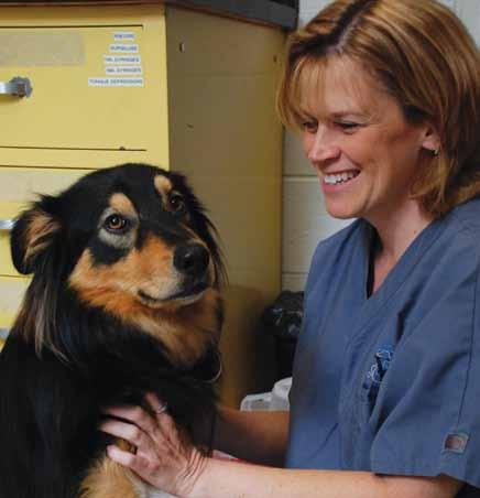
To create the owner assessment, focus groups were held, during which owners of arthritic dogs were asked a series of questions about their dogs’ behavior and evidence that their dogs were in pain.
Next, Dr. Brown and her team studied the transcripts from the focus groups.
1
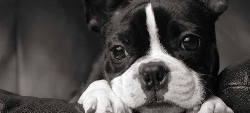
Two orthopedic researchers working to find outcome assessments and diagnostic tools that will work for all vets
“We looked for key words,” said Dr. Brown. “Were there common behaviors owners were noticing? Are there common themes through the conversations? We included owners of dogs that had surgery, as well as some that had not; we wanted a broad range to ensure the forms we develop are valid in a broad range of patients.”
After summarizing observations, general questions were drafted and vetted among a different cohort of owners whose dogs had arthritis.
“We wanted to look at how people answered the questions and look at which answers are most reliable,” said Dr. Brown. “It was an iterative process until we finally said, ‘I think this is it.’”
The current form of the assessment is being tested through the VCIC’s Arthritis Assessment Study, which is underway (see trial study information at right).
ONE LANGUAGE
For veterinary orthopedists, standardized client questionnaires and clinical assessment forms for function and quality of life that are validated are a logical approach for addressing the current shortcomings in study design and implementation.
In many cases, outcome assessments used in veterinary medicine have not been adequately evaluated for reliability and validity as opposed to assessments used in human orthopedics, in which the development and validation of outcomes instruments is well documented.
“This is an ethical undertaking as well as a scientific one,” said Dr. Brown. “And it is more and more important that veterinary orthopedists speak the same language and agree on the same methods for evaluating our techniques as the profession grows more quickly and aggressively. We owe it to our clients and to our patients to be able to validate our methods, procedures and recommendations for care and pain management.”
1 the
arthritis assessment study
PROMOTING THE PENNHIP METHOD
“The integrity of screening tests is paramount to the success of selective breeding to lower the incidence of hip dysplasia in dogs,” said Dr. Smith whose PennHIP method aims to accurately diagnose susceptibility in puppies so that breeders know which dogs should and should not be bred.
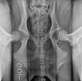
Canine hip dysplasia, or CHD, is defined by the radiographic presence of hip joint laxity or osteoarthritis with hip subluxation (laxity) early in life. A developmental disease of complex inheritance it is the most common orthopedic disease in large- and giant-breed dogs and causes pain and loss of mobility.
Currently, there are two screening methods used by breeders in the US: the Orthopedic Foundation of Animals (OFA) method, and the PennHIP method.
The traditional OFA screening method relies on conventional hip-extended (HE) radiographs, which Dr. Smith argues do not provide critical information needed to accurately assess passive hip joint laxity and therefore osteoarthritis susceptibility.
“We believe the insensitivity of the OFA method for detecting hip joint laxity is not the fault of the expert
radiologist interpreting the HE radiograph, but rather an inherent deficiency of the HE radiographic view,” said Dr. Smith.
In order to achieve genetic control of CHD and create a system that would have more accurate diagnostic outcomes, Dr. Smith developed the PennHIP method as a more effective way to evaluate whether a dog will develop canine hip dysplasia in its lifetime. His method has gone head-to-head with the traditional OFA hip-screening method, which was developed in the early 1960s.
Most recently, Dr. Smith offered a comparison of the PennHIP method against the OFA method in the September 2010 issue of the Journal of the American Veterinary Medical Association (JAVMA), where he illustrated that the PennHIP method called for more stringent breeding practices in order to lessen the prevalence of CHD.
PENNHIP VS. OFA
The PennHIP method quantifies hip laxity using the distraction index, or DI, metric, which ranges from a low of 0.08 to greater than 1.5.
Smaller numbers mean healthier hips.
The PennHIP DI has been shown in several studies at multiple institutions to be closely associated with the risk of osteoarthritis and canine hip dysplasia. It can be measured as early as 16 weeks of age without harm to the puppy.
The OFA grades hip joints in dogs in seven categories: excellent, good, fair, borderline, mild, moderate and severe hip dysplasia. The scoring system is a pass/fail one.
The PennHIP method is not pass/fail, but rather it quantifies on a continuous scale the “risk” of a dog acquiring OA. It considers a DI of less than 0.3 to be the threshold below which there is a near-zero risk of developing hip osteoarthritis later in life. In contrast, dogs having hip laxity with DI higher than 0.3 show increasing risk of developing hip osteoarthritis earlier and more severely, as the DI increases.
Comparing the overall results of the study, 52 percent of dogs OFA-rated “excellent,” 82 percent of dogs OFA-rated “good” and 94 percent of dogs OFA-rated “fair” fell above the PennHIP threshold of 0.3, making them all susceptible to the osteoarthritis of CHD though scored as “normal” by the OFA. Of the dogs the OFA scored as “dysplastic” all had hip laxity above the PennHIP threshold of 0.3, meaning there was agreement between the two methods on dogs showing CHD or the susceptibility to CHD.
The key feature of the PennHIP radiographic method is its ability to determine which dogs may be susceptible to osteoarthritis later in life. Because dogs are recognized as excellent models for hip osteoarthritis in humans, Dr. Smith’s PennHIP technology may be a viable option for human medicine.
milkyway
practice vets
“In humans, with appropriate studies of course, it is conceivable that mothers of susceptible children may adjust a child’s lifestyle, including diet, exercise and physical therapy to delay the onset or lessen the severity of this genetic condition,” Dr. Smith said.
OUTCOMES AND NEXT STEPS
In the meantime, Dr. Smith’s findings point to a weakness in current dog-breeding practices. If breeders continue to select breeding candidates based upon traditional scores provided by the OFA, then according to Dr. Smith, susceptible dogs will continue to be paired and hip quality in future generations will not improve.
Despite well-intentioned hip-screening programs to reduce the frequency of the disease, canine hip dysplasia continues to have a high prevalence worldwide with no studies showing a clinically meaningful reduction in disease frequency using mass selection.

“We’re sending mixed messages,” said Dr. Smith. “If we care about dogs then this is something that shouldn’t be ignored.”
PREVENTIVE MEDICINE
Knowing a dog’s risk for osteoarthritis early allows veterinarians to prescribe proven preventive strategies like weight loss to lower the risk of the genetic disorder. Also, dog breeders now have a better way to determine breeding quality to lower the risk of hip osteoarthritis in the future generations of dogs.
Dr. Smith urges veterinarians to take a proactive role in educating clients about this new school of thought in preventive medicine.
“Have a conversation about a pet with its owner when the pet is a puppy,” said Dr. Smith. “Educate the owner that the PennHIP procedure, performed, say, at the time of spay or castration, will permit assessing the probability of the dog’s developing the OA of hip dysplasia at some point in life. Then follow up by recommending known preventive measures, such as calorie restriction, to help the dog live a long, healthy, pain-free life.”
PennHIP is currently in common use by service-dog organizations such as the US Air Force, the US Army and numerous dog-guide schools. There are approximately 2,000 trained and certified professionals currently performing PennHIP procedure worldwide. For more information on becoming PennHIP-certified, visit www.pennhip.org.
small-animal
events

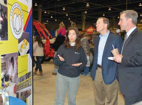
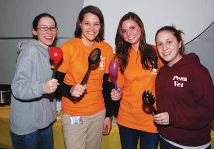
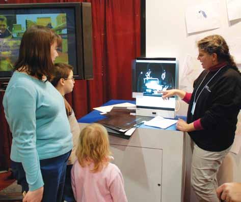

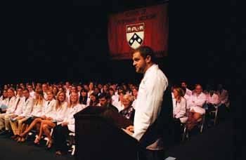
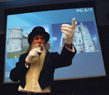
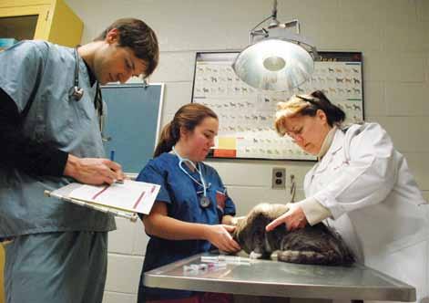
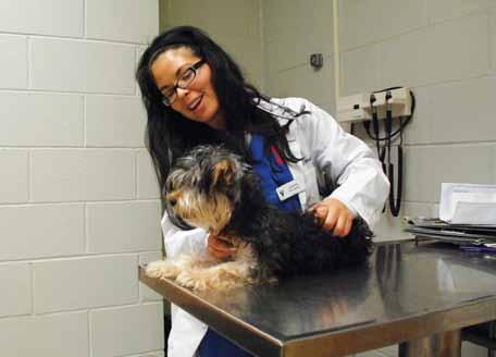
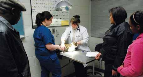
Pictured on these two pages: the annual Farm Show held in Harrisburg, PA provides an opportunity to showcase Penn Vet’s influence on ensuring public health and wellness; the White Coat Ceremony has become a tradition for students as they enter clinical rounds; Martin Luther King, Jr. Day of Service, which offers a free vaccination clinic to approximately 200 dogs and cats from local Philadelphia neighborhoods. Students, faculty, staff and alumni work together on this worthwhile outreach event; the annual SCAVMA auction, during which goods and services are auctioned off to the highest bidder in order to fund educational and conference opportunities for students; a SCAVMA-hosted chili cook-off to benefit local West Philadelphia Penn Vet students and an intern who were displaced from a significant apartment building fire; and The Westminster Kennel Club dog show event held at Madison Square Garden in New York City, which provides an opportunity to spend time with friends of the School.


events events
Changing Times, Changing Needs
Penn Vet addresses new trends in veterinary medicine with curriculum updates that speak to the times
This year marks a significant milestone for veterinary medicine: 250 years ago Claude Bourgelat established the first school of veterinary medicine in Lyon, France, thereby also establishing the profession.
The first veterinary students were required to take an array of classes that studied physiology, splanchnology (the study of internal organs), bandaging and proper use of medications. In addition to lectures, students were required to copy – verbatim – the information Bourgelat required them to learn and then recite the text without error to show they had learned the material.
Students also received assignments for animal care and facility maintenance, which required them to keep the dissection room, stables and forges clean. Students practiced skills under the authority of a teacher, performed consultations, monitored hospitalized animals and learned to prepare medicines.
Only two years after Bourgelat opened the first school of veterinary medicine, the profession earned the respect – and the financial backing – of the king. In 1763, a major cattle disease, rinderpest, broke out in France. Bourgelat sent educated students to combat the disease and soon it was under control. This success earned Bourgelat and his students the confidence of the king who then ensured the school’s funding indefinitely.
COMING TO THE AMERICAS
In North America, the earliest veterinary medical schools were established in the late 1800s. With an emphasis on agriculture at national and state/ provincial levels, important financial and popular support for colleges of veterinary medicine in the US and Canada came mostly from state and provincial departments of agriculture, respectively, thereby assuring the health of beef, dairy, swine and poultry populations raised for food and trade.
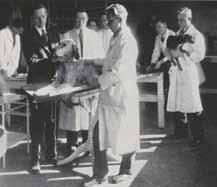
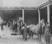
It was during that era, in 1884, that Penn Vet was established at the urging of the School of


Medicine whose leaders recognized that prevention and control of animal diseases had important implications for human health.
CHANGING LANDSCAPES
Over the years, the focus of veterinary medicine has changed. What started in the early 19th Century as a profession largely responsible for care of horses and agricultural animals has now become a profession that is important in improving sanitation and food safety as well as taking care of family pets, while still caring for agricultural animals and horses.
Today, with the globalization of food, goods and services, partnered with greater mobility and a worldwide population of nearly seven billion people, food production has gone from small-hold family farms to large agri-business. In addition to a greater need for safe food, pets in humans’ homes are becoming more and more commonplace in developed nations, making care for pet dogs and cats (and other species) a popular necessity.
Solo practicing veterinarians have increasingly joined with others to work in multiple-owner practices where partners can share knowledge and costly equipment more effectively and provide better all-around services to their clients.
As a result of the changing times, curricula for veterinary students must adapt to keep pace.
CHANGING TIMES, CHANGING CURRICULA
Today, there is a perfect storm of influence navigating the course for a veterinary medical education.
In 2008, the Association of American Veterinary Medical Colleges (AAVMC) established the North American Veterinary Medical Education Consortium (NAVMEC) to identify a cost-effective veterinary medical educational system that would produce graduates with competencies on day one
post-graduation that are required and valued by society, including the public and employers.
At the same time, the American Veterinary Medical Association’s Council on Education (AVMA COE), the body that accredits Penn Vet’s curriculum as well as the curricula of other vet schools in the US, set new requirements for clinical competency outcomes assessment (CCOA).
In addition to these two organized bodies, Penn Vet recognized the need for an update. While not a new phenomenon at Penn Vet – the faculty is constantly reviewing its curriculum for students to ensure each graduating class is ready and prepared for their futures and changes are made on a frequent basis – accreditation requirements set forth by the AVMA put special emphasis on this review.
EVOLVING EDUCATION AT PENN VET
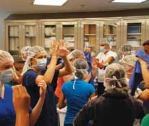
One of the most important educational missions of the School of Veterinary Medicine is to prepare students for multiple career opportunities.

The Penn Vet mission statement for Education includes the following:
• Training veterinarians for primary-care practice and preparing them for advanced study;
• Training veterinarians at post-doctoral levels for advanced clinical practice and research;
• Communicating advances to graduate veterinarians through continuing education; and
• Educating the public and the government about links between animal and public health. Currently, the Penn Vet curriculum emphasizes basic sciences in the first year; pathologic basis of disease in the second year; clinical aspects in the second and third years; and clinical rotations in the third and fourth years.
MORE CLINICAL, HANDS-ON LEARNING
One of the outcomes of these review processes has allowed for an increase in the efficiency of teaching in both the preclinical and clinical parts of the curriculum to allow students to be more involved with hands-on experience, case management, client communication and financial aspects of cases.
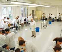
In the process of developing and implementing clinical competency evaluations, Penn Vet faculty recognized the need for earlier exposure to clinical experiences. The result is a new course for secondyear veterinary students – Introduction to Clinical Veterinary Medicine IV – that allows students to attend clinical rotations in the Ryan-VHUP wards during the mornings before classes and in the afternoons, in Ryan-VHUP’s Emergency Service and in the Community Practice. In addition,
students spend time on weekends in the George D. Widener Hospital for Large Animals at New Bolton Center.
In order to allow time in the curriculum for ICVM IV, course organizers of all second-year courses were asked to evaluate their courses in relationship to the rest of the curriculum and determine if hours could be reduced by either eliminating redundancies, consolidating materials or moving material into our web-based curriculum on Learn.Vet. As a result of these efforts, classes for the second-year students end on most days by 3PM, allowing them time to be in the clinics as well as to review online information.
Additional changes to allow students more individualized learning and supplementary clinical hours include:
• Increasing elective private practice experience rotations from four weeks to six;
• Allowing students to receive credit for private-practice rotations;
• Increasing the time and credit limits on externships;

• Reducing intramural rotation requirement from 70 credits to 65 for all majors.
NEW PROGRAM
In addition to increasing clinical, hands-on hours, a new program is being developed at Penn Vet: the VMD/MBA program. This program is a combined veterinary degree and master of business administration from the Wharton School at Penn. While the program has been informal for several years, Dr. David Galligan, a former member of the Education Committee and director of the program, is working out the details of this program with a committee from Penn Vet and Wharton.
ENGAGING ALUMNI TO ASSESS OUTCOMES
Penn Vet routinely surveys alumni and employers of alumni within the first three years after graduation to get feedback on changes alumni would like to see to the curriculum to ensure students are career-ready.
We are confident that we, as a profession and as a School, can meet the changing needs of the world today successfully, but it will require efforts not just by the School, but by our alumni, veterinary organizations, licensing boards, accrediting bodies and others.
If you have any suggestions for how we can better prepare our graduates or have thoughts on how we can meet these challenges, contact Dr. Tom Van Winkle, associate dean for education, at tomvw@vet.upenn.edu.
shedding new light on laminitis
Penn Vet continues to piece together this complex disease
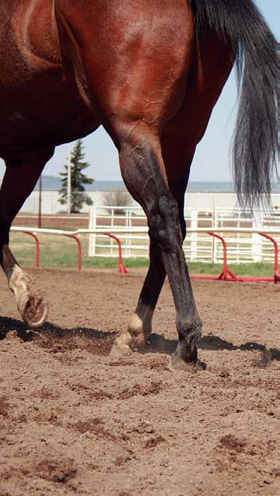
Laminitis — commonly known as “founder” — is a painful and life-threatening disease in which the tissue bonding the coffin bone of the foot and the hoof wall becomes inflamed. And it is a disease that has confounded horse owners and veterinarians for ages.

But it wasn’t until the untimely loss of legendary Thoroughbred Barbaro, the 2005 Kentucky Derby winner and hands-down favorite to earn Triple Crown status that year, that this painful disease drew international attention.
Since then, Penn Vet has been one of the leaders in laminitis research and discovery, and, in 2008 initiated the Laminitis Institute. In the past five years, Penn Vet’s leadership role has expanded to involve a collaborative, multi-disciplinary approach from the molecular level to the mechanical; from the understanding of the potential causes of laminitis and its prevention; to treating its clinical signs and minimizing its painful effects.
While there remain mysteries surrounding this complex disease, Penn Vet researchers, working collaboratively with equine researchers worldwide, have made significant strides to better understand laminitis.
Here, we’ll take a look at defining the disease as well as the projects currently underway, which continue to shed light on laminitis and give hope to horse owners and veterinarians, as well as novel treatment methods for consideration.
laminitis: what is it, exactly?
Afflicting approximately 15 percent of horses in the United States in their lifetime*, laminitis is an inflammation in the tissue that connects the inner wall of the hoof with the distal phalanx of a horse’s foot. Composed of an epidermal layer at the hoof wall, and a dermal layer associated with the bone, the laminae suspend the horse’s axial skeleton (See illustration, above right). When the laminae become
barbaro the legacy lives on
inflamed and separate, the stretching of the laminar nerves and increased pressure on the soft tissue of the horse’s foot cause severe pain and lameness. (See illustrations below and to the right.)

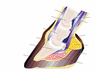
understanding the disease
Laminitis is typically secondary to another medical situation. It can be the result of metabolic syndrome or obesity, which are associated with impaired insulin function and share some similarities with pre-diabetes in people. It can also occur when an injury to one limb makes the horse favor another limb, putting added pressure on that foot. Horses with colic, pneumonia or a uterine infection are at much higher risk of developing laminitis.
Another mystery that remains surrounding the disease is its progression – something that Dr. Mary Robinson, lecturer in the Department of Clinical Studies at New Bolton Center, is studying by looking at the nitrous oxide signaling pathway and the response of blood vessels
Friends of Barbaro
From the moment Barbaro stole the international spotlight with his valor, athleticism and grace, he also stole our hearts. Thank you to the Friends of Barbaro who shared your resources with Penn Vet in honor and in memory of this brilliant Thoroughbred. On this anniversary we salute you for your support in helping to continue his legacy and to further our mission to find a cure for laminitis.
The following are friends who have made gifts in honor of Barbaro prior to March 8, 2011.
friends of barbaro
Joan C. Hendricks, V’79, GR’80, The Gilbert S. Kahn Dean of Veterinary Medicine
to inflammation. Dr. Robinson is interested in learning whether inflammation or lamellar pathology comes first. That is, does laminitis cause the inflammatory response or is the inflammatory response a symptom of the disease?
An acute episode of laminitis is associated with increased digital pulses, heat in the hoof and pain, all of which are suggestive of inflammatory disease. Activation of immune cells, especially macrophages, results in increased nitric oxide production, which is hypothesized to contribute to the pathophysiology of laminitis.
Dr. Robinson is also examining serum proteomics methodologies that, it is hoped, will aid in the identification of biomarkers of the developmental phase of laminitis. Though this stage is clinically silent, it is believed that this is the ideal time for intervention.
“There is nobody else in the world looking at this,” said Dr. Hannah Galantino-Homer, senior research investigator for the Laminitis Research Initiative at New Bolton Center. “This novel area of research could prove to have important implications with regard to etiopathogenesis, progression or treatment of laminitic disease in horses.”
Because the disease is rife with unanswered questions, it was clear that collaboration with other leading researchers in the field was essential.
And so, in order to better understand this disease and coordinate international research efforts, the Laminitis Institute at New Bolton Center, under the direction of board-certified surgeon Dr. James Orsini, associate professor of surgery, was created.
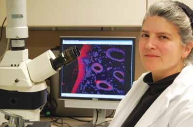
The goal of coordinating collaborative research has been attained. Since the Institute’s inception, there has been a marked increase in the numbers of scientific publications on the topic, featuring authors from more than one institution, as well as increased numbers of funded projects with co-investigators from multiple institutions.
“Many colleagues at Penn, University of Georgia, the Ohio State University, Texas A&M University and
friends of barbaro
University of Queensland, to name a few, have been working hand-in-hand to advance our understanding and management of laminitis,” said Dr. Orsini.
laminitis research initiative:
three major projects
Headed by Dr. Galantino-Homer, the Laminitis Research Initiative at New Bolton Center has three major ongoing projects:
1. The Laminitis Discovery Database
2. The Grayson-Jockey Club Research Foundation Project
3. Stem Cell Studies
“My hopes for these three projects are that the Laminitis Discovery Database will provide us and others with the samples and information needed to conduct collaborative research; that the Grayson project will increase our understanding of the disease process and disease biomarkers; and that the stem cell studies will provide a better understanding of normal and diseased lamellar biology as well as a laboratory model for testing hypotheses at the cellular and molecular level,” said Dr. Galantino-Homer.
The Laminitis Discovery Database
The Laminitis Discovery Database, established in 2008 and maintained through a grant from the Bernice Barbour Foundation, has been one of the most valuable tools for facilitating collaboration between scientists.
Most previously published research on laminitis uses experimental models, with very little investigation involving naturally occurring disease. But this database has the potential to change that.
The Laminitis Discovery Database banks tissue and serum samples collected from horses with naturally occurring laminitis and a control group of horses not afflicted by the disease that have died or been humanely euthanized for other reasons.
To date, there are approximately 40 sets of samples from each group, all processed in multiple ways for a variety of uses. Blood and tissues are banked for molecular studies and diagnostics; photos are taken along with a history and description of the horse’s body condition; and lamellar tissue and bone slices are sent to Penn Vet’s Pathology Lab at New Bolton Center.
Information obtained from these samples is compiled for equine disease research and is available worldwide. The Laminitis Discovery Database is a valuable provider of real-life samples and information not previously available, but necessary for Penn Vet and other researchers to conduct collaborative research to further the study of equine disease and laminitis.
friends of barbaro
Dr. Julie Engiles, assistant professor of pathology at Penn Vet, is one of the researchers on the New Bolton Center campus utilizing the database samples. Dr. Engiles uses micro-computer tomography technology to study the effects of laminitis on bone density and osteolysis, a prominent feature of laminitis that has not been previously investigated. The project is aimed at correlating laminitisassociated pathology that occurs at both the macro and micro-anatomic levels with pathology that occurs on the molecular level. She is also using the database to establish a scoring system for laminitis histopathology.
By using the samples, New Bolton Center boardcertified pathologist, Dr. Engiles, explained, “We have developed a very detailed histologic grading scheme, which specifically characterizes the micro-anatomic changes that differ between clinically normal horses and horses with laminitis.”
The Grayson-Jockey Club Research Foundation Project
Dr. Galantino-Homer’s collaborative Grayson-Jockey Club Research Foundation Project (begun in 2008 with the Laminitis Institute’s Drs. Orsini and Christopher Pollitt, Dr. Rebecca Carter and Dr. Neal Rubinstein of Penn Medical Center, as well as Dr. Bhanu Chowdhary, an expert in equine genomics of Texas A&M University) is a second ongoing project, which investigates the gene and protein expression profiles of two models of laminitis-the oligofructose model meant to mimic some forms of pasture laminitis caused by excess oligofructans, a type of sugar, and the hyperinsulinemia model intended to provide a model for endocrinopathic laminitis which is associated with insulin resistance and high circulating insulin levels.
“If we can identify proteins that are being produced or degraded during the early stages of laminitis, they might be used as biomarkers for the identification of at-risk horses or targets for therapeutic intervention,” said Dr. Galantino-Homer. “Since 2009, the availability of the horse genome sequence makes it possible to do a lot more than we could before.”
The proteomic studies are currently in the data analysis stage and should be presented this year.
According to Dr. Galantino-Homer, they provide insight into the proteins that are degraded in the laminitic foot, contributing to the biomechanical failure and loss of digital support. In addition, these studies reveal differences between insulin-related and oligofructose-induced laminitis in proteins that mediate inflammation. They have also provided quantitative data on the protein composition of the laminae, key to understanding the changes in keratin expression profile changes with laminitis, which, in turn, affect the biomechanics of the foot.
friends of barbaro
Stem Cell Studies
Dr. Galantino-Homer’s third project area is involved with the study of equine basal epithelial cells from the laminae and, in collaboration with Dr. Makoto Senoo, an epithelial stem cell biologist at Penn Vet’s Department of Animal Biology and Institute for Regenerative Medicine, is in the process of developing an in vitro culture system for the study of equine laminitis.
“Stem cell studies will provide a better understanding of normal and diseased lamellar biology and a laboratory model for testing hypotheses at the cellular and molecular level,” explained Dr. Galantino-Homer.
The project offers researchers a way to see what is happening to the cells at “ground zero” of laminitis: the epidermal basal cells that normally anchor the epidermal laminae to the dermal laminae.
Side-by-side evaluation, for example, of pharmaceutical agents for the treatment of laminitis, and investigation of trigger factors on isolated lamellar epidermal cells will also become a possibility. The epithelial stem cell study is also exciting for its potential as a therapeutic method: using stem cells for tissue regeneration in the laminitic hoof, much as it has been used in regenerative medicine for burn victims and corneal scarring.
related research
While the Laminitis Research Initiative is aggressively pursuing three projects that will directly impact the laminitic horse, there is other research happening at New Bolton Center that, while not being developed exclusively for laminitis, may be applicable to the disease.
One area includes pain management, a huge concern for horse owners and veterinarians when treating a horse that has foundered.
Dr. Kirsten Wegner, a board-certified anesthesiologist in the Section of Anesthesia and Critical Care at New Bolton Center, is working to refine an objective, quantifiable pain scoring system similar to that used in pediatric medicine. The clinical pain scoring system includes information such as the horse’s position in the stall, degree of alertness, whether standing or recumbent, frequency of weight shifting from limb to limb, and most importantly, how the horse responds to a caregiver prompting them to walk or lift a hoof.
By recording each observation and interaction, the horse’s behavioral responses and physiologic parameters, such as heart and respiratory rate, clinicians will have the ability to discern subtle changes in comfort level early on, and make appropriate changes in pain management strategy.
Working towards this goal, Dr. Wegner is developing an acute nociceptive (pain-causing stimulus) testing
friends of barbaro
device for use in freely moving horses, which may be adaptable to a laminitic horse.
in the meantime…
While the research component of the Laminitis Institute as well as independent research outside of the realm of specifically targeting laminitis is shedding new light everyday, horses continue to founder and, as a result, novel prevention, treatment and pain management techniques are also being discovered.
In 1916, a report from the USDA noted that the treatment of laminitis is probably more varied than of any other disease.
“Almost a hundred years later,” said Patrick Reilly, chief of Farrier Services at New Bolton Center, “we are faced with the same dilemma. There is a wide range of mechanical treatments for the laminitic hoof, but little scientific evidence to support any methodology.”
Cryotherapy
One potential way to prevent laminitis involves cryotherapy. It is hypothesized that cryotherapy, cooling the inflamed foot, would be effective in preventing a laminitic episode, but in the past such therapy has been limited to unwieldy methods such as hauling buckets of ice to the horse. It is labor-intensive and doesn’t last.
Through a collaborative effort, Drs. Orsini and Andrew van Eps, now at University of Queensland, have developed a chilling system that circulates icy cold water through a boot that envelops the hoof wall and pastern. It provides a dry cold, in high volume, at a very low temperature.
“It seems like cooling the vulnerable limb or limbs two to four days beyond the systemic condition can prevent a laminitic episode,” said Dr. Orsini. “We are looking to our new Equi-Assist home nursing care program (see page 20) to
friends of barbaro
provide data that will help us to identify those horses who may be at risk for a laminitic episode before it begins.”
Improving shoeing to support the laminitic hoof
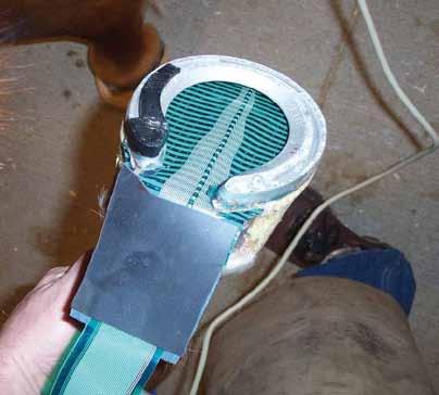
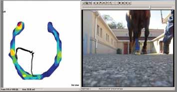
In addition to Dr. Orsini’s preventative cooling method, Reilly is working to develop an accurate in-shoe force measuring system to quantify the effects of the various treatment approaches relating to laminitis. In-shoe force measurements, with innovations developed at New Bolton Center, are proving valuable in quantifying the effectiveness of shoeing systems. The mobile in-shoe force measuring system sensor mat is cut to the shape of the foot and placed between hoof and horseshoe using the glue-on method of shoeing developed at NBC.
“Given the hundreds of years of anecdotal mechanical treatments,” said Reilly, “my hope is that measuring the effect of various shoeing techniques might enable us to better understand and improve our ability to support the laminitic hoof.”
Current areas of study include the effect of hoof capsule integrity, which is compromised during laminitis, and the effect of the shoe positioning in aiding the rehabilitation of the laminitic horse.
forward momentum
While much has been accomplished to better understand this debilitating disease, researchers at Penn Vet and beyond know there are still strides to make.
“As we focus on the challenges in front of us, it is important to recognize the efforts of the many trailblazers, researchers, owners, caregivers and colleagues who continually advance the understanding and treatment of laminitis through their dedicated work,” said Dr. Orsini. “We stand on the shoulders of their efforts for even greater future accomplishments with our vision to conquer laminitis.”
*Moore RM. Vision 20/20 – Conquering equine laminitis by 2020. Proceedings. 2nd AAEP Foundation Equine Laminitis Research Workshop, pp 11-15.
friends of barbaro
healing at home
Newly launched Equi-Assist program allows patients to recuperate in the comfort of their own barns while also allowing New Bolton Center researchers to gather important data

friends of barbaro
barbaro the legacy lives on
Eby has been through a lot. Complications following surgery to remove his diseased upper incisors left the 26-year-old retired Thoroughbred racehorse thin, in poor health and depressed.
But, since returning to his home turf at the end of December, Eby has been thriving in his familiar surroundings.
Because of his previously poor health, however, the horse needed continued careful monitoring of his weight, body condition and general demeanor to ensure a total recovery. And, while the barn manager where Eby is located is a competent, seasoned equestrian, providing day-to-day care for the many horses in the barn demands all of her time. The extra care and close monitoring that are crucial to Eby’s continued improving health are often too much for her busy schedule.
Eby’s case presents an ideal scenario for Equi-Assist.
home-field advantage
Equi-Assist is the innovative equine home nursing care program developed through the Laminitis Institute at the School of Veterinary Medicine’s New Bolton Center and the vision of Margaret Hamilton Duprey. The basic premise is to provide a high quality, clinical and posthospitalization nursing service to all equine patients at their home or lay-up facility, within a reasonable driving radius of the campus. Launched in December 2010, the program is the first of its kind anywhere.
The equine home nursing care program is grounded in the same principles that have caused an explosion in home nursing care for humans:
• Patients are more comfortable and more secure at home than in the hospital, and therefore more likely to recover faster;
• Home nursing care provides a savings over hospital costs;
• The technical skills of a home nursing professional are available at every stage of recovery.
Laminitis, or founder, is a painful and life-threatening disease in which the tissue bonding the coffin bone of the foot and the hoof wall becomes inflamed. When Margaret Hamilton Duprey, a well respected, lifelong equestrian in Chester County, PA, learned first-hand the time and skill required to care for a laminitic horse properly, she realized the need for a medical service that offers high-quality, skilled, compassionate and experienced professional care. This service would bridge the gap between hospital and home care for horse owners.
Mrs. Duprey approached Dr. James Orsini, associate professor and director of the Laminitis Institute at Penn Vet, to brainstorm the creation of a home care nursing program for horses and, once the idea took shape, she was instrumental in guiding the design of the program. Mrs. Duprey made a generous gift to Penn Vet to see the program completed.
“I’m thrilled to see a dream, a vision and a program setting quality-of-care standards for equine health, become a reality,” said Mrs. Duprey.
a wealth of care
When Eby first returned to his home barn following surgery, it was Equi-Assist nurse Jennifer Wrigley who noticed the gelding was not bouncing back to his normal self as quickly as he should. Wrigley quickly began treatment, starting fluids and giving the clinician a precise and detailed account of the horse’s clinical signs and response to therapy.
Now that he is completely recovered, she continues to check on Eby once a week. Wrigley monitors his
nutritional consulting for at-home care
general body condition, ensuring that he is maintaining the proper weight for his age and level of exercise and that his daily nutritional needs are met. With more than 15 years of clinical nursing experience, she runs her hand down his legs to check for heat or swelling, and feels his feet for heat or an increased pulse. She looks at his mucous membranes and observes his eyes, checks for nasal discharge, listens to his heart, lungs and bowel sounds and draws blood for periodic lab tests when indicated.
Wrigley also makes use of the latest pocket technology, taking digital pictures of Eby to share with the horse’s primary care veterinarian, the doctors at New Bolton Center and the owners. Videography is occasionally part of the clinical picture, used to document a horse’s behavior for analysis by clinicians at New Bolton Center.
In 2006, the average hospital stay for an ill horse was seven days. In 2010, it dropped to four days, a situation that makes the need for more skilled care for equine patients returning home even more compelling.
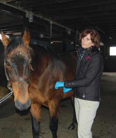
By taking on the role of the communication bridge between owner, primary care veterinarian, New Bolton Center clinician and the client’s farrier, the Equi-Assist nurse is able to optimize continuity of care. In addition to monitoring general health, the Equi-Assist nurse has the expertise and experience to clean and rebandage tricky wounds, provide post-operative care, tend to neonates, deliver difficult-to-administer medications, monitor pregnant mares and much more.
In the case of horses suffering from laminitis, home care might include pain management, assessment of nutritional needs and bandage changes. Trained in equine massage, the Equi-Assist nurse can also provide for stallbound patients the benefits of total body massage. Perhaps most importantly, the trained equine nurse is able to identify emerging issues quickly so that small problems do not become big issues.
benefits for the future
The costs and health benefits of early hospital discharge are clear. The average daily stay for a horse at the George D. Widener Hospital for Large Animals is variable, and can run into many hundreds of dollars. The cost for professional care through Equi-Assist is often a fraction of the in-house cost, based on a mileage charge and the length of time the home visit requires. It is believed that the non-tangible results will be even more valuable.
While health care professionals in the human world investigate the benefits of early discharge and continued care at home, parallel studies have not taken place in the animal world.
The program will also contribute to the development of evidence-based treatment protocols. Care at home, coupled with investigations at New Bolton Center’s labs, could have even greater short-term results.
“Ultimately we would like to identify clinical or biochemical markers enabling us to take preemptive action to prevent a laminitic event or other post-hospital complication,” Dr. Orsini said.
a model for care
As New Bolton Center veterinarians release their patients to the care of Equi-Assist professionals, and local veterinarians refer clients to the service, the shape and scope of the program will become more clearly defined. Additional nurses are in training, and it is envisioned that, as the program enjoys growing success, a new veterinary technology specialty in home care nursing will be defined.
“Our goal is for this program to become a model for the standard of care in the home environment,” said Dr. Orsini.
barbaro the legacy lives on
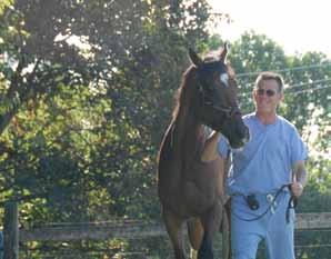
friends of barbaro
Jesse
Owens. Eric Liddell. Barbaro.
Athletes have long been inspirational icons for society, and Barbaro ranks with the best of them.
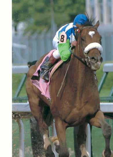

In 2006, the majestic, well-muscled Thoroughbred bay colt became one of only six horses ever to go into the Kentucky Derby undefeated and finish in the roses. At three years of age, he wowed the crowds with the largest winning margin seen at the Derby in more than half a century. He had the heart of a champion, pressing on to the finish line of his own will, carried by strength and determination without any urging from his jockey. That quality made him a favorite for the coveted Triple Crown, out of reach by any horse since 1978.
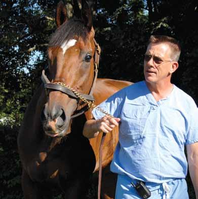
A devastating injury at the Preakness Stakes, the second race in the crown, shattered his right hind leg and all hopes of the highest honor in the world of racing. The injury brought him into the international limelight, allowing him to show the world that his heart was even bigger than could have been imagined. Barbaro spent the next eight months at New Bolton Center where he endured multiple surgeries to fix three bones shattered into more than 20 fragments. Miraculously, the fractures healed and Barbaro was able to walk again, until he developed laminitis. The devastating condition would prove to be the only competitor that Barbaro could not best.
“In my mind he was a very exceptional horse,” Dr. Dean Richardson, chief, Large Animal Surgery, said in an interview on CBS News in 2007. “He just kept such an amazingly positive attitude. So that would make him unforgettable even if he didn’t have any other characteristics that were memorable.”
The number of people who have come forward and continue to keep the memory of Barbaro alive is astounding and many show their admiration for this fine colt by joining the fight to find a treatment for his deadly opponent – laminitis – through gifts to Penn Vet’s Laminitis Research Fund.
And thanks to that support, Barbaro leaves much more than just a legacy of inspiration – he leaves a legacy that means a better life for horses that follow.
— Sally Silverman
remembering barbaro…
barbaro the legacy lives on

With less than two years remaining in Making History: The Campaign for Penn Vet, our loyal friends and supporters have helped us to raise more than $100 million toward our ambitious goal of $125 million.
Our goal of $125 million is not just a nice, round number – it is a number that represents a thoughtful strategy to provide top-flight facilities for our patients, students and faculty, and to advance cutting-edge research in veterinary medicine.
Specific priorities include:
❯ Investing in care and research
❯ Supporting students
❯ Leading the profession of veterinary medicine
❯ Expanding the frontiers of research
❯ Meeting our most pressing, current and future needs
Although we have so far benefited greatly from the generosity and dedication of our loyal alumni and friends, we have a large task of raising $25 million to realize our vision for the future.
Whether we are working to provide the finest in emergency and routine veterinary patient care for all species of large or small animals, we strive to better understand animal disease through research, and to be leaders in the area of animal health and productivity for the betterment of society. Achieving both objectives requires state-of-the-art facilities, and we are paving the way with capital projects on both our New Bolton Center and Philadelphia campuses. Here, we have highlighted just a few exciting developments and accomplishments in recent months.
Equine Performance Evaluation Facility
The Equine Performance Evaluation Facility is currently in development stages at the New Bolton Center, and we are pleased to report that two recent additional gifts have moved us closer to a construction date. Having achieved 80 percent of the funding necessary, we are hopeful that we will soon be able to get this project underway. This remarkable facility will allow faculty, staff and students to expedite a care plan for admitted equine patients and is a top priority for the School.
Updated Ryan-VHUP Lobby & Emergency Services
We have also made significant progress in developing plans for a comprehensive reconstruction of the first floor lobby and waiting area at the Matthew J. Ryan Veterinary Hospital (Ryan-VHUP) in Philadelphia to ensure clients have the best experience possible. Hospital and School administration have hired Cecil Baker + Partners, a Philadelphia-based architectural firm, to lead the renovations. Plans include a brighter, easily identifiable entrance with automatic doors, a greeting station and a client-friendly admissions area. These are only the first steps in enhancing our clients’ experiences.
This new, state-of-the-art lobby area will give RyanVHUP an appearance to match the superb level of care that Ryan-VHUP and its clinicians are known for, while also reflecting our commitment to excellence in customer satisfaction and experience.
CAMPAIGN REPORT $125 Million Goal, $100 Million Raised to Date
This attractive, comfortable and updated waiting room will include more private, separated areas, which will minimize stress for our patients and our clients.
The first-floor renovation project at Ryan-VHUP will also investigate the possibility of an after-hours entrance for Emergency Services, improve the Emergency Services waiting and intake areas and add a new Emergency Services procedure room. This organized layout for clinic workflow will also increase the amount of patient care space, thus enabling improved operational processes, such as patient flow, staff, materials and information. Improving the environment and processes will be an important step in decreasing wait time for our clients.
The first floor will also gain the outpatient pharmacy, a satellite facility to the main pharmacy on the third floor, and a retail sales area. As clients check out, they will also be able to pick up medications for their pet and have an opportunity to purchase necessary supplies and a keepsake. Our architects have begun schematic design of the interior spaces and the exterior approach from Spruce Street.
These crucial renovations will require approximately $1.5 million in funding for the interior renovations alone. Because of the generosity of our Friends of Ryan, we have $220,000 in-hand and ready to deploy for the earliest stages of this project. Another $365,000 has been pledged for future capital construction, leaving approximately $915,000 to be raised toward the transformational benefits of firstfloor renovations at Ryan-VHUP.
This transformation directly impacts Penn Vet’s key initiatives of enhancing customer satisfaction and matching our eminent reputation with modern, advanced facilities while also responding to the need to compete with our growing local competition.
Advanced Minimally Invasive Surgery Suite
In addition to first-floor renovations at Ryan-VHUP that will directly impact customer service and Emergency Services at the hospital, Penn Vet is also making strides in the construction of our minimally invasive surgery operating room (OR 1) within the Philadelphia hospital, which will provide the most technologically advanced surgical techniques in the profession. Endoscopic and arthroscopic procedures will enable our surgeons to minimize risk of infection to our patients, as well as lessen their pain and recovery times.
The total cost of the OR 1 project is just over $616,000. With generous gifts from Penn Vet supporters, we are ready to purchase the equipment boom, arms and lights, the endoscopy camera system upgrade to HD and the integration system. This equipment will provide clinical data and images for viewing and sharing from within the sterile field to local and remote locations. A July 2011 completion date for this impressive suite is expected.
friends of barbaro
Expanding the Frontiers of Research
Some of the brightest minds, most state-of-the-art equipment and cutting-edge knowledge reside in veterinary teaching hospitals, making them the ideal environment in which to perform clinical trials that determine which breakthroughs can actually change the way veterinary medicine is practiced. At Ryan-VHUP we have not only a wealth of opportunities for faculty to engage in groundbreaking research, but also the ability to translate our research into practice. Currently, some of the standout research being conducted by Penn Vet includes work in canine mammary tumors, cancer, pain management and gene therapy advances.
The VCIC
In 2005, Penn Vet launched the Veterinary Clinical Investigations Center (VCIC) in an effort to maximize the quantity, quality and efficiency of clinical trials. It was originally staffed with one clinical trials nurse whose sole responsibility was the collection of high-quality data from a single clinical trial. Today, the VCIC is directed by Dr. Dottie Brown, chair, Clinical Studies – Philadelphia, and staffed with full-time Associate Director Michael DiGregorio and five full-time clinical trials nurses certified in Good Clinical Practice, whose effort is entirely devoted to all aspects of clinical trial implementation, including study set-up, recruitment, data collection, data management and study closeout. By building a solid infrastructure for clinical trials management, we are able to efficiently and effectively run high quality studies in an academic teaching hospital. Currently, there are more than 12 clinical studies underway at the VCIC.
Incorporating Traditional Chinese Medicine with Western Medicine
Dogs that have been diagnosed with hemangiosarcoma (HSA) of the spleen traditionally have a grave prognosis of two to four months of survival. Researchers at Penn Vet are combining some principles of Traditional Chinese Medicine with the modern diagnostic tools of Western Medicine to investigate the health and welfare benefits of an herbal supplement derived from the Yunzhi mushroom, which may support the immune system while maintaining general fitness and overall quality of life. Surprisingly significant improvements in survival, with several dogs living over a year after diagnosis, have been documented to date.
As you can see, Penn Vet is a busy, changing place and we thank you – our friends and supporters – for being a part of this exciting time as we set the pace for the future of veterinary medicine. If you would like to learn more about our priorities, please contact Melissa von Stade, assistant dean for Advancement, Alumni Relations and Communication, at 215-898-1482 or via email at mstade@vet.upenn.edu.
friends of barbaro

friends of barbaro
The Board of Overseers serves as the advisory body to the Dean of the School of Veterinary Medicine and as a vital channel of communication with the University of Pennsylvania Board of Trustees on the activities of the School. Overseers also provide ready panels of professionals, experts and informed lay people who offer volunteer leadership and financial support to the areas they serve, as well as serving as ambassadors and spokespersons by linking the campus to the world.
“The contributed time and talent of the Overseers is a key component of the success of any school at Penn,” said Dean Joan C. Hendricks, The Gilbert S. Kahn Dean of Veterinary Medicine. “And it is a great delight to add these fresh, committed, impressive members to our Board. They are bringing new energy and renewed vision. And I believe I speak for all of us, including the long-serving Overseers, when I say it is tremendously enjoyable to share the wonders of Penn Vet with these new eyes and ears as they become acquainted with the breadth and excellence of our work.”
In the past year, Penn Vet has added six individuals to its Board of Overseers. Each of them will serve renewable, three-year terms. New members include:
Catherine George Adler of New York, NY. Ms. Adler was a vice president in investments for Cowen and Co., a member firm of the New York Stock Exchange. She has been a chairwoman of fundraisers for various organizations in Palm Beach, including the South Florida Science Museum, the YWCA Mary Rubloff Harmony House and the Children’s Home Society. She served on the Board of the Academy of the Palm Beaches and Ballet Florida, and was a member of the International Director’s Council for the Guggenheim Museum. She is currently a member of the Chairman’s Council of Conservation International and on the advisory board of the Palm Beach Theatre Guild.
Krista Buerger of Plymouth Meeting, PA. Ms. Buerger is executive vice president for Coventry, the leader in the secondary market for life insurance and the company that pioneered the life settlement industry. Ms. Buerger joined the company in 1999 and currently oversees the company’s operations, specializing in implementing key companywide initiatives that continue to strengthen Coventry’s distinctive corporate culture. Ms. Buerger plays a critical role in the continuity and development of Coventry’s operations by enhancing current processes and systems to continue to improve on the company’s existing platforms. Ms. Buerger is a graduate of Bucknell University.
Jay S. Fishman, W’74, WG’74 of Englewood, NJ. Mr. Fishman is chairman and chief executive officer of The Travelers Companies, Inc., a Dow 30 company offering property and casualty insurance products and services to businesses, organizations and individuals in the United States and selected international markets. Mr. Fishman is a member of the University of Pennsylvania Board of Trustees, the Industry Advisory Board of the Financial Institutions Center for The Wharton School and serves as chairman of the Travelers/Wharton Partnership for Risk Management and Leadership. He also serves on the Philharmonic-Symphony Society of New York, Inc. He is an active member of the Business Council, a vice chairman of the Kennedy Center Corporate Fund Board in Washington D.C., and member of the ExxonMobil Corporation Board of Directors.


John P. Shoemaker, C’87 of Wyndmoor, PA. Mr. Shoemaker is a managing partner with Milestone Partners, a Radnor-based middle market private equity firm. He currently serves on the boards of several Milestone portfolio companies including Safemark Systems, Miche Bag and Avure Technologies. Prior to entering the private equity business he was a corporate lawyer for Reed Smith Shaw & McClay in Philadelphia and an investment banker for Morgan Stanley, Inc. in New York. Mr. Shoemaker serves as a member of the Board of Trustees to the University of Pennsylvania where he is also on the Board of Overseers of the Athletic Department. He also serves on the board of the Children’s Scholarship Fund Philadelphia. Mr. Shoemaker holds a BA from the University of Pennsylvania and a JD from Boston College Law School.

Adam Silfen, C’98, WG’03 of New York, NY. Mr. Silfen is the founding partner of Wickapogue Advisors, LP, a real estate advisory and investment firm. Personally, Mr. Silfen rode horses competitively for many years at the national show level and was an avid polo player. He’s also an avid golfer and runner.
Lynne L. Tarnopol, CW’60, PAR’83, PAR’85 of New York, NY. Mrs. Tarnopol is a Trustee of the Tarnopol Foundation. As one of the founders of the Penn Club of New York, Mrs. Tarnopol served as its Founding President. At Penn, Mrs. Tarnopol currently serves on the Wharton Undergraduate Executive Board and is a member of the Trustees’ Council of Penn Women. In addition, she has served as a member of the Penn Alumni Board of Directors and was the fundraising chair for the 125th Anniversary of Women at Penn Committee. Mrs. Tarnopol was the recipient of the Alumni Award of Merit in 1995.


MA Y2011 alumniweekend PENN VETERINARY MEDICINE U N I VERS I T Y OF P ENNS YL VAN I A S CHOO L OF V ETER I NAR Y M ED I C I NE Friday, May 13 & Saturday, May 14, 2011 • Celebrating all Penn Vet classes ending in ’6 and ’1. Honoring all alumni. • www.vet.upenn.edu/pennvetalumniweekend2011 alumniweekend PENN VETERINARY MEDICINE friends of barbaro
Veterinary Medicine: Leading Social Change



Penn Executive Veterinary Leadership Program: Making an Impact as a Global Health Leader

June 6–9, 2011 ■ Philadelphia, PA
This leadership development program from the University of Pennsylvania’s School of Veterinary Medicine and the Wharton School is designed for veterinarians who seek to expand the profession’s impact on the crucial issues facing the world today.
Join fellow veterinary leaders in executive development sessions that will help you to discover new ways to increase your social impact and improve the well-being of animals and society.
For more information or to enroll, please contact Katrina Clark at execed@wharton.upenn.edu or call +1.215.898.1776.
ww w . Penn Vet Leadership . com
friends of barbaro

friends of barbaro

friends of barbaro
Are You a Member?
We are thankful to those that have made the ultimate commitment to Penn Vet as members of the Veterinary Heritage Circle of the Harrison Society, a program that allows friends and supporters to include Penn Vet in their estate plans.
It is, of course, through the generosity of alumni, supporters and friends that the University of Pennsylvania School of Veterinary Medicine is able to meet our mission of teaching, healing and research while leading the field in new treatment advances, ground-breaking research discoveries and educating the brightest young minds in the profession.
Veterinary Heritage Circle Members have made a commitment to helping Penn Vet reach our mission by including us in their estate plans through bequests, trusts, charitable gift annuities, retirement plans, life insurance and other arrangements. In doing so they have created a legacy that will have a lasting impact on the future of veterinary medicine.
To join the Veterinary Heritage Circle, or to learn more about the benefits of membership, please contact us at:
University of Pennsylvania School of Veterinary Medicine

Office of Gift Planning
215.898.6171 or 800.223.8236
giftplanning@dev.upenn.edu
www.makinghistory.upenn.edu/giftplanning
friends of barbaro THE VE T ERINARY H ERI T AGE CIR C LE
eyesight to Givingthe blind
Penn Vet ophthalmologists’ work in the lab restores vision to dogs and provides hope to human patients
You may remember a dog named Lancelot. In 2001, Dr. Gustavo D. Aguirre, professor of medical genetics and ophthalmology at Penn Vet, restored vision to the dog, who was born blind –and they both made national headlines.
Their next stop? Washington, DC.
“Because of publicity following Lancelot’s recovery of vision, he was invited to Capitol Hill to meet with members of Congress,” said Dr. Aguirre. “During the meeting, Lancelot laid down near the podium and looked carefully at all the members of the audience who asked questions. Unlike dogs that are blind but can hear, Lancelot fixed his stare on the people who asked questions.”

Dr. Aguirre’s goal in traveling to Washington with Lancelot was to get people to understand and appreciate the power of and need to continue to develop new therapy technologies for treating inherited blindness.
Their sojourn to Washington was a success and Dr. Aguirre’s point was made.
“They all wanted to adopt Lancelot,” said Dr. Aguirre, “so to me that meant they connected with the idea that this kind of work is important.”
Serving as lead researcher of the project at the time, Dr. Aguirre’s work began with Lancelot and a team that included researchers at Penn’s Scheie Eye Institute and University of Florida. Their goal was to restore vision to Lancelot and his littermates that were mixed-breed dogs of Briard origin. This is the first time that gene therapy was used to successfully restore vision to any living creature.
By injecting a genetically engineered virus carrying a healthy copy of the defective gene into the part of the dog’s retina that contained the retinal pigment epithelium, the cell
layer that is essential for the proper functioning of the light-sensing cells, Dr. Aguirre and his team were able to “infect” those cells with normal gene material. Within several weeks, cells with the healthy copy of the gene began to produce vitamin A in the proper form needed by the retina that, in combination with opsin, formed the visual pigment rhodopsin, allowing Lancelot to see.
This advance in veterinary medicine offers hope for curing a similar disease that blinds children at birth or in early childhood – Leber’s congenital amaurosis (LCA), an inherited retinal disease. And, because of the dog’s successful treatment, human clinical trials began in 2007 at several centers worldwide, including the University College of London, the Sheie Eye Institute and Children’s Hospital of Philadelphia.
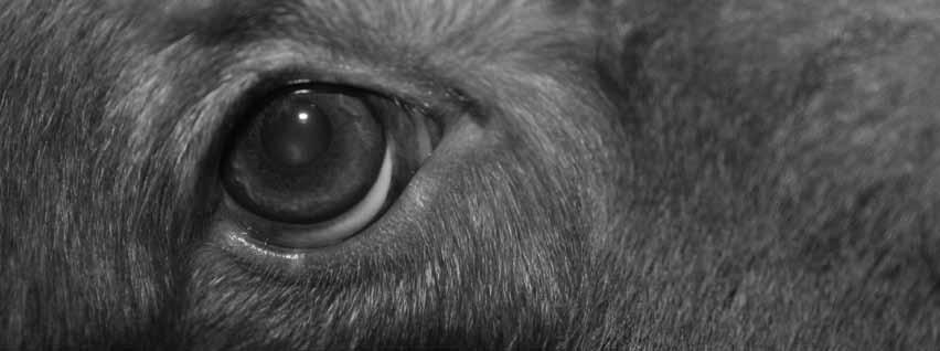
But Dr. Aguirre’s and his colleagues’ passion for restoring sight to the blind using gene therapy didn’t stop with Lancelot.
A SECOND MAJOR BREAKTHROUGH
Working hand-in-hand with researchers at Temple University, University of Florida and Cornell University, Dr. András M. Komáromy, assistant professor of ophthalmology – one of Dr. Aguirre’s colleagues – has made another major breakthrough in vision recovery with developing a gene therapy to treat achromatopsia in dogs.
Otherwise known as day blindness, rod monochromacy or total color blindness, achromatopsia affects the cones in the eye and makes daytime vision impossible for affected dogs.
Day blindness is rare in humans, affecting approximately one out of every 30,000 to 50,000, but because it affects the cones in the eye, Dr. Komáromy, lead author on the study, says it’s a perfect model to study other diseases affecting the cones, like macular degeneration in humans. Cones, responsible for day and color vision, are essential for central visual acuity and most daily visual activities.
The gene therapy developed by Dr. Komáromy is delivered directly to the cone cells in the retina and targets mutations of the CNGB3 gene – a mutation discovered by Dr. Aguirre’s research group several years ago. The CNGB3 gene codes for an ion channel that is crucial for normal cone function.
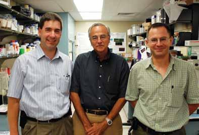
“We administer the virus carrying a normal copy of the CNGB3 gene underneath the retina,” said Dr. Komáromy. “The treated part of the retina is temporarily detached following the injection. The eye is an enclosed organ so you can inject high doses exactly where you need it.”
“It’s the same gene responsible for 50 – 80 percent of human achromatopsia,” said Dr. Aguirre. “This work can help researchers understand how to fight age-related macular degeneration in people, which is the leading cause of blindness in people over 60.”
“Dogs don’t have a well developed macula as in humans,” said Dr. Komáromy, “but because achromatopsia is a model cone disease, it allows us to answer questions for human medicine and consider important factors like age and allows us to follow up over a long period of time.”
Dr. Komáromy’s treatment cured younger canines regardless of the mutation that caused their achromatopsia and was effective for the 33-month span of the study. The successful restoration of cone function was documented through a technique called electroretinography. Researchers also measured the dogs’ ability to negotiate a short obstacle course in daylight.
“I remember the moment very well during the summer of 2006 when I recorded the recovered cone function for the first time by electroretinography,” said Dr. Komáromy. “I became very excited, but I repeated the recordings multiple times and kept replacing the electrode wires to make sure that I was not dealing with a technical artifact...the cone signal persisted!”
While younger dogs had tremendous success with this kind of gene therapy, dogs older than 54 weeks of age had less success.
“Because of the success in younger dogs, it makes sense to keep going,” said Dr. Komáromy. “We want to develop a treatment for older dogs.”
The results represent the second successful cone-directed gene replacement therapy in achromatopsia animal models and the first outside of mouse models.
The work done by Drs. Komáromy and Aguirre holds promise for future human clinical trials of cone-directed gene therapy in achromatopsia and other cone-specific disorders. It holds great promise to treat dog-specific diseases like the Progressive RodCone Degeneration (PRCD), a hereditary retinal disease that leads to blindness in dogs between the ages of three to seven years, and is the most common cause of inherited canine blindness.
More Work in the Lab
Veterinary Medicine: Influencing Animal and Human Medicine
What started in childhood as a love for animals and an intrigue with biology has become a launching pad for a budding career in translational research.
Veterinary medicine has become an invaluable foundation of comparative pathology and comparative medicine that enriched the research I performed while enrolled in the VMDPhD program at Penn Vet, influencing everything from the types of questions I asked to the interpretations of the answers as they applied to medical syndromes.
I pursued a PhD in neuroscience working with the Stress Neurobiology Group at the Children’s Hospital of Philadelphia Research Institute among researchers who explore neural pathways underlying stress and stress-related disorders such as anxiety, aggression and depression. Working under the mentorship of Dr. Sheryl G. Beck, I was drawn towards animal models of anxiety because so little is known about the disease process, despite its prevalence in both human and veterinary medicine.
TAKING ON ANXIETY
Collectively, anxiety disorders are the most prevalent psychiatric disorders in adult humans in the United States and tend to have an earlier age of onset than other mental disorders. Anxiety disorders often exist with other mental disorders and are associated with a wide range of other
medical conditions, including sleep disorders, interstitial cystitis, cardiovascular disease, chronic pain and irritable bowel syndrome.
Anxiety-related behavioral disorders are also prevalent in veterinary patients including dogs, cats and horses, among other companion animals. Frequent, repetitive actions and certain types of self-mutilation are behavioral disorders in equine patients for which social stress and social isolation can be contributing factors. Canine acral lick dermatitis resulting from over-grooming is often considered an animal model of obsessive-compulsive disorder with obvious parallels to the grooming behavior often seen in human obsessive-compulsive disorder.
Interestingly, these stress-related disorders in veterinary medicine are responsive to the same types of drugs that are useful in human anxiety disorders, suggesting a common biology and a mechanism rooted in conserved neural systems.
FOCUSING ON SEROTONIN IN THE BRAIN
To begin to tease out the mechanisms underlying anxiety, my thesis research centered on the serotonin system, which is conserved among mammals and has a role in steadying mood, appetite, vasomotor tone and panic behavior amongst several other behaviors and physiological functions. Serotonin is known to play a role in anxiety and is altered by anxiety-relieving drugs.
However, we now understand that there are several types of serotonin cells that interact with distinct brain regions,

VMD-PhD student LaTasha Crawford shares similarities found in animal and human anxiety and what the connection may mean for therapy options
function in different ways and may play different roles in anxiety. My project explored serotonin cells within the dorsal raphe nucleus of the midbrain (DR), which is made up of several subpopulations of serotonin cells.
The dense collection of serotonin cells in the ventromedial subregion (vmDR cells) are well characterized and known for their connections to the higher-level processing regions of the brain like the prefrontal cortex, a fear-processing region known as the amygdala and a learning and memory-processing region known as the hippocampus.
I characterized the physiology of a less dense, poorly understood subpopulation of serotonin neurons in the lateral wing subregion of the DR (lwDR). The lwDR subpopulation sends connections to many subcortical brain regions such as the periaqueductal gray, where release of serotonin minimizes panic behaviors, and the rostral ventro-lateral medulla, where release of serotonin dampens the sympathetic tone within the vascular system.
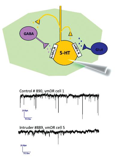

THE PROCESS
In addition to immunohistochemistry and confocal microscopy imaging, I became adept in in vitro electrophysiology, a technique that uses rapidly dissected brain slices to visualize, stimulate and record the electrical activity of individual living neurons. Because the local neural circuits remain intact in the slice, electrophysiology can also be used to characterize the signals from neurons that connect or synapse onto serotonin cells, including inhibitory signals from local GABA neurons. This led to several findings that differentiated lwDR cells from vmDR cells in the healthy brain of untreated mice: lwDR cells have a larger, more complex morphology and are more easily excited by electrical activity. However, the most intriguing difference between these two subtypes of serotonin cells was clarified using a model of anxiety.
Dr. LaTasha Crawford
I sought a mouse model that used social stress as an etiological agent with hopes of stimulating pathways that are consistent with naturally occurring disease states, for which social stress plays an important part.
Chronic social defeat is a type of model used in male rodents where repeated exposure to intimidation, bullying and threat of attack produces behaviors that are typical of anxiety and depression and that can be alleviated by anxiolytic and antidepressant drugs. Using behavioral tests, I characterized a five-day social stress paradigm that induced anxiety-like behaviors in stressed mice, such as increased stress-induced grooming and increased avoidance behavior where individuals avoid vulnerable areas of an apparatus and seek more enclosed, protected areas.
Though it was beyond the focus of my lab, I was also able to perform partial necropsies following the stress paradigm and identified several changes in peripheral organs of stressed mice, including increased bladder weight, potentially due to urine retention and bladder wall thickening, and increased heart weight, potentially due to increased blood pressure or direct sympathetic stimulation and consequent ventricular hypertrophy. The hypertrophy of the bladder and heart are likely sequelae (the negative aftereffects) of increased sympathetic tone, which is a common feature of human anxiety. The changes in behavior and peripheral organ systems underscored the utility of social defeat as a model to study anxiety and to examine the potential role that anxiety plays in the often comorbid clinical syndromes.
THE FINDINGS
I used the chronic social defeat model to investigate changes in serotonin cell function, and found that social stress caused distinct changes in the two subtypes of serotonin cells examined. Using electrophysiology in brain slices obtained from stressed mice and controls, I found that serotonin cells were capable of functioning normally; however the inhibitory input from nearby GABA cells was drastically altered by social stress.
Following social stress, vmDR serotonin cells received less inhibition from GABA inputs due to inactivity or death of GABA neurons and due to fewer GABA receptors on vmDR neurons. The loss of inhibition of vmDR serotonin cells could increase serotonin release in the cortex, amygdala and hippocampus in the anxious brain, as has been suggested in the literature. This would result in the enhanced fear processing and increased salience of fearful memories that are typical of anxiety disorders. A maladaptive reduction in inhibitory input to vmDR cells in anxiety could also explain the clinical efficacy of benzodiazepines, which act by increasing the potency of GABA signals and could theoretically counteract the pathological changes that disinhibit vmDR serotonin cells.
On the other hand, lwDR serotonin cells were affected in a distinct way following social stress: lwDR cells received more

potent inhibitory signals due to slower kinetics of the GABA receptor. This implies that in the anxious brain, fewer messages are traveling from the lwDR to the brainstem to prevent panic responses. This could then correspond to the exaggerated panic behaviors that typify anxiety disorders. In addition, because lwDR cells usually help to dampen sympathetic vasomotor tone, anxiety-induced inhibition of lwDR cells could lead to increasing blood pressure and consequent changes in heart musculature.
It is likely that stress-induced inhibition of lwDR serotonin projections to other brainstem regions could likewise contribute to increased sympathetic tone, urine retention and bladder wall hypertrophy, though this has not been shown definitively.
Collectively, these findings have contributed to a better understanding of the heterogeneity of serotonin cells, the way they are regulated by local inputs and the way they are dysregulated in anxiety.
WHAT THIS RESEARCH MAY MEAN
Experiments of this type lend clarity to the mechanisms of known anxiolytic drugs, and may one day aid in the development of novel therapies as well. Being better able to target pharmacotherapy to specific subsets of serotonin cells may also help us to one day target particular components of anxiety-related syndromes or to avoid undesirable side effects of drugs that modulate serotonin neurons.
For me, these findings have also piqued an interest in neuropathology and the interaction between the brain, the autonomic nervous system and visceral organ dysfunction. I have been exhilarated by the opportunity to incorporate my clinical background into my research and look forward to the unique perspective additional anatomical pathology training will grant me as I continue towards a career in research. With a better understanding of mechanisms of stress susceptibility and stress-related disorders, we can build on correlations observed on the clinical side of human and veterinary medicine to improve the diagnosis, treatment – and even prevention – of comorbid diseases. It is my hope that my pursuit of mental disease research with an eye for the sequelae seen in veterinary and human patients will be a step in that direction.
REFERENCES
Clemens et al., 2008 J Urol. 180(4):1378-82.
Kessler et al., 2005 N Engl J Med. 352(24):2515-23.
McDonnell SM, 2008 Anim Reprod Sci. 107(3-4):219-28.
Nicol C, 1999 Equine Vet J Suppl. (28):20-5.
National Institute of Mental Health “Anxiety Disorders” 2009 NIH
Publication No 09 3879.
Roth et al., 2006 Biol Psychiatry. 60(12):1364-71
Roy-Byrne et al., 2008 Gen Hosp Psychiatry. 30(3):208-25.
Stein et al., 1992 Compr Psychiatry. 33(4):274-81.
Woods-Kettelberger et al., 1997 Expert Opin Investig Drugs. 6(10):1369-81.
Mark has an outstanding mix of knowledge and experience leading successful pharmaceutical development efforts to help companies maximize portfolio potential and capitalize on growth opportunities,” said David Bupp, president and chief executive officer of Neoprobe Corporation, upon the appointment of Mark Pykett, V’91, GR’86 to the position of executive vice president and chief development officer for the company. (Neoprobe News, November 16, 2010)
Initially, after reading the above kudos from Mr. Bupp, one may be surprised to learn that Dr. Mark Pykett is a veterinarian. Indeed, he is one of Penn Veterinary Medicine’s VMD-PhD dual-degree program graduates. But instead of a traditional veterinary career in smallor large-animal practice, Dr. Pykett chose a career in biomedicine.
A Different Perspective
“I came to veterinary school with a different perspective than most other traditional students,” said Dr. Pykett. “I was always interested in fundamental basic science, but was more interested in the innovative research and the practical applications that a combined degree program supported in translational medicine.”
Translational medicine is focused on relating discoveries made in basic science research labs and using them to develop therapies or diagnostics useful for treating human and animal patients.
“After receiving a BA from Amherst College in 1986, I began looking for an advanced degree program that tied together an appreciation of comparative biology and comparative medicine,” he said. “Penn Veterinary Medicine’s VMD-PhD program offered thinking outside of the box. I was very impressed with the integration at Penn between the Graduate School and Schools of Medicine and Veterinary Medicine,” he said.
VMD-PhD students at Penn have access to more than 500 laboratories to conduct doctoral research, while also benefitting from two world-class veterinary hospitals for clinical training.
Opportunities to Explore
While at Penn Vet, Dr. Pykett completed his VMD coursework with his V’91 classmates, while doing double-duty at Penn’s Graduate School completing lab rotations and doctoral research with Donna George, PhD, in the Cell and Molecular Biology Graduate Group.
“Working with mentors like Dr. Richard Miselis, V’73, GR’73 (then head of the program) provided me with the guidance I needed to explore,” Dr. Pykett said. “As veterinary students we had the opportunity to explore the research side, as well as the small and large animal care experience.”
After finishing vet school requirements and completing his PhD in 1994, Dr. Pykett completed a postdoctoral program at the Harvard School of Public Health, and then earned an MBS from Northeastern University in 2000. He has served as an adjunct lecturer in cancer biology at Harvard University’s School of Public Health and on Northeastern University’s Center for Enterprise Growth Corporate Advisory Board, but the breadth of his career has included senior leadership positions at a number of biotechnology and diagnostics companies where he

Penn Veterinary Medicine hosted its 111th Penn Annual Conference from March 1 - 4, 2011 at the Sheraton City Center Hotel in Philadelphia.

With the theme “Partners in Care,” the four-day conference offered the signature PennHIP seminar, in addition to full-conference sessions, some of which focused specifically on feline medicine. In addition, interactive wet labs were held on the School’s Philadelphia campus.
Several special events were also hosted, such as the first-ever Keynote Luncheon Presentation on March 2 by Mattie Hendrick, V’78, pathologist. The prestigious Alumni Awards of Merit, given annually by the School to recognize distinguished graduates for their contributions that advance the veterinary profession and the School’s good name, were awarded at the luncheon to Eric Bregman, V’95; Pierre Conti, V’60; Charles Rupprecht, V’85; Raymond Stock, V’75; Donald Stremme, V’75; James Thomson, V’85 and Marilyn Weber, V’75
The VMAS Excellence in Teaching Award, selected by Penn Veterinary Medicine young alumni and fourth-year students, was presented at the Young Alumni Luncheon on March 3 to Patricia Sertich, V’83, director, Equine Endometrial Biopsy Service, associate professor, CE, of Medicine and Reproduction, and director of Clinical
Service, Georgia and Philip Hofmann Research Center for Animal Reproduction at New Bolton Center.
Thanks to this year’s Educational Committee for once again creating an outstanding conference, especially committee chairs Dr. Linda Baker, Dr. Eric Parente, Dr. Jon Palmer, Dr. Alexander Reiter and Michelle Traverse and Chair of the Conference Advisory Committee, Dr. Kathy Michel.
Penn Annual Conference Partners in Care 111th Tuesday, March 1, 2011 Penn HIP Seminar Wednesday & Thursday, March 2 and 3, 2011 Full Conference Sheraton City Center Hotel, Philadelphia, PA Friday, March 4, 2011 Wet Labs University of Pennsylvania, Matthew J. Ryan Veterinary Hospital
Dear Penn Veterinary Medicine Alumni:
As the Gilbert S. Kahn Dean of Veterinary Medicine, but moreover as an alumna of the School of Veterinary Medicine, I am keenly aware of the importance of a strong connection between the School and Penn Veterinary Medicine alumni. From our first graduating class in 1887 to our 124th class in 2011, the Veterinary Medical Alumni Society (VMAS) has existed to keep all graduates connected to the School long after graduation.
As a School, we have successfully navigated through two very challenging fiscal years, recreating ways to achieve our goals in an environment of shrinking resources. In all areas of how we teach our students, conduct research and provide care for our clients and patients, our alumni have been critically important in sharing ideas, sending to us the best and brightest students, supporting our projects and carrying our message to the world. Our alumni remain the living history of the School – our ears and eyes beyond Penn, and the biggest champions of our future. There are even more opportunities and challenges ahead for Penn Veterinary Medicine, and the time is ideal to ask how we can engage our graduates even more deeply in the pursuit of our highest priorities.
To intensify our connection with alumni, in January 2011 I created the Dean’s Alumni Council – a specific group of alumni volunteers with a robust demographic representation to help us move our projects forward and to serve as liaisons for the School to other alumni. As this group replaces the current VMAS Executive Board structure, it will allow the opportunity to involve a broader network of alumni leaders and ambassadors who are well-informed about Penn Veterinary Medicine today, who will bring the School fresh ideas while amplifying our voice to others, and who will find ways to interact with current students, faculty and administration meaningfully. I have invited current and former VMAS Board leaders to serve as the Founding Members of the Dean’s Alumni Council in recognition for their outstanding service to the School, in addition to other alumni who have expressed interest and enthusiasm in the School’s future.
My goal is to activate this initial core group of volunteers to work with me on innovative, flexible approaches to strengthen our alumni partnerships, presence in the community and interaction with students, in addition to other projects that alumni, students and faculty identify together. In the future, it is my hope to continue to evolve the group membership, which will allow a continuous flow of ideas and an opportunity for many to participate. I will be seeking new interest as the effort grows, but am grateful to those who have initially agreed to join me as we launch this new initiative.
I look forward to communicating again in the near future about the Council’s special projects, and continue to welcome your ideas on ways alumni can help the School, and the School can support its alumni.

Beloved Penn Vet Alumnus and Professor Emeritus Charles Raker (V’42) received the prestigious “Beyond the Call” award at the American Association of Equine Practitioners (AAEP) President’s Luncheon on December 7, 2010 in Baltimore, MD.
Edward Lemos (C’54, V’57) serves on the Board of Federal Savings Bank in Dover, NH The bank has served residents of the seacoast area of New Hampshire since 1890.
Produced by his daughter, Sandy, a news story about Bernard Levine (V’55) was shown on New Jersey Public Television to highlight how Dr. Levine saved the life of a Canada goose that had been illegally shot with a bow and arrow. The story became international news because the smart goose arranged for his own emergency medical care by choosing to land – of all places – on Dr. Levine’s waterfront property. Prior to Dr. Levine’s care, the bird was spotted by residents along the river in several towns, struggling with the arrow sticking straight out of its chest. See the video at http://www.youtube.com/ watch?v=f4qRKm2Xbao. Dr. Levine is also the father of Richard Levine (V’81)
Randy Aronson (V’80) co-owns P.A.W.S. Integrative Veterinary Center in Tucson, AZ. He hosts a radio program called “Radio Pet Vet,” which airs 8 – 10 a.m. each Saturday.
Metropolitan Veterinary Associates, owned by James Dougherty (V’80) in Norristown, PA, is celebrating 25 years of operation in 2011.
W. Boyd Henderson (V’82) of Henderson Veterinary Associates, Ltd. in Lancaster County, PA has a career in Embryo Transfer and other Assisted Reproductive Techniques (A.R.T.) He has been an extern instructor for the University of Pennsylvania School of Veterinary Medicine’s Department of Reproduction, a member of both the American and International Association for Embryo Transfer as well as the American Association of Equine Practitioners and the American Association of Bovine Practitioners. Dr. Henderson has been an author on more than a dozen published papers in A.R.T., most recently cloning. Currently, he is doing both bovine and equine embryo transfer for the practice as well as managing the stallion collection program.
Lawrence Gerson (V’75) is a veterinarian and founder of the Point Breeze Veterinary Clinic in Pittsburgh, PA, and hosts a biweekly column called “Pet Points” in the Pittsburgh Post-Gazette to educate pet owners.
William “Bill” Miller (V’76) DACVD of Cornell University College of Veterinary Medicine will be discussing “Update on Dermatology” at the May 5, 2011 continuing education lecture series for the Richmond Academy of Veterinary Medicine in Sandstone, VA.
Scott Palmer (V’76) received the 2010 AAEP President’s Award during the President’s Luncheon on December 7, 2010 in Baltimore, MD.
The US Department of the Interior, Bureau of Land Management released a report in December 2010, based on partial findings by Sarah Ralston (CW’73, V’80, GR’82) of Rutgers University, concerning the care and handling of wild horses and burros at three major gathers in the Western US in the summer of 2010. The report found “horses did not exhibit undue stress or show signs of extreme sweating or duress due to the helicopter portion of the gather, maintaining a trot or canter gait only as they entered the wings of the trap. Rather[,] horses showed more anxiety once they were closed in the pens in close quarters; however, given time to settle, most of the horses engaged in normal behavior.”
The AAEP has named Mary Beth Hamorski (V’86) as the January 2011 honoree of its Good Works Campaign in honor of her low-cost veterinary care to Mylestone Equine Rescue, a sanctuary for 34 abused, neglected and relinquished horses in Warren County, NJ.
Diane Deresienski (V’89) and Nick Sitinas (V’90) were featured on Animal Planet’s “Pets 101,” which aired in December 2010. They provided comments during several segments related to special species.
Peter Vogel (V’90) has been elected vice president of the Southern California Veterinary Medical Association.
Lance Bassage (V’93) has been named professor at the Ontario Veterinary College at Guelph. He will be working at the Large Animal Clinic providing both elective and emergency surgical care, as well as teaching OVC students.
Denise McAloose (V’93) serves as assistant clinical professor at the Albert Einstein College of Medicine of Yeshiva University, Bronx, NY and as an anatomic pathologist for the Wildlife Conservation Society – Bronx Zoo.
Karen O’Connor (V’04) opened Coastal Georgia Veterinary Care, her first practice, in December 2010 in Richmond Hill, GA, just outside Savannah. The practice will be dedicated to delivering full-service treatment in a spa-like, nurturing environment.
1939 James Howard Gillespie on January 10, 2011.
1950 Leonard B. Simmons on October 5, 2010.
1951 William A. “Doc” Kernick on January 27, 2011.
1952 Margaret L. Petrak on October 19, 2009.
1957 George Boyle on December 17, 2010.
1961 Andrew Cullen on November 17, 2010.
1972 Brian Silverlieb on February 24, 2011.
1979 John A. Burger on September 19, 2010.
Jill Abraham (V’03) DACVD, currently works at the InTown Veterinary Group/Massachusetts Veterinary Referral Hospital as a dermatologist. Before her position in Massachusetts, she completed a small animal rotating internship in medicine then worked for a year at Red Bank Veterinary Hospital in Red Bank, NJ in 2004. In 2007, Dr. Abraham completed her residency in dermatology at the Matthew J. Ryan Veterinary Hospital of the University of Pennsylvania in Philadelphia, PA. Dr. Abraham became board-certified by the American College of Veterinary Dermatology in the summer of 2007.
Cailin Heinze (V’04) has been appointed assistant professor in nutrition at the Tufts University Cummings School of Veterinary Medicine in Boston, MA. Dr. Heinze completed a three-year residency in nutrition at the University of California, Davis after graduation from University of Pennsylvania School of Veterinary Medicine and was recently certified by the American College of Veterinary Nutrition. Currently, she is investigating omega-3 fatty acid metabolism in psittacine birds, which include parrots, cockatiels and parakeets.
1983 Thomas E. Sooey on October 9, 2010.
1990 Cheryl Welch-Muller on February 23, 2011.
has managed product and corporate development efforts. Along the way, he started four private biotech companies and ran three others that are publicly held.
From stem cell research to cancer diagnostics to research for innovative neurological therapeutics to serving as a diversified developer of innovative oncology surgical and diagnostic products, VMD-PhD graduates play vital roles in our society both behind the microscope and in the board room.
Veterinarians Enable Change
“All veterinary students should have a keen appreciation of the options a veterinary degree can offer,” Dr. Pykett said. “As veterinarians, our unique contribution is that we have the ability to enable change and to bring critical thinking to an array of problems and challenges. The degree fosters broad thinking – veterinarians have the options for diverse career paths, including running a biotech company.”
Dr. Narayan Avadhani, chair, Department of Animal Biology, has been reappointed to this position for a third, three-year term.

Dr. Elise Boller, staff veterinarian at the Matthew J. Ryan Veterinary Hospital, has been named director of referring vet relations.
Dr. Ralph Brinster, professor of physiology, has been chosen as the 8th recipient of the annual International Society for Transgenic Technologies (ISTT) Prize for his contributions to the field of transgenic technologies. He will be presented the award at the annual TT meeting slated for October 2011.

Dr. Dottie Brown, chair of Penn Vet’s Department of Clinical Studies in Philadelphia and professor of surgery at Matthew J. Ryan Veterinary Hospital at the University of Pennsylvania (Ryan-VHUP), was recently invited to serve as the Oscar W. Schalm Lecturer at the School of Veterinary Medicine at University of California, Davis.
Ms. Maria Calabrese has been named director of customer service at the Matthew J. Ryan Veterinary Hospital.
Ms. Joy Crothers has recently joined the nursing team as a midnight nursing assistant.
Mr. Rob DiMeo, recruiter/counselor, and Ms. Rosanne Herpen, associate director, Penn Vet Admissions, received a 2011 Models of Excellence Honorable Mention for their VETS Summer Program, which invites high school- and college-aged students to participate in a week-long summer day camp that provides an overview of the field of veterinary medicine.
Ms. Kimm Dinsmore has joined Matthew J. Ryan Veterinary Hospital of the University of Pennsylvania School of Veterinary Medicine as referring vet liaison.
Dr. Tamara Dobbie has joined the Section of Reproduction at New Bolton Center as a staff veterinarian.

Dr. Michelle Harris, medicine resident at New Bolton Center, worked with a contributing author to Practical Horseman on an article about lower-leg swelling on horses, which ran in the publication’s March 2011 issue.
Ms. Kim Lewis, CVT has joined the Penn Vet team as a part-time nurse in the blood bank.

Ms. Jillian Marcussen has joined Penn Vet as director of stewardship and special projects.
Dr. Sue McDonnell, head of the Havemeyer Equine Behavior Lab at New Bolton Center, has been elected to serve a three-year term on the Animal Behavior Society’s Board of Professional Certification as well as to serve an eight-year term on the Organizing Committee of the International Symposium on Equine Reproduction, an international meeting of animal and veterinary scientists working on basic and applied aspects of equine reproduction. The next meeting is slated for 2014 in New Zealand.
Ms. Gloria Provost has joined the Sports Medicine and Imaging Team at New Bolton Center where she will work as a technician.
Ms. Helen Radenkovic has joined Penn Vet as director of development for Matthew J. Ryan Veterinary Hospital.

Dr. Virginia Reef, director of Large Animal Cardiology and Diagnostic Ultrasonography at New Bolton Center, is a charter diplomate of the new American College of Veterinary Sports Medicine & Imaging. She is also on the Credentials and Residency Training Committee.
Dr. Gail K Smith, founder/director of PennHIP and professor, orthopedic surgery, received the Blaine Award 2011, presented annually by Royal Canin for outstanding contributions to the advancement of small animal veterinary medicine or surgery. Dr. Smith was presented the award at the British Small Animal Veterinary Association Annual (BSAVA) Congress, held in March 2011.
Dr. Oriol Sunyer, associate professor of immunology, presented his lecture, “Novel discoveries into the immune system of teleost fish, and their impact into the future development of mucosal vaccines,” at the 6th International Symposium on Aquatic Animal Health held in Tampa, FL in September 2010.
Dr. Regina Turner has been promoted to associate professor of reproduction – clinical educator.

Alison Weltner, V’12 initially took a different career path than her father who was a veterinarian. But after working for several years in the pharmaceutical industry, Alison craved the veterinary experience of her father’s work in small-animal practice.
“I grew up watching my dad care for both animals and the people who love them,” she said. “As I advanced in my pharmaceutical career, I discovered that my true desire was to work on the front line of medicine, treating and caring for individual pets.“
As a non-traditional student, Alison sought an opportunity for mentorship upon entering veterinary school at Penn, which drew her to applying for the School’s Opportunity Scholarship Program.
“Mentorship through the Opportunity Scholarship award would fill many of my knowledge gaps regarding small animal practice and allow me access to the lifeknowledge of a practicing clinician,” she said.
St. George Hunt, V’86 had also been a nontraditional student, having worked for several years after college graduation as a counselor to children and adults with disabilities. Upon graduating from veterinary school, St. George owned a small animal practice where his love for his clients and their pets was rivaled only by his love for veterinary students and his family, which included his life-mate and companion, Suzi Robinovitz.
After St. George’s untimely death in 2008, friends, family and clients wanted to immortalize the verve and dedication to the profession that he loved. Through various gifts ranging from $25 to $500 from
more than 100 people, enough money was raised to support a $12,000 Opportunity Scholarship for a Penn Veterinary Medicine Student. Dear friend and classmate of St. George, Carla Chieffo, V’86, agreed to provide mentorship for the scholarship in St. George’s name.

Today, Alison is benefitting from that scholarship through mentoring and tuition support.
“The Opportunity Scholarship is unique in that it fosters a meaningful, long-term mentorship that begins early in a student’s education and continues throughout the four years of veterinary school,” said Alison. “Dr. Chieffo’s keen understanding of my current position in my career and my inspirational connection to Suzi have broadened my exposure to veterinary practice and reconnected me to why I changed my life’s work. I never had the honor to meet St. George, but it is through his inspiration that I am pursuing my own personal dream.”
Established in 1998, the mission of the Opportunity Scholarship Program at Penn Vet is to foster scholarship support and mentoring opportunities for future veterinarians trained at Penn. Through a $12,000 scholarship disbursed across four years, sponsors may honor a friend, relative or a family name, while simultaneously investing in the future of the School and the profession.
For more information, contact Coreen Haggerty, director of Alumni Relations, at 215.898.1481.
Penn Veterinary Medicine
3800 Spruce Street
Philadelphia, PA 19104-6008
MAY 2011
Tuesday, May 3, 2011
First Tuesdays Lecture Series at New Bolton Center, a free educational lecture series for horse owners and horse enthusiasts.
“You Think It’s Colic, But It’s Not”
New Bolton Center, Kennett Square, PA
Presented by Dr. Barbara Dallap, Emergency and Critical Care
Friday, May 13 & Saturday, May 14, 2011
Alumni Weekend 2011
University of Pennsylvania, Philadelphia Campus and New Bolton Center
We remember all Penn Vet alumni with special celebrations for classes ending in ’1 and ’6 including the 25th reunion Class of 1985 and the 50th reunion Class of 1961. Please contact Coreen Haggerty at haggertc@vet.upenn.edu or 215-898-1481 for details.
Thursday, May 19, 2011
Animal Lovers Lecture Series, a free educational lecture series for small animal owners.
“Care and Feeding: Basic Check-up”
New Bolton Center, Kennett Square, PA
Presented by Dr. Magi Casal and Dr. Kathy Michel
May 23 through August 5, 2011
Summer VETS Program
Week-long programs designed for college and high school students who are interested in learning about veterinary medicine. For more information, visit www.vet.upenn.edu and search “summer VETS.”
JUNE 2011
Thursday, June 2, 2011
Animal Lovers Lecture Series, a free educational lecture series for small animal owners.
“Caring for Older Pets”
Rago Arts & Auction Center, Lambertville, NJ
Presented by Dr. John Marcus and Dr. Scott Martens
Monday, June 6 through Thursday, June 9, 2011
Penn Executive Veterinary Leadership Program: “Making an Impact as a Health Leader”
The Wharton School, University of Pennsylvania, Philadelphia, PA
For more information and to register, visit http://executiveeducation. wharton.upenn.edu/ and search “Veterinary Medicine.”
Tuesday, June 7, 2011
First Tuesdays Lecture Series at New Bolton Center, a free educational lecture series for horse owners and horse enthusiasts.
“Get the Home Field Advantage: Advanced Equine Home Care for Your Horse”
New Bolton Center, Kennett Square, PA
Presented by Dr. James Orsini, Large Animal Surgery
Wednesday, June 15, 2011
Wednesday Exchange, bi-monthly interactive professional education opportunities for primary care veterinarians.
“Recent Advances in Cancer Diagnosis and Treatment: Molecular Diagnostics and Targeted Therapies”
Ryan Veterinary Hospital, Philadelphia, PA
Presented by Dr. Erika Krick, Oncology
JULY 2011
Tuesday, July 5, 2011
First Tuesdays Lecture Series, a free educational lecture series for horse owners and horse enthusiasts.
“Pre-purchase Examinations – The Role of the Veterinarian” New Bolton Center, Kennett Square, PA
Presented by Dr. Midge Leitch, Sports Medicine
Monday, July 18, 2011
American Veterinary Medical Association Annual Convention Penn Vet Alumni Reception St. Louis, MO
AUGUST 2011
Tuesday, August 2, 2011
First Tuesdays Lecture Series, a free educational lecture series for horse owners and horse enthusiasts. “Strangles – Prevention and Treatment in Your Horse”
New Bolton Center, Kennett Square, PA
Presented by Dr. Ashley Boyle, Field Service
Thursday, August 11 through Sunday, August 14, 2011
Pennsylvania Veterinary Medical Association
5th Annual Keystone Veterinary Conference
Hershey, PA
For information on any of these events, contact Darleen Coles, special event coordinator, at coles@vet.upenn.edu or 215-746-2421.
Nonprofit Organization U.S. Postage PAID
Philadelphia, PA Permit No. 2563


