Newsmagazine of the School of Veterinary Medicine
University of Pennsylvania
Summer 2000
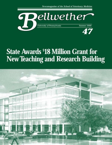
47 State Awards $18 Million Grant for New Teaching and Research Building
Newsmagazine of the School of Veterinary Medicine
University of Pennsylvania
Summer 2000
47 State Awards $18 Million Grant for New Teaching and Research Building