Penn’s Food Animal Program Penn’s Food Animal Program


The modern livestock producer is interested in the economic health of his operation and the welfare of his animals, and is willing to pay for professional services to correct issues that affect the operation’s profitability. To meet the needs of this emerging dimension of animal production the School of Veterinary Medicine has changed its curriculum to allow students the opportunity to acquire skills in production medicine. The School’s Center for Animal Health and Productivity (CAHP), established at New Bolton Center in 1986 to implement teaching, research, and service programs directed toward the improvement of health and herds and flocks, playing a vital role in shepherding the development of these courses.
The modern livestock producer is interested in the economic health of his operation and the welfare of his animals, and is willing to pay for professional services to correct issues that affect the operation’s profitability. To meet the needs of this emerging dimension of animal production the School of Veterinary Medicine has changed its curriculum to allow students the opportunity to acquire skills in production medicine. The School’s Center for Animal Health and Productivity (CAHP), established at New Bolton Center in 1986 to implement teaching, research, and service programs directed toward the improvement of health and herds and flocks, playing a vital role in shepherding the development of these courses.
1 45 45
University of PennsylvaniaSpring/Summer 1999 Newsmagazine of the School of Veterinary Medicine University of PennsylvaniaSpring/Summer 1999 Newsmagazine of the School of Medicine
(continued on page 4) (continued on page 4)
Dr. Ray Sweeney and two students consult on a cow in the Marshak Dairy. The cow is free to feed as she chooses. The loose boxes are visible in the back.
INSIDE From the Dean 2 Marookian Auditorium 3 Running on Empty? 6 Alumni Weekend 8 Alumni Awards 9 114th Commencement 10 School Honors Dr. Fagin 12 Scholarships 14 Symposia for Breeders &Owners 16Rosettes &Ribbons 22 Animal Crackers 24 INSIDE From the Dean 2 Marookian Auditorium 3 Running on Empty? 6 Alumni Weekend 8 Alumni Awards 9 114th Commencement 10 School Honors Dr. Fagin 12 Scholarships 14 Symposia for Breeders &Owners 16Rosettes &Ribbons 22 Animal Crackers 24
Dr. Ray Sweeney and two students consult on a cow in the Marshak Dairy. The cow is free to feed as she chooses. The loose boxes are visible in the back.
From the Dean
As we approach the end of the century it is time to look back and think about the significant advances that have shaped veterinary medicine in the past 100 years. This is relevant as the profession was in its infancy in 1900 and many predicted its demise with the decline of the horse. In this column I will mention two achievements that were vital to the growth of veterinary medicine.
The first is the Bureau of Animal Industry that took origin under the direction of Dr. Daniel Salmon at the end of the 19th Century and played an enormously important role in eradicating contagious diseases of domestic animals, protecting the nations livestock and improving the safety of foods of animal origin in the present century. Daniel Salmon, the first person to be awarded the degree of doctor of veterinary medicine in the U.S., is best known for his work in characterizing Salmonella bacteria, but his contributions as Director of the Bureau of Animal Industries are just as important. Under his leadership, the Bureau attracted a outstanding group of veterinarians and microbiologists and accomplished remarkable success in either controlling or eradicating bovine pleuro-pneumonia, Texas fever, foot and mouth disease, fowl plague, equine glanders, hog cholera, brucellosis, and bovine tuberculosis. The Bureau also established procedures for protecting the U.S. livestock industry from the spread of infectious diseases, especially along the Mexican border. Since their work, U.S. livestock has been remarkably free of epidemics of infectious disease.
The other great accomplishment of the Bureau was to bring public acceptance to the fledgling veterinary profession. When the Bureau was formed, detractors ridiculed the idea of Salmon “a mere horse doctor” as the head of a federal bureau. The achievements of the
Bureau changed this attitude and we owe Salmon and his colleagues an enduring debt of gratitude.

The second significant advance was the development of specialization in the profession. This advance started in the mid-fifties at a time when the nation was enjoying unprecedented prosperity. Goaded by Soviet advances in space technology and other scientific fields, the federal government increased exponentially its spending on research and higher education, including biomedical research and education. On a scale never before imagined, the National Institutes of Health and the National Science Foundation invited schools in all health professions to compete for research grants and for training grants and fellowships. As a full partner in one of the World's great biomedical research centers this School was uniquely equipped to compete for these funds to strengthen basic science faculties and research infrastructures, while developing veterinarian-scientists and clinical specialties.
Many of the first veterinary clinical specialists received rigorous training as veterinarian-scientists at the Medical School and Hospital of the University of Pennsylvania, and returned to the School of Veterinary Medicine to establish scholarly sections of radiology, dermatology, cardiology, ophthalmology, neurology, internal medicine, orthopedic surgery, oncology, medical genetics, and a variety of sub-specialties.
They conveyed great strength to the Department of Clinical Studies as they brought science and medicine together and bridged the traditional divide between clinical and basic science departments. Our teaching hospitals developed a science based curriculum and were equipped with the most advanced diagnostic equipment.
Penn was in the vanguard of this initiative which has transformed the education and practice of veterinary medicine in the United States and throughout the western world much to the benefit of the animal-owning public. Adirect outgrowth of this change in clinical education was the development of quality internship and residency training
programs, and the establishment of specialty colleges chartered to certify discipline specialists. As a result of this latter development, either corporate or privately owned specialty practices are becoming common in the U.S. and Board certification has become the standard of advanced clinical proficiency.
Both of these initiatives that have been so important to the development of veterinary medicine in the 20th Century, deserve close attention in the 21st. Iwonder what impact corporate practices will have on the structure of veterinary medicine and, after the brilliant contributions of the Bureau of Animal Industry, worry about the lack of investment in research and training on infectious diseases of animals today. Unlike the NIH budget that is burgeoning, the USDA research budget has not increased.
Free trade means increased movement of animals and animal products and with this goes the threat of spread of infectious disease. I sometimes wonder if the veterinary profession has been put into the same position as the medical profession in 1980 just before the start of the AIDS epidemic. I hope I am wrong.
Alan M. Kelly
The Gilbert S. Kahn Dean of VeterinaryMedicine
Marookian Scholarship Fund
Family members and friends of Dr. Marookian chose the occasion of the dedication to show their respect and admiration by making gifts toward a scholarship fund in Dr. Marookian's name. The E.R. Marookian, V.M.D. Research Scholarship Fund, established at the School through these contributions and contributions by Dr. and Mrs. Marookian, will be used to encourage and support veterinarians who wish to obtain a graduate degree (Ph.D.) in the field of basic research.
2
The E. R. Marookian,V.M.D. Auditorium
The afternoon of April 16 was a time for a very special celebration at VHUP. Friends and family of Dr. Edgar R. Marookian, V’54, joined faculty, staff and students for the dedication of the E.R. Marookian, V.M.D. Auditorium. Everyone assembled in the refurbished room B101, rejuvenated and equipped now with a new projection system. This was made possible by Dr. Marookian’s generosity and the School recognized him by naming the room his honor. Because Dr. Marookian’s gift also helps to pay for the critically needed new Research and Teaching Pavilion at 39th and Spruce Streets, plans are to transfer the name “E. R. Marookian, V.M.D. Auditorium” to a new space there once that building is completed.
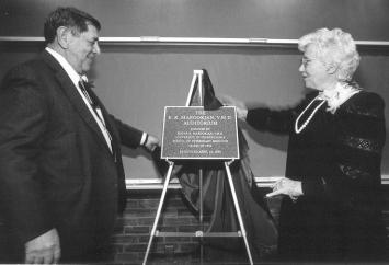
The School also established an annual endowed Marookian lecture in the field of animal biology. Dean Alan Kelly awarded Dr. Marookian the School’s Bellwether Medal for his long-time loyal support. The text of the citation follows:

For over forty years you have enjoyed the reputation as an immensely respected veterinarian and businessman. Ed, your contributions to the veterinary pharmaceutical industry are notable, enabling you to command immeasurable regard among your peers.
Even as a student it was evident that you would be successful as you found ingenious ways to sell lab coats, instruments and medicines to your classmates. Dean Mark Allam singled you out as the only student he had known to graduate financially better off than you entered the School.
As the founder of Clinton Labs in Frenchtown it wasn’t long before sales increased dramatically. Not content to ride on that success, you invested in a new suit to interview at Merck Sharp & Dohme and in two short years became head of your division, excelling in the sales of anthelmintics and other veterinary pharmaceuticals. Always looking for new veterinary products to enhance the field, Richardson Merrill, recognizing your talents, sent you around the world pioneering relations between business and veterinary medicine. Throughout these worthwhile endeavors, your devoted
wife, Myrval, has been by your side, vigorously supporting you in all of your achievements.

Believing that it was Penn’s School of Veterinary Medicine and the quality of instruction, particularly the science based education, that enabled you to interact with so many people in numerous walks of life, developing new ideas and products, you possess a rare and most cherished quality an appreciation of quality education with a sense of obligation sustained by supporting your alma mater so that others will benefit.
The University of Pennsylvania School of Veterinary Medicine has chosen the occasion of the dedication of the E.R. Marookian, V.M.D. Auditorium to pay tribute to you, Ed, and your myriad accomplishments. We applaud your challenge to colleagues to take pride in the rich educational fulfillment as a veterinary scientist and to advance both the stature of the profession and your alma mater. The E.R. Marookian, V.M.D. Auditorium is a perpetual tribute to your vision.
April 16, 1999
As Dean Kelly noted in his citation, Dr. Marookian’s financial savvy is of long standing. In making this very generous gift to the School of Veterinary Medicine he worked with officials of the School and the University to make a “planned gift.” Planned giving enables donors to avail themselves of certain tax advantages while also contributing a much larger sum than otherwise would be possible. For more information about planned giving call the Office of Planned Giving Programs at 1-800-223-8236.
3
Above:Dr. and Mrs. Marookian unveil the plaque for The E.R. Marookian,V.M.D. Auditorium. Right:Dean Kelly presents the School’s Bellwether Medal to Dr. Marookian; and lower right,Dr. and Mrs. Marookian during the ceremonies.
Penn’s Food Animal Program
(continued from page 1)
They emphasize an integrated, interdisciplinary approach and involve disciplines such as clinical nutrition, reproduction, health economics, and computer science in addition to conventional specialties in veterinary medicine. The focus of the teaching program is to maintain the physical and economic health in whole animal populations as well as the clinical treatment of individual sick animals. As part of the program, the Marshak Dairy, the swine unit, and the sheep flock are managed to emulate real on-farm production systems. The facilities are used for teaching purposes in many of the courses throughout the four-year curriculum.
The last five years have seen a very rapid evolution of the food animal program at Penn, a program which will continually grow as the needs of agriculture change. In addition to the excellent tradi-
tional medical and surgical training, new courses have been developed to emphasize areas of current importance. Veterinary students have the opportunity to train with the 38 faculty and academic staff for essential roles in modern food animal production through the very best multidisciplinary training available anywhere.
The eight-week course in Production Medicine for fourth year students focuses on evaluating the health of the production system on the dairy farm by integrating and evaluating information on facilities, milking, reproduction, nutrition, database
Food Animal Fellows Program
As technology grows and the role of the food animal practitioner expands, fewer students are pursuing careers in this essential aspect of veterinary medicine. More and more students matriculating in veterinary schools are from urban backgrounds. Having had limited exposure to food animal production systems, they often find it difficult to make informed decisions regarding career selection. The Food Animal Fellows Program at New Bolton Center is designed to encourage qualified veterinary students to pursue careers in food animal medicine. The program supports several activities to give students an understanding of what it takes to be a modern food animal practitioner and to better understand modern animal agriculture.
SummerFellowship Program: First and second year veterinary students are offered the opportunity to work with food animal practitioners for 10 weeks and to attend bi-weekly seminars on food animal production systems held at the Center for Health and Productivity at New Bolton Center. Students see a variety of animal production systems and get first-hand experience on the daily life of a food animal practitioner. They receive a stipend and course credit.
Students are accepted into the program based on essays submitted to the Food Animal Advisory Group. Since its inception in 1995 the program has had 29 students matriculated. The program also supports an externship for qualified students at the Miner Institute of Chazy.
Special Seminars: The Food Animal Fellows Program supports seminars on animal production issues presented by national and international speakers on issues germane to food animal production.
Food Animal Conferences: Students may accompany faculty on expense-paid food animal conferences. The annual Production Medicine Meeting and the Large Herd Conference of New York are often selected meetings. At these meetings, students are exposed to the most current issues facing animal agriculture from both the veterinary and producer perspectives.
Independent Study: There are also opportunities for independent study during the school year and summer months.
systems, and environmental issues. Taught by the faculty of CAHP, Field Service, and Reproduction, with guest lectures by nationally-recognized experts in production medicine, the course combines lecture, laboratory, computer lab, farm visits, and a two-week internship with a veterinary practice. The computer lab at CAHPis used extensively to train students in the evaluation of production records and in formulating solutions to
problems commonly encountered in dairy herds. Although the emphasis is on dairy, the principles are applicable to other herd/flock production systems.
The safety and quality of foods of animal origin is an area of mounting importance and concern, and the food animal practitioner has an important role to play in preventing and reducing the existence of chemical and microbial health hazards in the nation’s food supply. The Swine Production Medicine Program was initiated five years ago in response to the health care needs of the Pennsylvania swine industry. Participating veterinary students study pork production across many dimensions, including animal health and welfare, food safety, economics, and environmental assurance. Poultry Population Medicine involves the control of infectious diseases in flocks, which is an important concern to both human health and to the economic health of the poultry industry. The Food Safety and Quality Assurance course was established three years ago to introduce students to regulatory, diagnostic, and biological factors they will need to consider when evaluating potential problems in a herd or flock.
New and drastically changed courses have emerged as more and more food ani-

4
The free stall wing of the Marshak Dairy,used for group-based nutrition studies.
mal veterinarians rely on computers to stay progressive. Animal Health Economics is taught as an elective course and covers animal production budgets, financial analysis, decision trees, linear programming, dynamic programming, and risk analysis. Students may elect to take independent studies for more advanced exposure to some of these
Nutrition, Ration Formulation, Dairy Production Medicine, and independent studies.
Throughout the fourth year, students can take rotations in New Bolton Center’s Field Service practice, which includes dairy cattle, some beef cattle, small ruminants, and horses. Students travel to owners’farms with veterinarians in specially-equipped trucks, and acquire skills in palpation, surgery, and medicine. Field Service treated about19,000 animals last year.
topics. Dairy Ration Formulation and Evaluation are now taught in the computer lab in several courses: Advanced Dairy Cattle

The Food Animal Medicine and Surgery rotation is given on-campus and focuses on the care and treatment of individual animals (as opposed to production medicine or population medicine, which focus on the herd or flock). The rotation is taught as a cooperative effort by faculty in the sections of Large Animal Medicine and Large Animal Surgery and residents from both sections.
In 1997 New Bolton Center began an Aquaculture study in Harnwell Pond

The Marshak Dairy Facility at New Bolton Center
In 1996 the University of Pennsylvania School of Veterinary Medicine completed the construction of a state-of-the-art 200-head dairy barn for teaching and research. The Marshak Dairy Facility, named after former Dean Robert R. Marshak, is the only greenhouse-type barn in Pennsylvania. Agreenhouse barn is energy efficient, naturally bright, and easy to keep dry, all essential conditions for comfortable and productive cows. Also, it is cost effective in terms of manpower and building expense.
The Marshak Dairy Facility at New Bolton Center serves as a living laboratory for the School of Veterinary Medicine and as a research and teaching site in such fields as epidemiology and preventative medicine, nutrition, reproduction, infectious and chronic diseases, and dairy cattle health economics. In addition, the Marshak Dairy Facility provides the region a resource with potential for commercial applications and enhances the teaching environment for veterinary and graduate students interested in the medical and managerial aspects of dairying.
“In order to adapt to our climate we’ve made design modifications to reduce heat build-up,” Dr. David Galligan explains. “The shell of the building is pre-manufactured as a solar agriculture building, in essence, a plastic greenhouse.” In the summer, the sides of the barn are rolled-up to facilitate cross ventilation. The facility consists of an administration area that includes a room with view of the double ten herringbone milking parlor; four sections of forty freestalls each where cows can walk, stand, and lie down where they choose; and a space for 48 tie stalls.
The tie stall area accommodates 48 cows that can be tied-up at feed bins for nutritional studies. Each cow can be fed a different mix and monitored by computer. The tie stall area can be converted to a freestall-style barn, if needed. Manure from the entire barn is deposited into an eight month holding tank and is periodically and strategically spread onto fields. This reduces the need for chemical fertilizer, cutting the overall farm cost.
under the direction of Dr. David Nunamaker. This fish farming system for hybrid striped bass involves aerated cages placed in the pond. If it can be made cost-effective, it could become a source of income for properties with ponds. Elective courses in aquaculture are available to students during the third year.
The Food Animals Fellows Program was implemented to offer more exposure in food animal medicine to students in their first and second years. Students accepted as Fellows spend ten weeks during the summer working with food animal veterinarians and receive course credit and a stipend.
Both educational and social, the Food Animal Club has long been a part of the veterinary school experience at Penn. The club has become more organized and active in recent years, and includes lectures and activities emphasizing bovine, small ruminants, swine, poultry, and production aquaculture. Just as important, participation in club activities lets students see the wide range of options available to modern food animal practitioners.
Jeanie Robinson-Pownall Members of the Section of Animal Health Economics and Nutrition
and the Field Service contributed to this article.
There are 38 faculty and academic staff involved in the food animal program at Penn. Alist of faculty along with their area of interest will soon be available at the CAHPweb site at <http://cahpwww.nbc.upenn.edu/>
(continued on page 7)
5
The milking parlor in the Marshak Dairy. Each cow wears a computer chip that identifies her and tracks her production output.
Drs. Paul Pitcher,Jim Beach and Tom Parsons consult next to one of the fleet of six Field Service trucks.
Running on Empty?
By Eric Tulleners, D.V.M.
There are a number of serious breathing problems which can significantly affect a horse's performance. Typically, horses with problems which reduce the amount of air entering the lungs tire during maximal exertion, such as racing exercise, and these horses often make an abnormal breathing noise. The first step toward identifying the location of the obstruction is to have a comprehensive endoscopic examination performed by an experienced veterinarian. Many problems, such as vocal cord and flap lysis (left laryngeal hemiplegia or “roaring”), can be accurately confirmed based on the results of physical and resting endoscopic examination.
During the last ten years, it has become clear that many breathing problems only manifest themselves during high-speed exercise and can easily be completely missed or badly misdiagnosed during a resting examination. Atypical scenario is a horse that works 3/8 of a mile well in the morning, but perhaps makes a vague noise. During racing exercise the horse tires dramatically, often as early as the 1/4-pole in a six-furlong race. The jockeys comment that they felt they had “a ton of horse under them” and “were going to win for fun under a hand ride” but suddenly the horse began to make a breathing noise and choked down, backing through the field or at the very least hanging at the wire. Unfortunately, after the horse is pulled up and catches its breath, the noise immediately stops. The after-race scoping is uneventful with perhaps a little bit of mucus, and some reddening or pharyngitis noted by the veterinarian. What can be done to resolve this frustrating dilemma? The answer often involves scheduling a high-speed treadmill evaluation at New Bolton Center.
Before it goes on the treadmill, every horse receives a comprehensive physical
examination which includes endoscopic examination, examination of the heart and lungs with a stethoscope and with ultrasound, and a detailed lameness evaluation. If no abnormalities are detected, horses are first schooled to the treadmill to make them comfortable with the feeling of the ground being pulled out from underneath them. To get the most useful information from the test, it is important that the horses be fit and able to perform at near maximal exertion for their particular occupation.
The high-speed treadmill portion of the examination is then performed by a
team of experienced staff members and veterinarians who have performed over 1,200 clinical evaluations, more than any other facility in the country. During the evaluation, the horse's heart rate and rhythm is continuously monitored by telemetry and later evaluated for arrhythmias which can develop during strenuous exercise. The distance traveled and maximal speed are carefully controlled and adjusted according to the individual horse's level of fitness.
The horse's pharyngeal and laryngeal function is continuously monitored with a state-of-the-art videoendoscope. The scope allows the image to be seen in exquisite detail in full color on a 20-inch monitor, as well as recorded on standard videotape. Photographs of any abnormalities that are found can be made from the videotape for the owner, trainer, veterinarian, and the medical record. After the examination, the videotape can be scrutinized in super slow motion, frame-byframe, to detect even the most subtle abnormality.
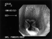
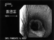
The most common problems, such as roaring and choking down caused by intermittent dorsal displacement of the soft palate, can usually be easily confirmed. Anumber of other problems which absolutely cannot be diagnosed without a high-speed treadmill evaluation have also been identified. One of these, termed axial deviation of the aryepiglottic folds (ADAF), results in severe narrowing of the airway just in front of the opening to the flaps, every time the horse takes a breath (Figures 1A and 1B). Dynamic collapse of this tissue into the airway usually causes the horse to make an abnormal respiratory noise and to tire during strenuous exercise. This problem has been diagnosed most commonly in Thoroughbred racehorses (80%) but has also been identified in Standardbred (13%) and in Arabian (7%) racehorses. Laser removal of this apparently flaccid or redundant tissue causing the problem usually can be done safely on the standing awake horse on a same-day outpatient basis. Dr. Eric Tulleners, Chief of Surgery at New Bolton Center, who developed the “no-touch” technique, performs the surgery using a 600µouter diameter laser fiber through the hollow channel in the videoendoscope with the image of the horse’s throat projected on a color monitor. The surgery is done with the horse sedated and the throat numbed with a topical anesthetic. As in arthroscopic surgery, traction on the tissue to be removed is necessary. Traction is placed using custom-designed long grasping forceps introduced up the horse’s other nostril. The laser quickly and efficiently cuts tissue and coagulates blood vessels. Bleeding is usually negligible. Anti-inflammatory medication is given after the surgery. Horses are typically restricted to shedrow exercise or small paddock turnout for two weeks
6
Figure 1A
Figure 1B
after surgery to allow for complete healing of this delicate area.
The results of a study of this problem were presented by Dr. Dana King, a surgery resident working with Dr. Tulleners at New Bolton Center, at the 1997 annual Scientific Meeting of the American College of Veterinary Surgeons in Orlando, Florida. Dr. King reported that complications were not encountered, and healing was uniformly unremarkable. Laser surgery definitely improved racing performance on 73% of racehorses and eliminated noise in 75% of horses. In comparison, horses with ADAF that did not undergo surgery and that were rested for less than two months usually did not improve in racing performance when training was resumed.
As an example, ADAF was diagnosed in a talented horse who was the winner of multiple races and had earnings in excess of $400,000. The trainer recognized a breathing problem after the horse atypically performed poorly in a race. Working carefully with the local track veterinarian, they were able to localize the problem to the throat region, but could not pinpoint the exact site. Ahighspeed treadmill evaluation at New Bolton Center performed by Dr. Eric Parente, a member of the Section of Sports Medicine and Imaging, confirmed the problem of ADAF. Standing laser surgery was performed to correct the problem, and the horse convalesced without complications. In the first start back the horse was back to usual form, winning handily.
While ADAF has been recognized only in recent years, it does illustrate the need for a high-speed treadmill evaluation in horses with suspected breathing problems.
“Acomprehensive high-speed treadmill performance evaluation is a timeconsuming, labor-intensive test which is not inexpensive to conduct due to the equipment and professional expertise needed. However, these caveats aside, I am convinced that the examination is an incredibly valuable diagnostic tool which is a worthwhile investment for owners and trainers with a horse which is not training up to its potential due to a suspected breathing or cardiovascular problem,” commented Dr. Eric Tulleners.
Continuing Education Opportunities for Graduate Food Animal Veterinarians
(continued from page 5)
TheUniversity of Pennsylvania School of Veterinary Medicine participates in a number of Food Animal Continuing Education (CE) programs. The content of these courses reflects the on-going change in food animal agriculture and the demands of veterinarians to address these emerging issues.
• Dairy Production Medicine
Certificate Program: This joint program sponsored by Penn State and the University of Pennsylvania involves ten threeday modules given over a period of three years, including nutrition (heifer, dry cow, and lactating cow), housing and facility design, mastitis control programs, reproductive management, farm finances, and herd expansion. Many experts from throughout the country, including faculty from both the University of Pennsylvania School of Veterinary Medicine and Pennsylvania State University, teach in this CE program which results in a Dairy Production Medicine Certificate.
• Software Development: Faculty members at the School of Veterinary
Medicine’s New Bolton Center are continuously involved in software development which can be used to teach veterinary students and post-graduate veterinarians.
• Penn Annual Conference: Two-day seminars have been given on Culling, Nutrition, Economics, Reproduction, Pregnancy Wastage, and Heifer Management with national and international speakers.
• M.B.A. Program: The School’s CAHPhas a joint MBAprogram with Penn’s Wharton School that integrates the underlying principles of animal health and economics in livestock production systems. Through the Wharton MBAprogram, basic fundamental skills and principles in economics, finance, cost accounting, and operations research are covered. Students also complete an application project that explores the use of these principles to a problem in animal production. (n.b. There is also a concurrent VMD/MBAdegree program at Penn.)
Veterinariae Medicinae Doctoris (V.M.D.) degree
It takes four years of graduate studies to earn a V.M.D. degree. The first two years are spent in lecture and laboratory, covering such basics as anatomy, biochemistry, physiology, embryology, pathology, and nutrition to lay the foundation for the clinical exposure in the third and fourth years. During the third year the students are increasingly exposed to clinical teaching and begin to have hands-on experience with animal patients. At the end of the third year each student selects one of five “tracks”: Small Animal, Large Animal, Large and Small Mixed, Equine, or Food Animal. During the fourth year students experience clinical rotations consisting of six foundation and 18 elective blocks.
For students interested in both veterinary medicine and business, the school of Veterinary Medicine and Penn’s Wharton School offer a combined course leading to the joint degrees of V.M.D. and M.B.A. This rigorous joint program involves five to six years of study. (There is also a M.B.A. program for graduated veterinarians; please see “Continuing Education Opportunities.”) Asix-to-seven year program of study leading to both a V.M.D. and Ph.D. degree is available for a small number of highly-motivated, highlyqualified students interested in in-depth research.
7
Alumni Weekend 1999
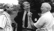
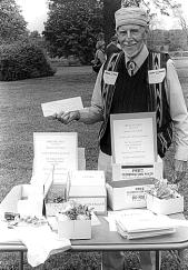

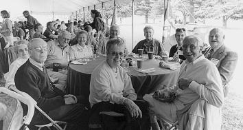


Alumni weekend festivities began on Saturday, May 15 when 28 alumni met at the Loch Nairn Golf Club for the third annual Alumni Golf Tournament. The team comprised of Dr. Roger Sembrant, V’74, and Cathy Sembrant, Dr. Sidney Mellman, V’49, and Dr. Arthur Richards, V’49, emerged as the winner of the day.
In the evening, alumni and their families joined the Dean and Mrs. Kelly for a cocktail party at the Allam House. The large crowd of 190 had a great time reminiscing and catching up on each other’s activities. Later the Classes of 1989 and 1949 got together for reunion dinners at the Allam House.
On Sunday more than 350 people converged on New Bolton Center for Alumni Day. During the morning meeting of the Veterinary Medical Alumni Society outgoing President Dr. Suzanne Smith, V’82, passed along the gavel of office to Dr. Robert W. Stewart, V’68, the incoming president of Penn’s VMAS. Dr. Eric Bregman, V’95, was elected president-elect. The five new board members are: Dr. Amy Bentz, V’97, Dr. Maureen Stokes Burdulis, V’79, Dr. Leonard N. Donato, V’96, Dr.Lawrence A. Rebbecchi, Jr.,V’90, and Dr. Raymond W. Stock, V’75.
VMAS honored three outstanding alumni for their achievements and contributions to the profession and Penn. The 1999 Alumni Award of Merit was presented to: Dr. David Meirs II, V’54, Dr. Michael Ratner, V’59, and Dr. Gail Smith, V’74.
Dr. Raymond W. Sweeney III, V’82, received the Excellence in Teaching Award presented by the VMAS Board on behalf of the members of the classes of 1995-1998 who selected the recipient.
The afternoon was devoted to food, catching-up-with-classmates, tours of various New Bolton Center facilities, and play for the many children who accompanied their parents. Late in the afternoon it was “good bye” and “until next year” for the alumni and their families.
8
The Veterinary Medical Alumni Society of the University of Pennsylvania Salutes
David A. Meirs II,V.M.D.,
Class of 1954
For your efforts to uphold a higher level of veterinary medicine and education through your membership on the New Jersey Board of Medical Examiners and your service on other boards.
For your contributions to veterinary medicine through the publishing of scholarly articles in publications of professional note that promote the University of Pennsylvania School of Veterinary Medicine.
For your service to your community through membership on civic and agricultural boards and commissions.
For your receipt of numerous honors that further advance the good name of your alma mater, among them the 1998 Governor’s Trophy, New Jersey Horse Person of the Year, and the 1997 Citation from the New Jersey State Board of Agriculture for Distinguished Service to Agriculture.
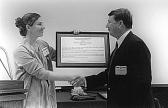
The Alumni Award of Merit is presented to you this 16th day of May 1999.
The Veterinary Medical Alumni Society of the University of Pennsylvania Salutes
Michael P. Ratner,V.M.D.,
Class of 1959
For your outstanding career, distinguished by dedicated service to the veterinary medical profession and your community at large.
For your service as a board member of the School of Veterinary Medicine’s Veterinary Medical Alumni Society and the State of Connecticut’s Veterinary Medical Association.
For your commitment to sharing your professional knowledge with students at the University of Bridgeport; for mentoring and encouraging young people through an outreach program at theUniversity of Connecticut.
For your service to youth and society through your leadership of the Boy Scouts of America, the Probus Club and the Rotary Club.
The Alumni Award of Merit is presented to you this 16th day of May 1999.
The Veterinary Medical Alumni Society of the University of Pennsylvania Salutes
Gail K. Smith,V.M.D.,Ph.D.,
Class of 1974
For your illustrious career, distinguished by your logical scientific approach and adroit teaching in the field of orthopaedic surgery.
For your commitment to sharing your exemplary scholarly contributions with students, colleagues and society as inventor and director of Penn HIP®at the University of Pennsylvania.
For your contributions to veterinary medicine through the publishing of prodigious scholarly articles that promote professional inquiry and distribution of knowledge.
For your numerous honors that advance the good name of your alma mater, among them the 1996 American Veterinary Medical Foundation and the AVMACouncil on Research “Excellence in Research Award” and the 1994 SmithKline Beecham Award for Research Excellence.
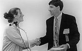


The Alumni Award of Merit is presented to you this 16th day of May 1999.
Correction:
In the story about the Pennsylvania Veterinary Medical Historical Society in Bellwether 44, we stated that Dr. Otto Stader's son, Robert, had graduated from Penn's School of Veterinary Medicine in 1990. In fact, it was Dr. Stader's grandson, Dr. Alan Stader, who graduated from Penn in 1990. Dr. Robert Stader, also a veterinarian, graduated from Ohio State University Veterinary School in 1946.
9
Dr. Suzanne Smith presents the 1999 Alumni Award of Merit to Dr. David Meirs.
Dr. Suzanne Smith presents the 1999 Alumni Award of Merit to Dr. Michael Ratner.
Dr. Suzanne Smith presents the 1999 Alumni Award of Merit to Dr. Pamela McKelvie who accepts it for her husband,Dr. Gail Smith.
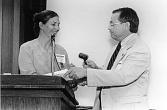
Dr. Ray Sweeney accepts the Excellence in Teaching Award from Dr. Suzanne Smith. Passing the gavel.
The graduating Class of 1999, 80 women and 25 men, marched triumphantly onto the stage of the Zellerbach Theater on May 17 for the 114th Commencement Exercises of the School. The auditorium was packed with families, friends and faculty, all ready to cheer on a favorite graduate.
Dr. Alan M. Kelly, The Gilbert S. Kahn Dean of Veterinary Medicine, opened the proceedings and provided an overview of the history of veterinary medicine and Penn’s special place in that
Class of 1999
Sarah Smith Adams
Amy Lynn Anderson
Amy Lynn Bader
Vered Bar
Nancy Louise Bathurst
Joyce Ellen Bendokas
Jennifer Michelle Bevilacqua
Jennifer Leigh Bitman**
Kenneth Dean Bixel
Kathryn Rodgers Blanch
Dipa Pushkar Brahmbhatt
Rebecca Vaill Christie**
Jennifer Jeitles Clarke***
Diane Pamela Cordray
David Croman
Patricia Curran
Elizabeth Kathryne Daniel
Pandora Lynn Davis***
Alysia Deaven***
Erin Page DeTurck
Suzanne Donahue
John Eccher, Jr.*
Aimee Suzanne Eisenberg
Julie Fishman Ekedahl***
Marla Mozelle Ellman
Robyn Denise Engelman
Vicki Nicole Estes**
Brian Gregory Fenchak
Ellen Elizabeth Fitzgibbon
Dana Frederick
Avra Ingrid Frucht*
Heather Rose Galano
Emily Austin Graves*
Bradley Scott Gray
James Francis Hagan
Barbara Hasnain
Wayne J. Hassinger II***
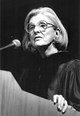
history. The Commencement Address, given by Claire M. Fagin, M.A., Ph.D., Professor Emerita and Dean Emerita, School of Nursing, University of Pennsylvania, and former acting president of Penn, is reprinted elsewhere in this issue.
The presentation of diplomas and hooding was done by Dean Kelly, assisted by Dr. James B. Lok, 1999 Lindback Awardee for Distinguished Teaching; Dr. Cynthia Ward, 1999 Carl J. Norden Distinguished Teacher Awardee; and Associate Dean Dr. Charles D. Newton.
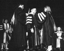
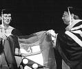
Dr. Robert W. Stewart, V’68, president of the Veterinary Medical Alumni Society, presented the class flag to Dr. Emily Graves, V’99, class president. The Veterinarian’s Oath was administered by Dr. Amy L. Hinton, president of the Pennsylvania Veterinary Medical Association.
Once the graduates left the stage as newly minted VMDs, the focus shifted to a celebration in the Annenberg School’s court yard. Here the graduates posed for pictures for their families and said “good bye” to classmates and teachers.
Donatella Elizabeth Hecht
John Francisco Heller
Melissa Margaret Hobday
Mason Francis Holland*
Catherine Bisque Jackson***
Matthew Stephen Johnston
Stacey Wanda Kent*
Danielle Karyn Kessler
Justin Ian Kirchhofer
Kathryn Leigh Kirstein
Julie Elizabeth Kyle
Bernadette Hutchinson LaMonte
Anna Marie Lange
Albert Sze-Yen Leung
Kristen Mary Lohr
Kelly Elizabeth Longenecker
Melissa Marie Lutz
Patrick Anthony Mahaney
Cassandra Ash Mahoney
Courtney Jones Manetti
Richard David Marchetti
Robin Michele Mazin
Mira Lea McGregor
Kathryn Mary McPherson
Stephen Charles Meister
Claire Alice Morissette
Jennifer Pollock Morris
Deborah Lynn Murtha
David John Nebzydoski
Marjorie Jean Elizabeth O’Brien
Andrew Obstler
Mark William Paradise
Erica Clarice Parthum
Kristen Su Pelletier
Don James Petersen**
Christine Polaneczky
Caryn Leah Porter
Mary Elizabeth Powers
Clive Laurel Rahamut-Ali
Jacqueline Alane Rapp
Danielle Marie Reinhardt
Mark Stephen Restey
Jennifer Ann Rumbold
Michele Ann Saletros
Akiko Sato
Laura Schmitt
Carlton Benner Seybolt
Suzanne Shalet
Shannon Devon Shank
Renee Elizabeth Simpler**
Jennifer Ann Smelstoys
Corrina Sue Snook
Stacey Solovei
Benjamin J. Spitz*
Sandra Springer
Tracy Ann Springer
Robert William Stewart, Jr.
Wendy Jo Taft
Janet Madenford Triplett
Jacqueline Hazel Vockroth
Jenny Suzanne Vodenichar
Franciszek von Esse*
Diane Simpson Wagner
Karen Wallace
Mary Hayes Wallace*
Ellen Bart Wiedner
Jessica Zeman
Dara Marie Zerrenner
***Summa Cum Laude
**Magna Cum Laude
*Cum Laude
10
114 TH C OMMENCEMENT
Award Recipients
Leonard Pearson Prize
Catherine Bisque Jackson
J.B. Lippincott Prize
Pandora Lynn Davis
1930 Class Prize in Surgery
Pandora Lynn Davis
Auxiliary to the American Veterinary Medical Association Prize
Emily Austin Graves
Auxiliary to the Pennsylvania Veterinary Medical Association
Prize — Small Animal Award
Erica Clarice Parthum
Auxiliary to the Pennsylvania Veterinary Medical Association
Prize — Large Animal Award
Stacey Wanda Kent
1956 Class Medal forAchievement in Pathology
Jennifer Jeitles Clarke
James Hazlitt Jones Prize in Biochemistry
Vicki Nicole Estes
American Animal Hospital Association
Award
Tracy Ann Springer
Merck Awards
Small Animal Award
Amy Lynn Bader
Large Animal Award
Julie Elizabeth Kyle
George M. PalmerPrize
Nancy Louise Bathurst
Everingham Prize forCardiology
Donatella Elizabeth Hecht
Large Animal Surgery Prize
Clive Laurel Rahamut-Ali
Large Animal Medicine Prize
Corrina Sue Snook
Morris L. Ziskind Prize in Food Animal Medicine
Wayne J. Hassinger II
Morris L. Ziskind Prize in Public Health
Kathryn Mary McPherson
Hill’s Award
Robyn Denise Engelman
Pharmacia & Upjohn Awards
Small Animal Award
Robin Michele Mazin
Large Animal Award
Julie Fishman Ekedahl
Faculty/SCAVMAPrize
Mason Francis Holland
American College of Veterinary Surgeons Prizes

Small Animal Surgery Prize
Renee Elizabeth Simpler
Large Animal Surgery Prize
Alysia Deaven
American Association of Feline Practitioners Award
Diane Pamela Cordray
Field Service Prize
Alysia Deaven
Phi Zeta Award
Mary Hayes Wallace

Anatomy Prize
Julie Fishman Ekedahl
American College of Veterinary Radiology Award
Julie Fishman Ekedahl
Iams/VECCS Award forExcellence in Veterinary Emergency and Critical Care Medicine
Erica Clarice Parthum
Charles F. Reid Sports Medicine and Imaging Award

Diane Simpson Wagner
Large Animal Reproduction Award
Jacqueline Hazel Vockroth
Lynn Sammons Food Animal Award
Erin Page DeTurck
11
114 TH C OMMENCEMEN T
School Honors Dr. Fagin
Commencement Address
by Claire M. Fagin,M.A.,Ph.D.
Congratulations colleagues and let me add my personal congratulations to you and everyone who has helped sustain you and encourage you as you have pursued this important degree. It’s a thrill to be here with you today, and with so many of my colleagues on your faculty, as we celebrate you and the last graduation of the 20th century.
You have chosen this profession at a particularly interesting time and that choice tells us a great deal about you. You know that you have not made an easy choice for your future and that the rewards, both financial and otherwise, will be more complicated then for your predecessors. Therefore we have to hope that your commitment to the practice of veterinary medicine is very deep. Your willingness to confront this different future gives us reason to believe that you will participate with others in reshaping and redefining the delivery not only of veterinary health care, but through that, of human health care, in the coming decades.
In preparing to talk with you today I reviewed speeches and papers written 100 years ago, as your leaders were setting the agenda for the new century. I wanted to know what these leaders saw as the essential elements necessary for the continuing development of the field and how they would differ from the essential elements leaders might highlight today. Speakers were exhortatory in asking the graduating classes to do whatever was required to bring the university and the field up to its potential. Its potential included raising capital and investing it wisely, seeing to it that the field was independent, lengthening the courses in veterinary medicine, building the research and science enterprise, building their organizations, and advancing the status and conditions governing the profession. The future of veterinary education was believed to be tied to ensuring that veterinarians had a broad point of view with regard to their domain in clinical work and science and that students
had a right to expect the full measure of laboratory and clinical facilities in which to learn no matter whose pockets the money came from to support these innovations. Only in this way could veterinary medicine achieve the high status which the public was ready to accord it. In his 1899 address to the American Veterinary Medical Association James Law remarked, “We stand at the parting of the ways and the future of veterinary education, and of the veterinary profession, depends on our ability to secure the means which will provide a modern scientific education.”
These goals were met to a phenomenal degree during the 20th century and your School and its faculty were responsible for meeting and exceeding many of them. The leaders of 1899 seemed to be visionaries, however even they could not have envisioned the developments in this school alone which will shape your future and the future of the profession.
You will share in continuing to build and ensure the viability of your school and profession through some of the same avenues that the leaders talked about at the turn of the century: contributing to the capital enterprise, building the science, continuing to advance veterinary education in the clinical and biological spheres, and integrating research into your clinical practice. You will have several other goals to work on as well. One that was not discussed 100 years ago and which is necessary for all professions is seeking to further develop a peer review process for your practitioners which will include ethical considerations and clinical competence in practice. Another which you will have to wrestle with will be how you deal with the increasing domination of the for-profit corporate model in your work.
When you take a quick scan at what our veterinary school has offered you and the opportunities which they have created for you and other veterinarians in biological science, (and I know you shared my pride in the prominent mention of Dr. Karen Overall and the School in the recent New Yorker Magazine), in animal-human interaction, in food safety, in animal rights, in aquatic science, in
equine sports medicine, and I could go on you can draw three messages at least: your education has been the richest in the field, your future is guaranteed to be as diverse and challenging as you want to make it, and your future is unbounded by today’s achievements. These guarantees could not have been made to graduates 100 years ago.
There is another major difference between you and your predecessors. Speakers 100 years ago addressed their audience dramatically. They called on sons of Penn, Gentlemen, (stated repeatedly), men of singular aim, my brothers, men of the graduating classes there were only he’s, and him’s in all the references and they certainly did not give me any doubt that there was not a professional woman in the room, or at least, if there was, they ignored them completely. Now, I don’t want to offend those of you who are men but it is clear that veterinary medicine in the 21st century will be preponderantly a woman’s profession.
That fact creates an imperative for both the men and women graduating today. While it is wonderful that women have options and opportunities today unknown to our predecessors, it is also true that the dominance of women in the field will require you to be active in fighting against the economic inequities which still burden us. I know very well some of the problems you will face. Men and women, have always chosen this profession more for the love of the work than for its monetary rewards. That is particularly wonderful in this day and age. However, you should pay careful attention to the inequities in income between fields dominated by women and those dominated by men, and those who choose fields based on their love for the work and those whose choice is initially made with money in mind.
Further, as corporations come to dominate the practice world, their offers will include a style of work that may be more attractive to women but also to men it promises no necessary start-up loans, more controlled working hours, and a guaranteed salary, and benefits package which brings more security to family life. However, be sure that corporate
12
114 TH C OMMENCEMENT
dominance will impact on the control of your practice and personal initiative. The corporations’connections to pharmaceutical and equipment companies will reduce your freedom to do comparative appraisals and judgments. It may be easier, perhaps; but satisfying questionable. There is such an enormous demand
for veterinary services that there will be no compelling reason for you to accept restrictions on your freedom nor non competitive incomes. This will require your own thoughts about strategies and actions and the advocacy of your associations so that they are proactive on your behalf.
Claire M. Fagin,R.N.,Ph.D.,F.A.A.N.
Leadership Professor Emeritus and Dean Emeritus of the University of Pennsylvania School of Nursing
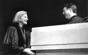
Claire M. Fagin, for over forty years you have been an internationally celebrated leader of the nursing profession and commanded immeasurable respect among your peers.
Your contributions to the field are notable, beginning with the baccalaureate nursing program that prepared nurses for primary care practice. You have been honored with myriad nursing awards, among them the Distinguished Scholar Award from the American Nurses Foundation and the Hildegard E. Peplau Award presented by the American Nurses Association. Your legacy includes a strong commitment to humanity. Your contributions were recognized by many, most notably by the Institute of Medicine, National Academy of Sciences, as a Scholar-in-Residence, with the Philadelphia Women’s Way “Women of Courage” Award, the University of Pennsylvania’s Alumni Award of Merit, the Distinguished Daughter of Pennsylvania Award and, most recently, the New York University President’s Medal.
As the first woman to serve as Chief Executive Officer of the University of Pennsylvania and the first woman to serve a term as Interim President of any Ivy League University, you brought honor and pride to the entire University and you provided pivotal support at a crucial time in the history of the School of Veterinary Medicine.
Your service to humanity continues through your incomparable leadership. Although retired as dean of the University of Pennsylvania School of Nursing, you continue to teach and remain active with many professional and corporate organizations. Now returning to New York where you were born, raised and educated, we applaud your compassion and contributions to society through your forthright advocacy of universal health care and your involvement with New York City’s West Side Urban Renewal Project.
The University of Pennsylvania School of Veterinary Medicine has chosen the occasion of their 1999 Commencement to pay tribute to you and your scintillating accomplishments by awarding you the Bellwether Leadership Medal. We celebrate your rich achievements in education and the promotion of humankind by advancing the stature of women in all of the health professions.
As corporatization is likely to be in your future, I would like to share with you my views on the parallel trend in human health care. To put it simply, I believe that in human health care, the for-profit, market approach, is inappropriate, and immoral.
The sine qua non of market disciplines is that people can vote with their feet when they don’t like the product. Alternative options are not available to most users of the health care system.
Markets are amoral in general; that is, sentimentally neutral, but in health care this general amorality has the potential to become immoral. The buyers, industry and government, want to reduce costs. The sellers, the managed care organizations, must reduce costs to remain competitive and provide profits to shareholders. Caregivers become implicit and explicit rationers of care who often benefit directly from rationing decisions, a factor that is unique in the American system, and exists nowhere else in the industrialized world.
More importantly, denying care to the sick diminishes us as human beings and as a society. Further, and perhaps most dangerous, a for-profit dominated system, grounded in price competition, forces the not-for-profits and the public sector to join in the same behaviors.
Universal health care must be on our policy agenda. Universal health care is inevitable. It is only a question of when. It will eventually succeed because the market approach is doomed to fail.
Let me highlight some signs of trouble that lead me to this conclusion.
1. The job market is changing rapidly and dramatically. In the next century we will find that employment and health benefits cannot be linked as they were in the past. We are seeing rising numbers of uninsured Americans, many working, who cannot afford the health insurance options offered by their employers.
2. Managed care, once seen as the solution to our escalating cost problems, is on a slippery slope and showing signs of deep strain and overreaching, particularly in relation to Medicare and Medicaid. Companies have blamed their
(continued on page 14)
13
114 TH C OMMENCEMEN T
Dr. Fagin’s Commencement Address
coverage of the medicare market for their huge losses last year and this year’s second quarter profits are attributed to rasing fees and getting out of medicare.
3. Labor Department economists noted that “Health insurance costs are picking up for the first time in three and a half years.”1 Out of pocket spending for premiums, coinsurance and copayments grew 5.3% to $187.6 billion last year, the first time since the late 1980s that out-of-pocket growth outstripped costs for private health insurance.
Managed care or managed cost, has neither expanded the populations who receive care nor saved money for individuals and families. In fact, as I have stated, the market has shifted costs to these consumers through greater out of pocket costs, higher deductibles, higher copayments, and dehospitalization, leading often to great personal burdens and costs for families losing work days to care for their sick at home. If anyone has benefitted by the market incursion into health care it would appear to be large corporate buyers, the pharmaceutical companies, some shareholders, and some hospitals.
Now what does all this mean to you?
It’s hard for me to believe that there are not direct parallels between what we see in for-profit human medicine and what you will experience in animal medicine. I have several reasons for saying this.
First, many of your scientific and clinical innovations have a direct fit to human medicine and human health care.
Second, you will have less opportunity to innovate when your practice is not under your own control. You have been very lucky in this respect. Yes, your incomes, in general, have not been on the same level as your colleagues in human medicine. You have not had the insurance advantages that they have had. But you have had a loving and respectful human and animal clientele who have not been separated from you by third party payers and now by the restrictive practices of companies, which are threatening all but the healthy, with sharp reduction in services. Third, and no less important, as respected clinicians and scientists, you have a great influence on how all of us as citizens are affected by what goes on
(continued from page 13)
in the broad health field. Your voices count more than those of many of us who are directly involved in human health care. You are seen as the quintessential health care provider. The curer who is also the carer. The health carer who does not and can not separate the family priorities and concerns from the care and the treatment of the patient. Your closeness to the patient and family is paramount. Keep it that way. If those
Even those of you who don’t, will find that work that you enjoy and that absorbs you carries you through all sorts of life crises that might be impossible to handle otherwise. I share the writer Jane Howard’s view on work: That “work,” [even] “with its inevitable drudgery and tension, is what allows most of us to transcend and redefine what... [We may have thought] were our limits. ...Work, for many of us, is the way we meet the people we most esteem and cherish. Work is what distracts us, at times for weeks on end, from life’s incessant chaos and uncertainty. Even when it is menial, [and don’t kid yourselves]...
of you in clinical practice move away from the caring role it will not only change your practice but it will change your image in our society. Think hard about developmental role changes that contradict what people value about you and remember always your historic mission. But also use your voices and your influence to bring your clinical and scientific knowledge into advocacy in the total health care scene today. Make your voices count for all of us.
In closing let me comment briefly about work. Until my experience at Penn which started in 1977, I had changed my career within nursing many times and, early on, so frequently that I thought I had a maximum two-year longevity per job. The job changes were always in nursing, a field which has given me the opportunity for learning, satisfaction, change, and commitment. I have loved my work.
Because work is so important, whatever you do, make sure that you don’t let any of the negative parts of it dominate your life. If they do make a change. Most of you will have to earn a living.
Everybody’s often is, work confers some degree of pattern, purpose and continuity.”
But what is most important is that you keep in sight your “vital powers” the Greeks spoke of, and challenge yourself on whether your are exercising them along lines of excellence. Penn has prepared you to do that and your wonderful school and its faculty will always be here to remind you of it.
In a great victorian novel, Middlemarch, George Eliot described what she called “The other side of silence.” She wrote, “If we had a keen vision and feeling of all ordinary human life, it would be like hearing the grass grow and the squirrel’s heartbeat”... Because of the work you have chosen, you have the opportunity few in our society have to develop that keen vision and feeling for all ordinary life because of this choice you have made you will reach the other side of silence, on occasion, and your whole being will be enriched in the process.
Congratulations to all of you.
1 Nasar, New York Times, 7/31/98
14
114 TH C OMMENCEMENT
Commencement Address
by Dr. Emily Graves,V’99
May 17th. We have thought about this day for weeks even years in some cases. Over the past few weeks, we have been celebrating, but we have also been interviewing, working, planning our moves to various places, renting new places, buying new homes, buying cars, selling cars... the list goes on. At the same time, many of us have become weary of entering the REALworld of veterinary medicine. Doubts and anxieties abound. DO WE ACTUALLYKNOWENOUGH? The national and state veterinary boards think so. Our fellow colleagues at both hospitals think so. So should we.
To my classmates, consider the astounding volume of information we have studied. I will list only a few for all of you. We have learned the difference between granulation and granulomatous. We know the difference between first and second intention healing. We know how to properly place a bone screw in lag fashion.
Scholarships
uncommon,” actually does have a use.
Remember this and know that each one of us has accomplished a great deal. And remember our education continues from here.
Success
by Ralph Waldo Emerson
How do you measure success?
To laugh often and much.
To win the respect of intelligent people. And the affection of children.
To earn the appreciation of honest critics, And endure the betrayal of false friends.
To appreciate beauty.
As an incoming student, I marveled at the variety of people’s backgrounds. If you ask enough of us, you will realize that we have done it all. We followed many paths to this point; we will undoubtedly take many different roads through our professional careers. During our time together, I learned that we also have some very important qualities in common. First, we all have a considerable support network of family, friends, significant others, and of course, our pets. We sincerely thank you all for being there. Second, we share a strong desire to be successful. Success means something different to each one of us because our priorities and motivators are unique; but that common desire helped all of us make it here today.
To find the best in others.
When palpating a mare, we can now tell the difference between a fecal ball and an ovary. We have observed the seemingly endless number of techniques to repair a torn cranial cruciate ligament. We understand the difference between lavage and gavage. We know the meaning of countless acronyms, including OCD, LSA, PIE, EIPH, HIE, OSA, and of course the NBE AND CCT. Finally, we have realized the phrase, “it is NOT
To leave the world a bit better, Whether by a healthy child, Aredeemed social condition, Or a job well done.
To know even one other life
That has breathed because you lived. That is to have succeeded.
In closing, I wish success to all of my classmates however you choose to define it. Thanks for an incredible four years. Thank you also to our faculty and staff members who have helped us achieve our goal of becoming veterinarians. Continued success to all of you. Lastly, I want to share a poem about success. I dedicate this to my mom. Thank you, Mom, for sharing these words with me.
Dr. M. Josephine Deubler Dean’s Scholarships were awarded to Katherine Wentworth, V’01,Laney Baris, Elena Sawickij, Sabrina Nye, and Michael Engler from the class V’00. The ScheringPlough Animal Health Sales Corporation has awarded a scholarship to Sonja Chou, V’00. The recipient of the Ethel H. Mitchell Dean’s Scholarship was Danalyn Dess, V’02. The Dr. J.E. Salsbury Scholarships were awarded to four members of the class V’00: Jason Brooks, Cheryl Gross, Krista Kasperinski, and GaborCapodanno. The Lois F. Fairchild Scholarship in Veterinary Public Service was received by Justin Stull, V’01. Meredith Borakove, V’00 received the Charles S. and Phyllis H. Wolf Dean’s Scholarship. The Ethel G. and Allen H. Carruth Dean’s Scholarship was awarded to Tara Trotman, V’00. The new Anne Linn White Dean’s Scholarship was awarded to Caroline Soter, V’00. Ian Spiegel, V’02, was the recipient of the Dr. W. Edward McGough Dean’s Scholarship. The Dr. John Baxter Taylor Dean’s Scholarship was awarded to Elsa Campos, V’01. JenniferSorowitz, V’02 received the Harry B. Roshon Dean’s Scholarship. The Hill’s Dean’s Scholarships were awarded to Brennan McKenzie, V’01, and Jennifer Buccholz, Elvira Tate, and Kimberly Hammer, from the class V’00. The recipients of the Samuel T. and Emily Rawnsley Dean’s Scholarship were Dorian Haldeman, V’01, and JenniferHamlet, V’00. Dawn Fitzhugh, V’00, received the Westminster Kennel Foundation Scholarship. The J. Maxwell Moran, Sr. Dean’s Scholarships were awarded to Raymond Reiners, V’01, and Juliene Throop, V’01. The Erna, Herbert and Eberhardt LeSchin Founders Scholarship was awarded to Teri Drean, V’02. Traci Holder, V’01, received the Henry S. McNeil, Jr. Dean’s Scholarship.
15
Mark Your Calendar! Alumni Weekend in 2000 May 20 Golf Tournament May 21 Alumni Day May 22 Commencement 114 TH C OMMENCEMEN T
Symposia for Breeders &Owners
The 29th edition of Penn’s Annual Canine Symposium for breeders and owners was held on January 30, 1999 at VHUP. On March 6th cat breeders and owners came for the 22nd Annual Feline Symposium. Both events were generously supported by Pedigree®, and the feline event received additional support from Mrs. Robert V. Clark, Jr. and Mrs. Edith Young.
At each symposium the first morning session was devoted to New Diagnostic Options at VHUP, showcasing new equipment that aids in the treatment of companion animals. Later sessions were more specifically aimed at either dogs or cats.
Following are the summaries of the seminars. Please note that we placed the New Diagnostic Options at VHUP at the beginning of the section.
CTScan Technology and Its Use in Small Animal Practice
Addressing the Canine and the Feline Symposium audiences, Dr. Mark Saunders, associate professor of radiology, said, “a cat scanner” is a very expensive piece of equipment. He was referring to the imaging device more technically known as a computed tomography (CT) scanner and the technology it employs explains the hefty price tag. The CTscanner uses a highly collimated x-ray beam and computer processing to mathematically reconstruct a cross-sectional image of a body area. The final image is simply a picture of the varying tissue densities and therefore varying absorption of the xray beam as it passes through the body. The varying degrees of x-ray absorption are reconstructed by the computer into thin tomographic images representing a particular area of the body. The major advantage of CTover conventional radiography is greater contrast resolution which allows subtle differences in
New Clinical Laboratory Methods
Technology is truly a double-edged sword” said Dr. Raquel Walton, clinical pathologist at VHUP. In the clinical laboratory, exhaustive techniques and protocols are followed in a meticulous way to ensure accuracy and reliability. The laboratory must produce results that the clinician can trust and rely upon.
Point-of-care tests are often hand-held or countertop devices that give immediate results in critical care or emergency situations. However, quality control is difficult to implement with this type of testing. Point-of-care tests may be useful for providing quick guidelines, but, they should not replace the clinical laboratory.
The new hematology analyzer at VHUPuses light and lenses to magnify objects that cannot be seen with the naked eye. Arepresentative sample of blood is passed through a stationary laser beam. The beam of light is deflected
according to the size and complexity of the cells it encounters and an overall image of the sample's cellularity is produced. The analyzer then produces a graphic representation of the different cells called a scatterplot. Each cell type produces a characteristic scatter image. All mammalian blood samples produce the same characteristic patterns representative of the cells present. The system can also use depolarized light to distinguish between neutrophils and eosinophils. Platelets and red blood cells are also plotted, albeit by an older technology called impedance which is based on electricity and only allows cells to be differentiated by size. Lasers allow to distinguish between cell types by their size as well as their complexity.
The information produced must be interpreted appropriately and accurately by the clinician. For example, steroids
tissue density to be depicted on the image.
The new CTscanner at VHUP employs the most recent scanning technology. Slip ring technology allows the x-ray tube to execute a continuous, non-interrupted rotation around the patient and makes it possible for both axial and helical scanning modes to be performed. The VHUPCTcan perform up to 60 revolutions per scanning run with fine adjustments to timing and thickness of images down to 1mm being possible. Helical scanning may allow a pet’s entire chest to be imaged in 20 to 30 seconds. This helical imaging eliminates the distracting movement artifacts created during normal breathing as occurs when using axial format. It also means that more patients can be scanned during any day than can be imaged with conventional axial scanning. And it will be capable of performing studies not possible with
will affect the blood cells in a very specific manner and will produce a scatterplot of cells that tells the clinician the animal is ill, under stress or is receiving exogenous corticosteroids. Inflammation and leukemias also produce characteristic blood scatterplots. The plots provide information about what is happening in the patient's blood without having to perform a manual count. The cellular differential as it is known can now be accomplished in seconds with laser technology. This is an extremely important time and money saver for clients especially when their animals are having blood drawn and analyzed regularly.
These technologies will ultimately provide benefits to the client and patient, however, responsibility will always rest with the clinician who must interpret and apply the information with intelligence and insight. M.R.
16
“
Symposia for Breeders &Owners
conventional scanners, such as CT angiography.
An accompanying CTwork station will permit image manipulation and 3D image generation while other patients are being scanned. The digital image format allows for the selective removal of tissues not necessary to the evaluation of the condition such as removal of portions of the skull image in order to see the brain.
Endoscopy
Endoscopy
surgical intervention. The endoscope can be used to take tissue biopsies, remove foreign bodies and project images of the intestinal mucosa for evaluation. With this information the clinician may be able to diagnose cancer, strictures, and ulcers involving any portion of the GI tract as well as abnormalities of the urinary system and airways.
Computed Dental Radiography
M.R.
The CTscanner will allow veterinary radiologists to image and diagnose abnormalities in anatomic locations previously inaccessible to conventional radiography and ultrasound. For example, the brain and spinal cord, surrounded by the dense bone of the skull and vertebra, can be imaged using CT. The CTstudy is usually accompanied by the injection of contrast media into the blood to help identify vessels. Derangements in vascular integrity will allow contrast media to back into abnormal aveos, helping to identify the lesion. With properly trained technicians to operate the CT unit and veterinary radiologists to interpret the images, VHUPcan now offer a wide range of diagnostic and therapeutic options to veterinary patients.
quite literally translates as looking within and may be considered as another diagnostic imaging tool for recording and characterizing anatomic structures,” said Dr. Robert Washabau, associate professor of medicine. The endoscope provides a non-invasive way to diagnose and treat a variety of disorders involving the gastrointestinal, airway and urinary tracts. It is also possible to view the abdominal and chest cavities with an endoscope.
At present there are two types of endoscope fiberoptic and video chip that employ different image producing technologies. The earlier scopes use fiberoptics and are somewhat limited by the fragility of their fibers which can be damaged through improper handling. The more recent video chip cameras are less fragile, give improved images and have greater versatility and are better suited for instructional use.
Before endoscopy was available, the GI tract was commonly evaluated using radiography and contrast studies, and abdominal surgery was often necessary for a definitive diagnosis and/or treatment. An endoscope is a versatile diagnostic tool allowing the clinician to look within the animal without the need for
Endoscopic airway studies may involve the trachea, mainstem bronchi and bronchioles. Once inside the airway a brush sample can be taken to evaluate the cellular material present and to diagnose diseases such as chronic bronchitis or asthma. In addition to this, foreign bodies may also be retrieved and the instrument may be used to perform biopsies from masses or bronchial lymph nodes that can be sent to the laboratory for cytologic evaluation.
Endoscopy has a wide variety of noninvasive diagnostic applications that are easily performed with the patient under general anesthesia. After an endoscopic exam the animal is expected to recover without complications and the clinician may be able to arrive at a diagnosis without the cost, time or possible complications of surgery. This makes endoscopy an attractive and practical application of technology for the veterinary clinician. H.R.
Intraoral radiographs are taken to aid in the determination of a diagnosis in nearly every dental patient at VHUP according to staff dental hygienist Bonnie Miller. They are used to visualize the extent of bone loss in periodontal disease, to observe the pulp cavity prior to and during endodontic therapy. Assessment of resorptive lesions, tooth development, malocclusions, oral tumors and jaw fractures are additional applications for intra-oral dental radiography in veterinary medicine.
At VHUPeach patient receives a complete oral examination. Dental radiographs, which require general anesthesia, are necessary to detect abnormalities
not clinically visible in the mouth. Computed dental radiography (CDR) utilizes a receptor that is placed into the mouth, rather than film, that is connected by a fiber optic cable to the computer. Instead of storing the image on the receptor once it is exposed to the x-rays, the image is sent back to the computer where it is stored as digitized pixels and is immediately available for viewing.
In contrast to conventional dental radiography, there is no film to develop (therefore, saving up to 5 minutes per image), no chemicals are required, and, there is a 90% reduction in radiation exposure to the patient. The image on the computer screen can be modified to enhance magnification and contrast. Calibrated measurement and coloring of the image are also possible. With the use of electronic mail, the radiographs can be
sent immediately to consultants or referring veterinarians. The CDR saves time along with the associated reduction in the expense of additional anesthetics and processing solutions; however, the cost of the system may be prohibitive to many practitioners. The software alone costs $10,000 and the receptors cost $5,000 each. Currently, the image quality of the digital radiograph is less than that of the conventional radiograph, although, the comparison of resolution is improving as the technology is constantly improving. Since the digital image can be manipulated, digital radiographs may not be acceptable as legal evidence in the courtroom.
The new technology used in Computed Dental Radiology brings VHUPinto an era of improved dental diagnostics to enhance the treatment of our veterinary patients.
M.R.
17
“
Symposia for Breeders &Owners Feline
Vaccine-associated Feline Sarcomas
In the late 1980s and through the early 1990s, Dr. Mattie Hendrick, associate professor of pathology, noticed an increase in incidence in feline fibrosarcomas seen in the pathology department of the School. Pennsylvania had recently enacted a law that made rabies vaccinations mandatory for all cats, and new, powerful rabies vaccines as well as feline leukemia vaccines had just been introduced. Aretrospective analysis of the cases found that these sarcomas had occurred at sites typically used for injections, and a microanalysis of some of the tumor sites revealed measurable amounts of aluminum hydroxide, a commonly used adjuvant in vaccines surrounding the tumors. Clinicians worldwide were made aware of the possible connection between vaccines and feline sarcomas.
Many vaccines cause a lump or an inflammatory reaction in cats one to two weeks after vaccination. Dr. Hendrick said that it has been hypothesized that in some cases, something during the inflammatory reaction causes the local cells at these reaction sites to “change from normal cells that proliferate in response to injury or wounding to become tumor cells.” Some investigators are looking at the role of growth factors and oncogenes in the proliferation of the sarcomas.
Feline vaccine-associated sarcomas have occurred in cats that received all different types of vaccines, including those with and without adjuvants. The clear majority of the tumors in one study, however, was associated with the FeLVand rabies vaccines. The effect of vaccines has been found to be additive, that is, the more vaccines injected simultaneously into one site, the greater the tumor risk.
Although the cats that experienced these sarcomas were of all ages, the mean age was eight years and most of the tumors appeared two months to 10 years after vaccination. No breed of cat was particularly susceptible. Females and males were equally affected. Neither
Feline Birthing Problems
Dystocia or feline birthing difficulty is an uncommon, yet serious problem in cats when it occurs. Dr. Cynthia Otto, assistant professor of critical care and a clinician in VHUP's Emergency Service, discussed feline dystocia and presented the report of a retrospective study of 110 cases seen in ES from 1986 to 1996.
The normal gestational period in domestic cats is 64 to 69 days, and pregnancy is followed by three stages of birth. In the first stage, which typically lasts two to 12 hours (or as much as 36 hours for a primapara), there are relatively few signs of outward labor, including restlessness, nesting behaviors, vocalization, and increased respiration. Evidence of dystocia is very rare in first-stage labor. Most dystocias occur in second-stage labor, when the kittens move into the birth canal and are born, a period typically lasting from 30 minutes to one hour per kitten. The third stage of birth, the delivery of the fetal membranes, happens anywhere from immediately following delivery of a
feline leukemia virus (FeLV) or feline immunodeficiency virus (FIV) infections were factors in incidence. To approximate an accurate rate of prevalence, 2,000 cats vaccinated in one practice were followed for five years. Five developed sarcomas all at rabies vaccination sites.
One study showed that vaccine-associated sarcomas have a recurrence rate as high as 62%; most tumors recurring two to five times in the period six months to two years after the initial occurrence. Metastasis, however, is uncommon.
The tumors themselves are characterized as grey-white, firm, and usually well demarcated, often with central areas of necrosis. One of the reasons that the sarcomas have such a poor prognosis and high rate of recurrence is that this characteristic demarcation can be extremely deceptive. There are often tumor “fingers” that stretch out along the fascial plane and can be very difficult to excise completely. Excision of the tumor
kitten to 15 minutes later.
Some queens experience what Dr. Otto calls “interrupted labor,” a normal delay between births. Sometime confused with dystocia, this delay can last 12 hours or more. In contrast with a cat in birthing distress, an animal in interrupted labor will engage in normal behaviors such as caring for kittens already born or eating.
Some of the causes of feline dystocia include uterine inertia, in which the uterus is unable to contract or contracts weakly, fractured pelvis, narrow birth canal, fetal-maternal disproportion, or malpresentation (usually breech).
Of the 110 queens in the VHUPstudy, 80% were domestic shorthairs, and among the purebred cats, 10% were Persian. Most of the cats were indoor/outdoor animals and were not receiving the highest level of care; many of the owners were unaware of the date of the conception, or even of the existence of the pregnancy, and were consequently not looking for signs of labor. This was the first litter for half of the cats, and the majority were two years old or
“fingers” is vital and provides the cat with the best chance of prolonged survival. “Most people believe that aggressive surgery with wide margins is the best treatment,” said Dr. Hendrick, “and ‘aggressive’is the important part.” The results of radiation and chemotherapy remain largely unclear.
In 1996, the Vaccine-Associated Feline Sarcoma Task Force (VAFSTF) was formed. One of the first achievements of the VAFSTF was the issuance of a list of recommendations to veterinarians, recommending, among other things, that veterinarians keep complete vaccination records, vaccinate at separate sites, and keep those sites distal. The group has also funded a number of ongoing studies in the epidemiology, etiology and pathogenesis, and treatment of vaccine-associated feline sarcomas. The ultimate goal of all research is, as Dr. Hendrick pointed out, prevention or cure of this condition.
P.C.
18
Symposia for Breeders &Owners Feline
younger. Afull 60% of the cats in this study had concurrent health problems, including tachypnea (rapid breathing), elevated temperature > 103% F, poor pulse quality, anemia, dehydration, hypothermia, purulent vaginal discharge, sepsis, and decreased responsiveness.
Queens in the study received three kinds of treatment. The first and least invasive type of treatment was digital manipulation of the fetus. The second type involved administration of oxytocin, a drug that stimulates uterine contractions, usually resulting in birth within 30 minutes. Oxytocin injections were successful in half the cats treated in this
Feline Ringworm
Dermatophytosis or ringworm is a common infection in cats. Dr. Kevin Byrne, lecturer in dermatology at the School, discussed the etiology, pathogenesis, diagnosis and treatment of feline ringworm.
Feline dermatophytosis may be caused by any of three different fungi; the most common is Microsporum canis. Cats become infected by the arthrospores reproductive structures of M. canis through direct contact with infected cats or contaminated environmental components, such as bedding, clipper blades or insect vectors. Quite hardy, these spores may persist in the environment and be spread between cats indirectly through contaminated household ventilation systems.
Ringworm infection is more prevalent and severe in young cats. Severity of infection may also be increased in cats that are immunocompromised due to disease (particularly FIVor FeLV), poor nutrition, steroid treatment, pregnancy or lactation. Ringworm lesions are generally limited to alopecia and crusting, and typically occur on the head, neck or ears. They may, however, be generalized. The remaining hair of an affected cat appears brittle. Less frequent sequelae of ringworm infection include fungal mycetoma, miliary dermatitis and eosinophilic plaques.
study. Abdominal radiographs should be taken before administration of oxytocin, to rule out structural abnormalities that may be preventing delivery, and to prevent subsequent uterine rupture. The third type of treatment was surgical. Caesarean sections were performed in 11% of the queens in the study, for conditions such as fractured pelvises or abnormally small birth canals.
All queens seen at VHUPreceived a thorough examination and diagnostic work-up, including tests for anemia and dehydration and evaluations of serum glucose levels and kidney function. Feline leukemia virus (FeLV) and feline
immunodeficiency virus (FIV) are associated with fetal malformation and stillbirth, and should be tested for in queens experiencing dystocia. EKGs will show the cat's heart rate and rhythm and pulse oximetry measures the amount of oxygen reaching the blood. Fetal ultrasound can be used to show the viability and heart rate of the kittens.
Dr. Otto explained that one of the most important elements in treatment of the animals in this retrospective analysis was educating the owners about the responsibilities attendant with pet ownership, particularly regarding spaying and neutering. P.C.
Cats are unique in that their immune response to ringworm is minimal, with little or no inflammation. This mild response, Dr. Byrne explained, “probably enables these organisms to persist on some cats and maybe lead to the development of a carrier state.”
About half of the strains of M. canis fluoresce under Wood’s lamp, which can be a useful screening tool for dermatophytosis. Microscopic examination of broken hairs may reveal grape-like clusters of arthrospores. The “gold standard” for diagnosis of ringworm is fungal culture.
Acombination of oral and topical treatments may be effective in treating ringworm. Oral systemic therapy is necessary to decrease the duration of infection. Oral therapies include griseofulvin and itraconazole. Careful monitoring is advocated with the use of these drugs, as liver enzyme elevations may result. Topical treatments include lime sulfur dip, chlorhexidine and miconazole shampoo. New oral and topical therapies are currently under investigation.
Careful clipping the hairs around lesions may decrease the spore burden on the haircoat and slow the spread of infection. Total-body clipping is recommended only for generalized infections.
Further spread of infection can be prevented by separating cats into treatment groups based on presence and severity of lesions, and culture results, and by treat-
ing the environment. New cats should be quarantined for three weeks and have two negative fungal cultures (at least one week apart) before being introduced into the resident cat population. The M. canis vaccine has been shown to decrease the duration and severity of infection, but not its occurrence. Dr. Byrne sometimes recommends the vaccine in catteries, but not in one- or two-cat households. J.C.
Feline Vaccinations a Discussion
Vaccination protocols have prompted considerable controversy in recent years, as veterinarians have begun to reevaluate vaccination strategy on a riskbenefit basis. “We began to see that there were some adverse effects to vaccination that these vaccines weren’t as benign as experts have led us to believe,” said Dr. Diane Eigner, president-elect of the American Association of Feline Practitioners (AAFP).
New data and fresh reanalysis of key therapeutic issues have prompted the AAFPto revise its feline vaccination guidelines. Dr. Eigner, who operates the Philadelphia-based veterinary practice, The Cat Doctor, discussed the key issues regarding vaccination and presented the current vaccination guidelines.
To address the issue of adverse
(continued on page 25)
19
Symposia for Breeders &Owners
The Geriatric Dog Signs of Trouble That Cannot be Ignored
Dogs are living longer today than ever before. But those extra years come at a price. Dr. Meryl Littman, associate professor of medicine at the School, lectured on health problems in the geriatric dog and discussed preventive measures that can help to preserve the quality of life in these animals.
“Veterinary medicine has reached a point at which we don’t have to worry as much about those infectious diseases that animals often succumbed to in the past,” said Dr. Littman. “Now we put our expertise into prolonging life and preventing the diseases that make older animals sick.”
“Old age” in dogs varies according to the size of the individual. Small dogs (<20 lbs.), who tend to live longer than larger dogs, become geriatric after about 10 to 12 years of age. Large dogs reach their senior years at eight or nine years old.
Signs of canine aging include graying of the muzzle, clouding of the ocular lenses, joint stiffness, alterations in fat and muscle distribution, lethargy and behavioral changes. Among the more common old-age diseases in dogs are arthritis, cancer, kidney and heart failure, diabetes, prostate disease, and cataracts. Signs such as weight loss, vomiting/diarrhea, drinking or urinating more, nosebleeds and growing masses are just a few of the changes seen in aged dogs that should not be ignored.
One of the most common and critical disease manifestations is polyuria/polydipsia (PU/PD), which is present in a dog that is urinating 50 ml/kg/day, drinking 100 ml/kg/day, and has a urine specific gravity <1.025. Some causes for PU/PD include kidney disease, diabetes mellitus, Cushing’s disease, liver disease, and hypertension. The work-up for PU/PD is quite extensive, as it involves a number of ruleouts. The top differential for PU/PD is kidney failure. The kidneys excrete metabolic waste products, maintain the salt, water and acid-base balance, and regulate the levels of certain vitamins, minerals and hormones. Once 67% of their kidney function is
lost, dogs begin to show PU/PD. Serum abnormalities may not be seen until 75% of kidney function is lost.
Other clinical signs of chronic renal failure (CRF) include weight loss, decreased appetite, vomiting, oral/gastric ulcers and anemia. Management for CRF involves hydration (fluids/electrolytes) and possibly antibiotics (if infection is present), aluminum hydroxide (phosphorus binder), antiemetics, antihypertensives and dietary modification (low protein and salt; high carbohydrate; and vitamins).
Vigilance on the part of the owner can
perhaps forestall renal and other geriatric problems in the dog. Dr. Littman advocates baseline testing for dogs at six to eight years of age, just prior to their geriatric years. Geriatric baseline tests include routine bloodwork, urinalysis, dental exam and, if heart problems are suspected or a breed predisposition exists, heart/lung diagnostics, including EKG and chest radiographs. Anutrition consultation should also be done at this time. Thereon after, she said, healthy geriatric dogs should have an annual or twice-yearly veterinary checkup. J.C.
Vaccinations Should We Change the Protocol?
Toomuch of a good thing is bad?
This statement seems custom-made for the subject of vaccination. Dr. Nicola Mason, lecturer in medicine at the School, presented relevant immunological principles and made a compelling case for the conservative approach to canine vaccination.
The aim of vaccination, Dr. Mason explained, is to prime the immune system to respond rapidly and efficiently to antigenic challenge. The first puppy and kitten vaccinations will stimulate the primary immune response that is characterized by a relatively slow increase in antibody titers. The puppy and kitten booster vaccinations and thereafter annual vaccinations stimulate an anamnestic response during which the antibody titers rise more rapidly and to a greater extent than is seen with the initial introduction to the vaccine antigen.
Vaccines may contain either killed or attenuated antigen. Killed vaccines require parenteral administration and usually contain high levels of antigen together with an adjuvant. Killed vaccines will not revert to virulence. Modified live vaccines (MLV) are generally more potent, and are formulated in lower antigenic doses without the necessity for adjuvants. MLV’s may be administered locally (i.e. intranasally) and reversion to virulence is possible.
Whether they are administered subcu-
taneously, intramuscularly, intranasally or orally, vaccines are not entirely benign. The most immediate adverse effects of vaccination are anaphylaxis and anaphylactoid responses. These responses are very uncommon, but when they do occur the effects are often seen within minutes to hours of vaccination and appear to be more frequently seen with the use of killed vaccines.
Dr. Mason also addressed the topical issue of vaccine-induced hemolytic anemia. “Retrospective studies have demonstrated a temporal relationship between vaccination and the onset of immunemediated hemolytic anemia. Although suggestive, a true cause and effect relationship between vaccination and immune-mediated hemolytic anemia has not yet been demonstrated,” Dr. Mason said.
The potential adverse effects of vaccination underscore the need to tailor vaccination protocols on an individual basis. Canine “core” vaccinations include rabies, distemper, adenovirus, parvovirus and parainfluenza. Use of “non-core” vaccines such as Lyme, leptospirosis, coronavirus and broadtail should be determined based on the risk-to-benefit ratio of administration for each individual patient. Dr. Mason did not support the use of the Lyme vaccine stating that most cases of “Lyme disease” in the dog are often self-limiting or readily responsive to antibiotic therapy (Lyme
20
Canine
Symposia for Breeders &Owners
Pancreatitis is a highly destructive disease that often progresses quite rapidly. Dr. Rebecka Hess, senior lecturer of medicine at the School, described normal pancreatic function, as well as the pathogenesis, diagnosis and treatment of acute pancreatitis in the dog.
The pancreas is a “v”-shaped organ located in the cranial abdomen adjacent to the stomach, duodenum and liver. The endocrine portion of the pancreas secretes hormones, such as insulin and glucagon. In its exocrine role, the pancreas secretes enzymes that digest food.
“They chop up proteins into amino acids, fats into fatty acids, and carbohydrates into sugars so they can then be absorbed,” Dr. Hess explained.
In the case of pancreatitis, these digestive enzymes become overactivated, with serious sequelae. “Instead of attacking food, they attack other proteins, fats and carbohydrates in the abdomen. In so doing, they damage nearby abdominal organs,” such as the stomach and intestines.
Through the release of these enzymes, the pancreas also digests itself and becomes very inflamed and enlarged. With inflammation, more digestive enzymes are released, setting up a vicious cycle. Additionally, pancreatic enlargement may obstruct normal flow from the bile duct into the duodenum, resulting in icterus.
Any dog can develop pancreatitis. The disease, however, is seen more commonly in middle-aged (5-9 years old) and older (7-10 years old) dogs, obese dogs and Yorkshire terriers; intact female
nephropathy was the exception).
Typically canine vaccination protocols call for DA2LPPi immunizations every 3-4 weeks between the ages of 6-8 weeks and 16 weeks. Rabies vaccines are usually administered at 13 weeks of age. The frequency of booster vaccinations is controversial. Most of the U.S. veterinary schools advocate annual revaccination with DA2LPPi; however,
dogs, Labrador retrievers and miniature poodles carry decreased risk. Predisposing medical factors include diabetes mellitus, hyperadrenocorticism (Cushing’s disease), hypothyroidism, prior gastrointestinal disease and epilepsy. Dietary indiscretion (high fat ingestion) and treatment with L’asparaginase and sulphonamide antibiotics are also implicated in the etiology of pancreatitis.
Diagnosis of pancreatitis, often challenging, is based on history, clinical signs, physical examination findings and diagnostic tests. Clinical signs of acute pancreatitis include anorexia, vomiting (+/- blood), diarrhea and weight loss. On physical examination, affected dogs often are febrile and exhibit abdominal pain, bleeding in some form (i.e. epistaxis, ecchymosis, hematoma) and, perhaps, icterus. Bloodwork may show elevated pancreatic amylase/lipase and white blood cell count, decreased platelets, lipemia, and serum chemistry changes reflective of liver and kidney dysfunction. Radiographs may show loss of abdominal detail, and ultrasound may reveal an enlarged, hypoechoic pancreas. On histopathologic examination, pancreatic tissue is inflamed and fat necrosis is often present.
Treatment, which can be quite extensive, is primarily supportive, said Dr. Hess. It includes fasting “to let the pancreas rest,” intravenous fluids, and perhaps antibiotics, antiemetics, analgesics and plasma. The prognosis for recovery is very variable, ranging from excellent to very poor.
J.C.
Arthroscopy
The School now has state-of-the-art equipment to perform arthroscopic diagnostics and surgeries on joints of dogs. The acquisition of the equipment was made possible by a generous gift from John and Margrit McCrane and the McCrane Foundation.
Arthroscopic equipment, used for quite a few years in human and equine medicine, is beginning to be utilized for dogs because the instruments are now small enough to be useful for canine patients and their smaller, tight stifle and shoulder joints. Dr. Gail Smith, professor of surgery, discussed the applications of the equipment and the advantages of arthroscospy over conventional surgery.
When an arthroscopy is performed, the trauma to the joint is less because it is not opened as it would be during a conventional arthrotomy. Arthroscopy instruments are inserted into a joint through three small incisions. The arthroscope contains a chip and a light and an image of the joint is transmitted to a video screen. The clinician can view the joint and determine whether there are lesions, loose fragments of cartilage, foreign bodies or whether the joint is infected. Fragments or foreign bodies can be removed with instruments introduced into the joint through one of the channels in the arthroscope. The joint can be debrided, osteophytes can be excised, a synovectomy or a capsular release can be performed. The joint can be irrigated or flushed. All these proceduces cause very little trauma and the animal has much less pain and a faster return to normal function.
some are now recommending booster vaccinations every three years.
“Although vaccination protocols and vaccines are under increasing scrutiny with respect to adverse effects, we should not forget that the frequency of adverse effects is extremely low and because of vaccination, we are able to protect our pet population from a number of potentially fatal diseases,” Dr. Mason said. J.C.
In addition to treatment, Dr. Smith explained, the equipment is used for diagnostics to determine what is wrong with a joint or to obtain a biopsy of cartilage. It will also be used for research purposes to study joints in dogs. He stated that the procedures are technically difficult and take just as long as a full arthrotomy because canine joints are small and the set up and take down take considerable time. However, the advantages for the patient are less pain and faster return to function.
21
Pancreatitis in the Dog Canine
Dr. Gail Smith, V’74, professor of surgery, was appointed Interim-Chair, Department of Clinical StudiesPhiladelphia. In May, Dr. Smith traveled to Copenhagen, Denmark to present a PennHip®training seminar to the Danish Small Animal Veterinary Association. Dr. Smith, Dr. Hilary Fordyce, V’96, and Tom Gregor, taught 190 Scandinavian veterinarians, the largest group ever to attend a PennHip®seminar.
Dr. David Holt, associate professor of surgery, has been appointed Acting Chief of Surgery.
Dr. Sydney Evans, V’77, associate professor of radiology, was a scientific reviewer for three projects for the Department of Defense, United States Army Medical Research and Material Command, Radiological Sciences/ Radiologic Sciences study section, Norfolk, VA. Dr. Evans was also a scientific reviewer for the National Institutes of Health, Radiation Study section. She presented an invited lecture at a conference in Philadelphia on Intracellular pO2 during hypoxia.
Dr. H. Mark Saunders, V’81, associate professor of radiology, spoke at the British Small Animal 1999 Congress in Birmingham, UK in April. Dr. Derek Hughes, lecturer in critical care, and Dr. Colin Harvey, professor of surgery and dentistry, also made presentations there.
Dr. Karen Overall, V’83, director of the behavior clinic, was a guest speaker at the centennial celebration of the Norwegian Kennel Club in Oslo, Norway in October. While there, she also lectured at the Norwegian College of Veterinary Medicine in Oslo. Dr. Overall was a participant in an NIMH panel on animal models of psychiatric illness and new drug development at a meeting in June.
Dr. Kevin Byrne, lecturer in dermatology, and Dr. Daniel Morris, assistant professor of dermatology, received a grant from the American College of
Veterinary Dermatology to study the clinical usefulness of a new immunosuppresive drug, mycophenolate mofetil, in the treatment of canine pemphigus foliacecus, an autoimmune skin disease that occurs in humans and animals.
Dr. Eric Parente, assistant professor of sports medicine, received the Dean’s Award for Leadership in Clinical Science Education. Dr. Richard Miselis, V’73, professor of animal biology, received the Dean’s Award for Leadership in Basic Science Education.
Dr. Sherrill Davison, V’83, has been promoted to associate professor of avian medicine and pathology.
Dr. Robert Eckroade, associate professor of avian medicine and pathology, traveled to Casablanca, Morocco to give a talk on Biosecurity and Best Management Practices for the Prevention and Control of Infectious Diseases at the Poultry Nutrition Management and Marketing Seminar held there.
Dr. Eric Tulleners, Lawrence Baker Sheppard Professor of Surgery, spoke on upper respiratory tract surgery and laser surgery at the Norwegian College of Veterinary Medicine in Oslo in October. He also presented lectures on upper respiratory dysfunction, laser surgery, and food animal orthopedics at the VIII National Congress of the Spanish Veterinary Surgical Society meeting in March, held in Caleres, Spain. Dr. Tulleners was elected to a three year term on the Board of Regents of the American College of Veterinary Surgeons.
Dr. Donald F. Patterson, director of the Center for Comparative Medical Genetics, received a grant from the AKC Canine Health Foundation and Ralston Purina for a research project entitled “Integrated Map of Canine Gene and Microsatelite Loci.” The co-investigator is Dr. Elaine Ostrander of the Fred Hutchinson Cancer Research Center, Seattle, WA.
Donna Oakley, VHUP’s director of nursing, was appointed to the Committee on Veterinary Technician Education and Activities of the American Veterinary Medical Association.
Dr. James Ferguson, V’81, associate professor of nutrition, was inducted into the Lacrosse Hall of Fame.
Dr. Gary Smith, professor of epidemiology, has been appointed a consultant expert by the European Union to participate in an international panel that has been given the task of performing a BSE risk assessment for individual countries.
Susan Barrett, associate director of development, received her second Masters Degree from Penn. The degree is in education.

Dr. Stephen J. Peoples, V’84, was named vice president, Worldwide Clinical, Regulatory and Quality Systems at the DePuy Group of Johnson and Johnson. Dr. Peoples is responsible for worldwide oversight and coordination of clinical research, regulatory affairs and quality management for the DePuy Group companies which manufacture and sell joint replacements, sports medicine, trauma, patient care, OR, and spinal and neurosurgery medical devices.
Dr. Gregory Bossart, V’78, received the Dean’s Junior Clinical Research Award from the School of Medicine, University of Miami.
Dr. ChristopherHunter, assistant professor of pathobiology, received a Burroughs Wellcome Award to study immunity to malaria.
Dr. Leslie King, associate professor of critical care medicine, is the editor together with Dr. Richard Hammond of British Small Animal Veterinary Association Manual of Canine and Feline Emergency and Critical Care, published by the British Small Animal Veterinary Association.
22
Dr. Ina Drobrinski, assistant professor of reproduction, was invited to the Western College of Veterinary Medicine, University of Saskatchewan, Saskatoon, SK, Canada as the D.L.T. Smith Visiting Scientist in March. Dr. Dobrinski presented two lectures.
Dr. James Serpell, Marie A. Moore Associate Professor of Humane Ethics and Animal Welfare, received a grant from the Humane Society of United States to conduct a study of cultural factors influencing the problem of stray dogs in Taiwan. Dr. Serpell visited Taiwan with his post doctoral student, Dr. Yuying Hsu, in April to initiate the study. Dr. Serpell received a third and final year renewal of the Provost’s Grant for the series of interdisciplinary seminars on “Human Relations with Animals and the Natural World.”
Mr. Barry Stupine, associate dean for administration and director of VHUP, was elected vice president of the Abington, PAschool board.
Dr. Ronald N. Harty, assistant professor of pathobiology, received a University of Pennsylvania Research Foundation grant for his project “Budding Domains within the VP40 Protein of Ebola Virus.” He also received an American Cancer Society Institutional Research Grant from the University’s Cancer Center.
Dr. Charles H. Vite, post doctoral fellow in neurology, received an NIHsponsored KO8 grant to develop magnetic resonance techniques to study
disease progression of neurodegenerative disorders and to quantify response to therapy of these disorders, The research is performed in collaboration with Drs. John Wolfe, Mark Haskins and Tom van Winkle at the School; Dr. David Wenger,Jefferson Medical Center; and Drs. Jerry Glickson, Robert Lenkinski and Joseph McGowan of Penn’s School of Medicine.
Dr. Susan Volk, V’95, an intern at VHUP, received her Ph.D. in pathology.
Dr. Gerhard Schad, professor of parasitology, was a guest of the James Baker Institute at Cornell University and presented a microbiology seminar. Dr. Schad presented two seminars at the McGill University Center for Tropical Disease and the Institute of Parasitology in Montreal, Canada. Dr. Schad was named as the teacher of “the best doctoral course taken at Penn” by Ph.D. graduates. Dr. James B. Lok, associate professor of parasitology, received the same honors.
Dr. ArthurBickford, V’60, was installed as president of the Western Veterinary Conference in February. Dr. Bickford is professor of clinical diagnostic pathology at the School of Veterinary Medicine, UC Davis, and associate director of the California Veterinary Diagnostic Laboratory System.
Dr. Cynthia Bossart, V’78, and her top-winning rough collie, Ch. Glenhill Argent Quantam Leap, are featured in a Purina ad that appears in many dog magazines.
New Leadership for Board of Overseers
The University's Board of Trustees appointed Ms. Christine Connelly (left) chair of the School’s Board of Overseers to replace Mr. Bill Schawbel who stepped down from the board. Ms. Lynda Barness (right) was appointed vice-chair, a position held by Ms. Connelly.
Ms. Connelly, a member of the board since the early 1980s, served as co-chair of the equine committee during the School’s Second Century Campaign and was instrumental in raising funds for
New Bolton Center’s Connelly Intensive Care Unit/Graham French Neonatal Section.
She is president of Bright View Farm, Inc., a Thoroughbred racing and breeding farm in New Jersey. Ms. Connelly is chairman of the Philadelphia Chapter of Operation Smile, serving as logistics coordinator organizing medical missions to Kenya and Nicaragua. She is a member of the Board of Trustees of the Academy of Natural Sciences and a trustee of the Connelly Foundation.
Ms. Barness, a University Trustee since 1994, joined the School’s Board of Overseers in 1995. She serves as chair of


Dr. Lili Duda, V’90, lecturer in radiology, joined the editorial board of Penn’s OncoLink. She provides information for the veterinary cancer menu on the website. Access to it has doubled since Dr. Duda joined the board.
Dr. HeatherSwann, V’93, lecturer in surgery, has passed the ACVS Diploma examination.
Dr. Ralph E. Werner, V’68, has been appointed assistant professor of physiology at the Richard Stockton College of New Jersey. Dr. Werner sold his practice three years ago and began teaching at the college as an adjunct. He then was appointed a visiting professor and now holds a tenure-track position.
Dr. Meryl Littman, V’75, associate professor of medicine, presented a paper on “Wheaten Terrier PLE-PLN” at the ACVIM National Meeting in Chicago, ILin June.
Dr. Ralph Brinster, V’60, Richard King Mellon Professor of Reproductive Physiology, received the George Hammel Cook Distinguished Alumni Award from Rutgers University.
Dr. WilburAmand, V’66, was awarded the 1999 Exotic DVM of the Year Award by the Exotic DVM Magazine at the First International Conference on Exotics held in May at Delray, FL.
Dr. Linda Mansfield, V’86, has been appointed associate professor of microbiology at the College of Veterinary Medicine, Michigan State University.
the Companion Animal Committee.
Ms. Barness is president of the Barness Organization, a real estate company in Warrington, PA. She serves on the board of directors of a number of organizations, among them: Homebuilders Association of Bucks/Montgomery Counties and Pennsylvanians for Effective Government. Ms. Barness is a member of the Finance Committee of Philadelphia 2000 and a member of the Trustees’Council of Penn Women.
23
Hot Weather
Heat stroke can develop in just a few minutes when a dog is left in a car with the windows rolled up on hot, humid days. Even if you leave the motor and air conditioning running, something can go wrong. Adequate ventilation, shade and drinking water are essential in hot weather. Treatment of heat stroke requires lowering the dog’s body temperature with ice or cool water. Seek veterinary attention immediately.
Fleas and ticks can be a year-round problems but more so in the summer. They can be avoided by monthly use of products developed in the past few years.
Heartworm preventatives should be considered, especially in areas where mosquitoes are prevalent and the dog is outside at night.
Dogs can swim, but should be watched while in the water. Panic or exhaustion can result in drowning. Swimming pools should be fenced and dogs should never be left unattended in pool enclosures.
Up-to-date rabies vaccination is important, particularly because of the danger of exposure to infected wildlife. The usual protocol is vaccination at three months of age and one year later. Thereafter boosters are given every three years or annually, depending on the type of vaccine used. Keep the vaccination certificate available.
“Hot Spots” are skin lesions which may appear when the dog scratches. Reddened, moist areas may appear overnight. Your veterinarian can recommend a preparation to have available at the first sign of trouble. If the problem persists, seek help to determine the cause.
CommonDisorders of Dogs and Cats
Arecently published study reported the most common disorders reported for dogs and cats examined at private veterinary practices. The study suggests that nearly 32% of US households owned at least one dog and 27% owned at least one cat. The estimated population of dogs in the United States was 53 million. This was exceeded by 59 million cats. While 85.3% of dog-owning households visited a veterinarian at least once during the year studied, 67.7% of cat-owning households sought veterinary attention.
About 7% of dogs and 10% of cats examined were considered healthy. For cats and dogs, the most commonly reported disorders were dental calculus and gingivitis. Many disorders were common to dogs and cats (flea infestation, diarrhea, vomiting, conjunctivitis). Dogs were likely to be examined for lameness, anal sac disease, pyoderma and other skin problems, arthritis, and otitis externa. Cats were likely to be examined for cystitis, feline urologic syndrome, and loss of appetite.

Of course there are emergency situations which require immediate attention. The conditions listed above represent prevalent disorders where veterinary advice can be helpful. There is a long list of other problems where veterinary attention and advice are needed.
Popular Dogs
The registration statistics of the American Kennel Club indicate that Labrador retrievers, with 157, 936 individual dogs registered in 1998, can be considered a “most popular” breed. This is the eighth consecutive year that the Lab has been in first place. Golden
retrievers moved into second place, followed by German shepherds and Rottweilers. Poodles were highly favored in the ‘60s and still are in the top ten. Dachshunds, beagles, Chihuahuas, Yorkshire terriers, and Pomeranians also are high in the standings.
There have been big gains in registration of some breeds such as the Cavalier King Charles spaniel and Jack Russell terrier. The border collie does extremely well in obedience and agility and is becoming quite popular although it is not a breed for everybody.
For those looking for a family dog, it’s important to study all the characteristics of a breed before making a decision. Television and movies have produced “fad” breeds which just don’t fit into every household.
When a dog is registered with the American Kennel Club, it becomes eligible to compete in dog shows, obedience trials, agility trials and other events. AKC sponsors more than 13,000 dog competitions each year and records the results of competitive events held under its rules, in effect supporting and promoting the sport of purebred dogs.
V.M.D. or D.V.M.?
There are 27 Colleges of Veterinary Medicine in the United States which are accredited by the American Veterinary Medical Association. The University of Pennsylvania grants a V.M.D. (Veterinariae Medicinae Doctoris) degree. Graduates of the other schools are D.V.M.s. University of Pennsylvania graduates can be recognized by their degree. The V.M.D. has been awarded to 5,527 graduates, beginning with the first class in 1887. The degree has been awarded to 1,605 women.
It might be pointed out that if “Dr.” is used before a name, the academic
24
degrees are not included after the surname. To be grammatically correct, the name should be John Doe, V.M.D. or Dr. John Doe, never Dr. John Doe, V.M.D.
Another error of semantics is using the word veterinary as a noun (it is an adjective). Aveterinarian practices veterinary medicine.
Manatees
The Florida manatee, with an estimated population of 2,300, is an endangered marine mammal facing extinction because of human activities. The largest human-related mortality factor is collision with boats. Unrestricted development is another serious threat. Federal, state, private and industry groups are working to save the manatee. They are protected by the Endangered Species Act. Boat speed regulations are enforced. Unfortunately, manatees are not considered as “important” as other endangered species such as the great apes, giant pandas, and dolphins. This leads to the question:how important is the manatee’s ability to help keep waterways clear by consuming vegetation?
The Florida manatee is a member of the order Sirenia. In folklore, Sirenia were mythical mermaids. Manatees are intolerant of cold weather. They can move between salinity extremes and can live in fresh or salt water. Adults may reach a length of nine to 10 feet and weigh between 900 and 1,200 lbs. They have a low reproductive rate. Acalf is produced only one in three to five years. The gestation period is about 13 months and calves are dependent on their dams for about two years. Calves nurse underwater for three to five minutes every one to two hours.
Manatees appear remarkably resistant to natural disease and research indicates this may partially result from remarkable efficient and responsive immune system.
Arecent study indicates that manatees can co-exist indefinitely with humans if boating and other regulations are completely enforced and effective. It seems that the manatee has gotten in the way of our lifestyle.
Student Government Teaching Awards
Students, faculty and staff gathered at the Academy of Sciences on April 10 for the Annual Student Government Teaching Awards Ceremony. The award recipients are selected by the individual classes. The Norden Award, won by Dr. Cynthia Ward, assistant professor of medicine, is presented on the basis of the vote of the entire student body. The Lindback Distinguished Teaching Award is presented by the University to outstanding teachers on the faculty. There is a limited number of these awards, so not every school is lucky enough to have a faculty member selected. This year, Dr. James Lok, associate professor of parasitology at the School, was a recipient of this prestigious award.
The Class of 1999 presented its Faculty Award to Dr. Rebecca Hess and its Resident Award to Dr. Patricia Kull. Dr. Kim Casey and Dr. Chick Weisse received the Intern Award. The class honored the following technicians: Jo Graugh, New Bolton Center; Tracy Mansueto and Joe Rogosky, Philadelphia.
The Class of 2000 presented its award to Dr. Cynthia Ward. Dr. Tom Van Winkle was honored by the Class of 2001, and the Class of 2002 presented its
Feline Vaccination
award to Dr. Olena Jacenko.
Harcum students presented the Veterinary Technician Award to Carla Garcia, Philadelphia, and Colleen Klein, New Bolton Center. The nursing staff presented Senior Student Patient Care Awards to Diane Cordray, V’99, New Bolton Center, and Erica Pathum, V’99, and Dana Frederick, V’99, Philadelphia.
Colleen Klein received the Gretchen Wolf Swartz Award for Outstanding Nursing at New Bolton Center. Dr. Bonnie Burke received the Jules and Lucy Silver Animal Bedside Manner Award.
The Resident’s Award for Outstanding Teaching by a Faculty Member was presented to Dr. Kenneth Drobatz.
The Interns’Mentor Award was given to Dr. Matthew Beal. Dr. Brett Dollente received the Boucher Award. The VMSG Commendation Award was presented to Kathleen Aucamp, Richard Aucamp and Barbara Grandstaff.
Dr. Richard Miselis was the recipient of the Dean’s Award for Leadership in Basic Science Education. The Dean’s Award for Leadership in Clinical Science Education was presented to Dr. Eric Parente.
(continued from page 19)
reactions to vaccines, the AAFPmodified its recommendations regarding which vaccines to administer and the frequency at which they should be given. Core antigens were defined as those for which the consequences of infection are severe, public health issues are involved, and infection is prevalent. The AAFPlisted the following as core antigens: rabies, feline panleukopenia (FPL), feline viral rhinotracheitis (FVR) and feline calicivirus.
The AAFPclassified as non-core antigens: feline leukemia virus (FeLV), feline infectious peritonitis (FIP), chlamydia and Microsporum canis, and recommended that FeLVand FIPbe given only to at-risk cats.
Based on clinical studies that revealed durations of vaccine immunity to exceed one year, the AAFPrecommended that vaccinations not be given annually, as has been the convention. They advocated vac-
cinating kittens for the three core antigens, and revaccinating at one year of age and then every three years thereafter (annually in high-risk populations, such as breeding colonies and cats being boarded). The rabies vaccine should be administered at three months of age, one year of age, and then every three years thereafter, unless local law mandates greater frequency. For FeLV, at-risk cats should be vaccinated according to manufacturers’recommendations (generally annually).
The AAFPalso made suggestions regarding vaccine type (killed vs. attenuated), composition (single antigen vs. multivalent) and administration route.
Dr. Eigner encouraged owners to learn about vaccination issues and participate in making decisions regarding the vaccination of their cats. “We want people to look at the benefits as well as the risks.”
25
J.C.
Special Gifts to the School
Philadelphia Campus
The following have made gifts to the Small Animal Hospital in Memory of Mr. William Boyer:
Mr. Robert C. Bohnenberger
Mr. John Brouse
Ms. Sherry J. & Mr. Tom Brown
Carpenters’Health & Welfare Fund of Philadelphia & Vicinity
Carpenters’Pension & Annuity Fund
Mr. & Mrs. Paul Cervellero, Jr.
Mr. & Mrs. Albert Ciccone
Friends Independence Blue Cross H&W
Independence Blue Cross
Mrs. Jacquelyn M. Dracup
Ms. Joan R. Eiding
Electrical Contractors, Inc.
Mr. and Mrs. Nicholas Fainelli
Ms. Kathryn Farley
Ms. Gina Hanna
IBPATDistrict Council 21
Mr. & Mrs. Terry Jones
Ms. Karen G. Lessin
Ms. Maryann S. Miller
Mr. and Mrs. William A. Mottin
Ms. Elizabeth O’Neill
Mr. & Mrs. James Oliver
Ms. Rosemary A. Park
Philadelphia Building & Construction
Ms. Judith Ann Pollins
Mr. Perry J. Pogany
Mr. Robert M. Poore, Sr.
Ms. June Pruchincki
Mr. & Mrs. Robert Richardson
Mr. and Mrs. Jon C. Rothschild
Mr. George L. Sell
Sheet Metal Worker’s Pension Fund
Mr. and Mrs. Carlo Simone
Ms. Katherine G. Songin
Union Local 35 AFL-CIO United Paper Workers International
The following have made gifts to the Friends of the Small Animal Hospital in Memory of those listed:
Endot Industries in memory of Ms. Tabatha Jones Sukonick
Stanton J. Salkin, C.P.A. in memory of Ms. Tabatha Jones Sukonick
Mr. and Mrs. Ray Leonard in memory of Israel Magid
The following have made a donation to Clinical Studies/Behaviorin Memory of George L. Hartenstein III, V.M.D.: Dee Stelmach, D.P.M.
The following made gifts to the Josephine DeublerGenetic Testing Laboratory in Memory of those listed:
Mr. Walter F. Goodman in memory of Ms. Adelaide Riggs
Mr. Walter F. Goodman in memory of Mr. James Higgins
The following have contributed to the Raja IyengarMemorial Lecture Fund in Memory of Dr. M. Raja Iyengar: Arun K. Iyengar
The following have made gifts to the Friends of the Small Animal Hospital in Honorof those listed:
Ms. Susan Broadnix and Aid Association for Lutherans, Branch 5854 in honor of Ms. Peggy Frischmann
Ms. Anne Brown in honor of Dr. Lily Duda
Mr. Arthur Blumenthal in honor of Mrs. Ina Blumenthal
Mr. Barry and Ms. Margo Friedman in honor of Ed Molesworth, V.M.D.
Ms. Joan Kistler in honor of Dr. Dan Rosenberg
Ms. Joan Kistler in honor of Dr. Lori Cabel
The following have made gifts to the Friends of the Small Animal Hospital in Honorof Mr. Alan Glickman:
Mr. & Mrs. Steven Glickman
The following have contributed to Student Scholarship in Honorof those listed:
Mrs. Margaret M. Bazarian in honor of Dr. Edgar R. Marookian
Mr. & Mrs. Thomas Cookson in honor of Dr. Edgar R. Marookian
Mr. & Mrs. Edward Elmasian in honor of Dr. Edgar R. Marookian
Mr. & Mrs. Martin H. Ravitch in honor of Mrs. Shelley Obert
The following have contributed to the Friends of the Small Animal Hospital in Memory of a special pet:
Mr. and Mrs. Curt Adams in memory of “CORDELIA”
Mr. and Mrs. William Arnold, Jr. in memory of “KELLY”
Mr. Jake and Ms. Jem Auten in memory of “MOZART”
Ms. Linda Barnett in memory of “SEAN”
Mrs. Elaine Brinster in memory of “HERSHEY”
Mr. & Mrs. James R. Brower in memory of “RAGS”
Mr. Wayne J. Carey in memory of “BARKLEY”
Ms. Jeannine H. Chesler in memory of “MARIE”
Mr. B. David Crew in memory of “SEAN”
Ms. Joanne M. D’Errico in memory of “SMOKEY”
Ms. Jean Flanagan in memory of “MOLLY”
Mr. Barry and Ms. Margo Friedman in memory of “SAMANTHA”
Ms. Susan Friel in memory of “MORGAN”
Mr. Kenneth Galler & Ms. Hilary Ingalls in memory of “LUCY”
Mr. and Mrs. George Geller in memory of “MADDY”
Ms. Nancy and Mr. Spy Giacumbo in memory of “CHET”
Ms. Linda Graycar and Ms. Cindy Ayres in memory of “MEGGIE”
Mr. and Mrs. David B. Grayce, Jr. in memory of “PHOEBE”
Mr. Steven Haak and Mr. Ross Starkey in memory of “COUS COUS”
Mrs. Diane Hayes-Schofield in memory of “SNUGGLES”
Ms. Carolyn J. King in memory of “FROSTY”
Ms. Joan Kistler in memory of “NIKKI,” “PEPPER KISTLER” & “MAGGIE”
Mr. Kris T. Klufas in memory of “NUMA”
Mr. and Mrs. Christian Korfist in memory of “SLY”
Ms. Diana Kruglick in memory of “EDGAR”
Ms. Christine M. Lally in memory of “MAUREEN”
David S. Landay, Esq. in memory of “RHODY”
Ms. Andrea and Mr. Michael Lankester in memory of “RAGS”
Ms. Joanne M. Lemelin in memory of “BENJI”
Ms. Wendy S. Mace in memory of “SANDY SPOHN”
Ms. Lila Matlin in memory of “SWEETPEA”
Ms. Anita Moore in memory of “BARNEY” and “SOPHIE”
Ms. Lois W. Morgis in memory of “ARGUS,” “MOOSE” & “MOUSE”
Ms. Norman Morris in memory of “LILLIE”
Ms. Patricia Antoinette Neff in memory of “DANNY”
Ms. Despina F. Page in memory of “PEACHES,” IKE,” “DELPHIE” & “BUNKER”
Ms. Patricia Paul and Ms. Alta Paul in memory of “KRISTY”
Dr. Brenda Pekins in memory of “NELSON”
Mr. Neal Frank & Dr. Janet Remetta in memory of “SHERMON”
Mr. and Mrs. Charles Romeo in memory of “COTÉ”
Mr. & Mrs. Maurice Serret in memory of “KIRBY”
Mr. & Mrs. Paul E. Seymour in memory of “LADY”
Ms. Innis Howe Shoemaker in memory of “DARCY”
Ms. Diane C. Smith in memory of “PEPPER”
Mr. and Mrs. Robert D. Smith in memory of “MIKI”
Mrs. Alleyne Tanham in memory of “BUFFER”
Ms. Marilyn and Mr. Anthony Trusso in Memory of “WALLY”
Ms. Vicki A. Unger in memory of “FAFNIR”
Ms. Jo Anne Venice and family in memory of “BUSTER”
Ms. Helma Weeks in memory of “FRITZ” & “SARALEE”
Ms. Judy and Mr. David Zeft and Family in memory of “POOCH”
26
The following have contributed to the Humanitarian Fund in Memory of a special pet:
Ms. Helen J. Smith in memory of “MISTY”
The following have made a donation to the Oncology Department in Memory of a special pet:
Winding Hill Veterinary Clinic in memory of “DELBERT”
The following have donated to the Class of 1966 Lecture Fund in Memory of a special pet: K. Leroy Irvis, Esq. in memory of “LADY” & “FAWN”
The following have contributed to the Feline Genetic Research Fund in Memory of a special pet:
Ms. Lesley Wechsler Mueller in memory of “ELMO”
Mr. Sanford A. Bristol and The Bristol Fund in memory of “MISS KITTY”
New Bolton Center
The following gifts have been made to Friends of New Bolton Centerin Memory of PeterTurnerStine, D.M.D.:
Ms. Joan Aprahamian
Mrs. Sandra Aprahamian
Mr. & Mrs. John R. Bartholdson
Ms. Kylene T. Bertoia
Ms. Sheila Bodine
Mr. & Mrs. Robert O. Dietz
Mr. & Mrs. Charles M. DiMarco
Mr. Adam C. Drum
Mrs. Mary Kathryn Farrell
Mr. & Mrs. David Gale
Mr. & Mrs. R. Jeffrey Galli
Ms. Ronne Charen Goldstein
Mr. & Mrs. Richard Gross
Mr. and Mrs. Dick Guelich
Ms. Margaret E. Harron
Dr. Stan Hart
Mr. Robert D. Holman
Mrs. Jean E. Humphreys
Ms. Anne Kasprinski
Mrs. Catherine Kasprinski
Mr. & Mrs. Thomas M. Kenny
Mr. & Mrs. Scott Killinger
Rev. Frank L. Lamson
Mr. & Mrs. Joseph Leibman
Mr. & Mrs. Harry C. Lewis
Ms. Lynn E. Long
Dr. & Mrs. Jerry Markowitz
Ms. Beverly Anderson McCausland
Mrs. Norman Nickerson
Mr. & Mrs. Stephen P. Novak
Mr. & Mrs. Norman L. Norris
Penn Annual Conference
The 99th Penn Annual Conference, held January 27 and 28 at the Adam’s Mark Hotel, drew almost 800 veterinarians and technicians from the Mid-Atlantic region for two days packed with continuing education
seminars. This year again a number of companies supported the Penn Annual Conference as sponsors and patrons. Associate Dean Charles Newton presented a plaque to representatives of the companies; see pictures below.
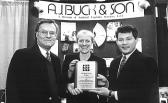


Ms. Constance T. Otley
Mr. & Mrs. H. Donald Pasquale
Ms. Elena T. Perri
Ms. Helen G. Perri
Mrs. June M. Pulido
Dr. & Mrs. William H. Ramsey
Mr. & Mrs. Dennis C. Reardon
Dr. & Mrs. R. Scott Scheer
Mr. & Mrs. Boyd Lee Spahr III.
Mr. & Mrs. Anthony Spallone
Ms. M. Kathryn Taylor
Dr. & Mrs. James A. Tatoian, Jr.
Mr. & Mrs. Thomas C. Test
Mr. & Mrs. Peter F. Waitneight
Linda & Floyd Walters
Mr. & Mrs. George Wausnock
Mr. & Mrs. Kenneth C. Weaver
Mr. & Mrs. Thomas Yeakle
The following gifts were made to New Bolton Centerin Memory of a beloved animal:
Dr. Edward Mersky in memory of “J.T.”
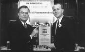
The following gifts were made to Friends of New Bolton Centerin memory of a beloved animal:
Ms. Lesley A. Davis Arnold in memory of “DAISY”
Mrs. Barbara M. Bauer in memory of “WOODSTOCK”
Ms. Carol L. Griffin in memory of “VESZTA”
Ms. Carroll N. Griffin & Ms. Mary G. Griffin in memory of “VESZTA”
Ms. Amy McKenna in memory of “MABEL”
Dr. Edward Mersky in memory of “ANNIE ROSE”
Agift was made to Laminitis Research in Memory of a special pet:
Mrs. Anne F. Thorington in memory of “MAPLE”
Agift was made to the Imaging/Heart Station Building in Memory of a special pet:
Mr. & Mrs. Keith Adams in memory of “MONTAGE”
27
DVM Pharmaceuticals,a sponsor.
A.J. Buck and Son,a patron.
Novartis Animal Health,a patron.
The Iams Company,a sponsor.
Join Us for the American Gold Cup
September 16-19 at the Devon Show Grounds,Devon,PA
Fourdays of Olympic calibershow jumping, culminating in the Gold Cup competition on Sunday, Sept. 19 at 2:30 PM.
Saturday, Sept. 18 is Family Day with activities for childrem, including an art show, a celebrity dog show and a M*A*S*Htent where injured, stuffed animals are brought back to health by Penn’s veterinary students. Children under 12 who bring a drawing or painting of their pet receive free admission that day.
Areserved seat, including admission to the show, is $5 for Thursday, $10 each Friday and Saturday and $20 on Sunday.
Admission and a reserved seat for children under 12 is $5 on Sunday. General admission for any day without a reserved seat is $5.
Calendar
August 4 to 7, 1999A Weekend in Old Saratoga, Saratoga, NY
August 7, 1999An Evening in Old Saratoga Gala, Saratoga, NY
To benefit New Bolton Center
September 16 to 19American Gold Cup, Devon show Grounds, Devon, PA
To benefit the School and New Bolton Center
Find
University of Pennsylvania
School of Veterinary Medicine
3800 Spruce Street
Philadelphia, PA19104-6008
Address correction requested
Bellwether is published by the School of Veterinary Medicine at the University of Pennsylvania.
EditorPhotographers
Helma WeeksAddison Geary
Writers Doug Thayer
Joan Capuzzi, V.M.D. New Bolton Liaison
Patricia ConnellyJeanie RobinsonDr. Josephine DeublerPownall
Mark Restey, V.M.D.
Jeannie Robinson-Pownall
We’d like to hear your praise, criticisms, or comments. Please address your correspondence to:
Helma Weeks, University of Pennsylvania School of Veterinary Medicine, 3800 Spruce Street, Philadelphia, PA 19104-6010; (215) 898-1475.
None of these articles are to be reproduced in any form without the permission of the editor of Bellwether.
©Copyright 1999 by the Trustees of the University of Pennsylvania.
The University of Pennsylvania values diversity and seeks talented students, faculty and staff from diverse backgrounds. The University of Pennsylvania does not discriminate on the basis of race, sex, sexual orientation, religion, color, national or ethnic origin, age, disability, or status as a Vietnam Era Veteran or disabled veteran in the administration of educational policies, programs or activities; admissions policies; scholarship and loan awards; athletic, or other University administered programs or employment. Questions or complaints regarding this policy should be directed to Anita Jenious, Executive Director, Office of Affirmative Action, 1133 Blockley Hall, Philadelphia, PA 19104-6021 or (215) 898-6993 (Voice) or (215) 8987803 (TDD).
Philadelphia, PA
Permit No. 2563
Printed on recycled paper.
Nonprofit Organization U.S. Postage PAID
™
the School on the internet at
www.vet.upenn.edu
45
45


