Since 1984, proton therapy has been used at the Center for Proton Therapy at PSI for the treatment of patients with eye tumors. The treatment is carried out using the OPTIS irradiation system.
The irradiation setup, known as OPTIS, involves a patient seated on the treatment chair. To prevent any head movement during the treatment, the head is secured with a plastic mask and a bite block.
We treat:
• Uveal melanomas
• Hemangiomas (vascular growths in the eye)
• Metastases in the eye
• Melanomas of the conjunctiva and iris
By the end of 2023, we at PSI had successfully treated over 8,000 patients with eye tumors. The results of proton irradiation for these tumors are excellent. In more than 98% of cases, tumor growth was definitively halted or the tumor was eliminated. In over 90% of cases, the affected eye was preserved.
Why Treat Eye Tumors with Protons?
Proton beams release their energy primarily where it is needed – within the tumor. This precision is achieved because the penetration depth of protons can be calculated accurately. There is
no radiation beyond the tumor. Additionally, the healthy tissue along the path from the surface of the body or the eye to the tumor is only slightly irradiated.
How Does Proton Therapy for Eye Tumors Work?
If you have been diagnosed with an eye tumor and your doctors have recommended proton therapy, here is what you can expect.
Preparations for the Treatment
Before the final decision for proton irradiation is made, all patients are thoroughly examined once more by a specialist.
Most patients come to us after an examination at the Jules Gonin Eye Hospital in Lausanne and are referred from there. However, patients also come from other clinics, such as the Eye Clinic in Innsbruck or the University Hospital Zurich Eye Clinic.
Once it is determined that proton therapy is the best treatment option, patients undergo a minor surgical procedure at their respective clinics. During this procedure, four to six small tantalum clips are sewn onto the eye under anesthesia. These clips make the tumor visible on an X-ray image, allowing for precise radiation planning.
Only after this marking procedure can millimeter-accurate radiation therapy be performed at the PSI Center for Proton Therapy.
The Jules Gonin Eye Hospital in Lausanne, where Dr. Ann Schalenbourg prepares patients for the treatment of eye tumors at the PSI Proton Therapy Center.
First Treatment Day at PSI
A few days after the tantalum clips are sewn onto the eye, patients arrive at PSI for a detailed briefing on the treatment procedure and to plan the therapy.
The radio-oncologist responsible for eye treatments at the PSI Center for Proton Therapy is Dr. Alessia Pica.
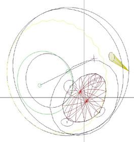
Geometric model of the eye and the tumor for the purpose of treatment planning. On the left, a view from the radiation perspective; on the right, a side view.
Collimators are apertures custom-made at PSI. They are mounted in an apparatus before the treatment, through which the proton beam is directed to the eye. The aperture, shaped to match the tumor, ensures that the proton beam only targets the eye tumor and spares the surrounding healthy tissue. At PSI this technique is specifically used for eye tumors.
After an initial discussion with the treating radio-oncologist,the radiation therapists make a custom plastic face mask and a bite block with a dental impression for each patient. These are used to immobilize and stabilize the patient’s head during the week of therapy.
Subsequently, X-ray images of the affected eye are taken. These images are needed for therapy planning and to align the patient in front of the irradiation device before each treatment. The planning images are taken from three to four different eye directions. To ensure the eyeball is not covered by the eyelid, lid retractors are used. At this point, the patient’s first day at PSI is complete.
Before the actual start of irradiation, a few days later, additional important planning preparations are carried out in the absence of the patient. This includes the creation of treatment
plans by our clinical medical physicists. The plans are developed based on instructions from the radio-oncologists, discussed with the ophthalmologists from the referring clinics, and modified as necessary. A treatment plan specifies, among other things, how the eye should be positioned to ensure the beam optimally targets the tumor, for example, avoiding the iris whenever possible, and the appropriate radiation dose.
Preparation for patient treatment also includes the fabrication of collimators. These are metal apertures shaped to match the size and form of the tumor. The collimators are custom-made for each individual patient in the workshops at PSI.
Collimator aperture
Crosshair for indicating the rotation axis
Tumor
“Clips” Optical nerv
Tumor
Conducting the Irradiation
The following week, the series of irradiation sessions begins at PSI, which is usually performed on an outpatient basis and consists of 4 or 5 treatments on consecutive days.
On the first day of the treatment week (usually a Monday), a test session or simulation is performed, which lasts about 20 minutes. The simulation checks whether the patient’s positioning and the treatment setup work exactly as planned. If adjustments are needed, the simulation may take longer, so patients should allow for this in their schedules.
During the simulation, lid retractors are again used, and the mask and bite block are applied, checked, and adjusted as necessary. Additional X-ray images are taken, and final corrections are made. Once this test session is complete, the patient can leave.
Clinical medical physicists at PSI review the treatment plan one last time to ensure that everything is ready for the next day.
The first actual irradiation session begins on Tuesday. Each treatment session, including preparation and follow-up, lasts about 20 minutes. The patient is seated on the treatment chair, and the mask, bite block, and lid retractors are applied. His or her eye is positioned with millimeter precision in front of the opening from which the proton beam is emitted. X-ray images are taken again to verify correct positioning. The actual radiation lasts between 40 and 60 seconds. The typ-
ical daily dose is 15 Gy (RBE), and the treatment is administered over four consecutive days, for a total dose of 60 Gy (RBE).
During the irradiation, the patient must fix his or her gaze on a yellow light spot. The patient’s position is continuously monitored using cameras both in the treatment area and in the control room.
The radiation process is painless. After each session, the patient can return home or to his or her accommodation. Patients are provided with instructions on how to care for their eye, which includes a flyer titled “Eye Care.”
The entire treatment duration, including the initial surgery, is usually around two to three weeks. Follow-up examinations are conducted at the clinics that referred the patients to PSI’s Center for Proton Therapy.
During the irradiation, the patient must look at a light spot with the affected eye (visible here on the long left pointer). This ensures that the proton beam precisely targets the tumor. The patient’s gaze is continuously monitored using multiple cameras to confirm correct positioning. In the center, you can see the copper-colored collimator, which is shaped to match the tumor.
PSI in brief
The Paul Scherrer Institute PSI develops, builds and operates large, complex research facilities and makes them available to the national and international research community. The institute’s own key research priorities are in the fields of future technologies, energy and climate, health innovation and fundamentals of nature. PSI is committed to the training of future generations. Therefore about one quarter of our staff are post-docs, post-graduates or apprentices. Altogether PSI employs 2300 people, thus being the largest research institute in Switzerland.