Surgical Excision of Bilateral Xanthelasma Palpebrarum: Clinical Insights and Definitive Surgical Management
Intralesional immunotherapy by Vitamin D3 for treatment of Periungual Warts: A Case Report
EXECUTIVE EDITOR & PUBLISHER
Dom Daniel CORPORATE OFFICE
22, Shreeji Bhavan, 275-279, Samuel Street, Masjid Bunder (W), Mumbai-4000 03, INDIA.
EMAIL: info@residerm.com
TEL: + 91 98209 81556
Printed, Published, Edited and Owned by Dom Daniel Printed at Swastik Printer, Gala No.9 & 10, Vishal Industrial Estate, Bhandup (West), Mumbai- 400078. Published at 22 Shreeji Bhavan, 275/279, Samuel Street, Masjid Bunder (West), Mumbai - 400003. India.
“Residerm ” takes no responsibility for unsolicited photographs or material
ALL PHOTOGRAPHS, UNLESS OTHERWISE INDICATED, ARE USED FOR ILLUSTRATIVE PURPOSE ONLY.
Views expressed in this Journal are those of the contributors and not of the publisher. Reproduction in whole or in parts of texts or photography is prohibited. Manuscripts, Photographs and art are selected at the discretion of the publisher free of charge (advertising excluded). Whether published or not, no material will be returned and remains the property of the publishing house, which may make use of it as seen fit. This may include the withdrawal of publication rights to other publishing houses.
All rights reserved. Reproducing in any manner without prior written permission prohibited.
Published for the period of February 2026
FROM CLINICAL OBSERVATION TO ACADEMIC INSIGHT
We are pleased to present the latest issue of Residerm, a journal committed to fostering academic curiosity, critical thinking, and clinical competence among dermatology residents. As dermatology continues to evolve with advances in diagnostics and therapeutics, the importance of sound clinical judgment grounded in real-world experience remains paramount. This issue emphasizes the pivotal role of well-documented clinical case presentations in sharpening diagnostic accuracy, refining therapeutic decision-making, and broadening overall clinical perspective.
The current edition features carefully curated case reports that highlight both surgical and immunotherapeutic approaches in contemporary dermatologic practice. A detailed account of bilateral xanthelasma palpebrarum managed with surgical excision provides valuable insights into definitive management, surgical planning, and outcome-oriented care, reinforcing the relevance of procedural dermatology in everyday practice. Complementing this, a case report on intralesional vitamin D3 immunotherapy for periungual warts explores an emerging, minimally invasive treatment modality, reflecting the growing interest in immunotherapeutic strategies for recalcitrant cutaneous conditions.
Together, these contributions underscore the importance of evidence-based practice and clinical innovation. They encourage residents to engage actively in scholarly pursuits through meticulous observation, thoughtful documentation, and critical analysis of clinical encounters. We hope this issue inspires young dermatologists to transform routine patient care into meaningful academic learning and to cultivate a lifelong commitment to clinical excellence and research-oriented thinking.
We look forward to your contributions for the next edition.
Hope you have a great read!
Thanks & Cheers
- Dom Daniel Executive Editor & Publisher
Surgical Excision of Bilateral Xanthelasma Palpebrarum: Clinical Insights and Definitive Surgical Management
Dr. Shantanu Gupta
3rd Year Resident (2025)
Department of Dermatology, Venereology and Leprosy Government Medical College, Surat
Dr. Yogesh Patel
MD (Dermatology)
Associate Professor
Department of Dermatology, Venereology and Leprosy Government Medical College, Surat
Intralesional immunotherapy by Vitamin D3 for treatment of Periungual Warts: A Case Report
Dr. Amit Murkute
MBBS, DDVL, DNB
Consultant Dermatologist, Cosmetologist and Hair Transplant Surgeon Founder of Elite Skin and Hair Clinic, Pune
Surgical
Surgical Excision of Bilateral Xanthelasma Palpebrarum: Clinical Insights and Definitive Surgical Management
Dr. Shantanu Gupta
3rd Year Resident (2025)
Department of Dermatology, Venereology and Leprosy
Government Medical College, Surat
Dr. Yogesh Patel
MD (Dermatology)
Associate Professor
Department of Dermatology, Venereology and Leprosy
Government Medical College, Surat
Introduction
Xanthomas are localized accumulations of lipidladen foam cells in the skin, tendons, fascia, and occasionally the periosteum, arising from macrophages that uptake lipoproteins or phagocytose aggregated LDL particles. Clinically, they appear as soft to firm macules, papules, plaques, or nodules with a yellow to yellow-orange
hue, reflecting deposited lipids. Their development is closely linked to systemic lipid metabolism disorders, often paralleling early atherosclerotic processes. Xanthomas commonly indicate inherited or acquired dyslipidemias, including familial hypercholesterolemia........ (LDLR mutations), familial defective apolipoprotein B-100 (APOB mutations),
Surgical Excision of Bilateral Xanthelasma Palpebrarum: Clinical Insights and Definitive Surgical Management
PCSK9-related ................ hypercholesterolemia, ...... severe hypertriglyceridemia, and primary dysbetalipoproteinemia. ... They may also signal rarer metabolic disorders such as cerebrotendinous xanthomatosis (CYP27A1 mutations) or familial β-sitosterolemia (ABCG5/ ABCG8 mutations). Beyond indicating lipid dysregulation, xanthomas can reflect increased risk for metabolic syndrome, premature cardiovascular disease, and, occasionally, lymphoproliferative ......... malignancies.1
Xanthomas are clinically classified into several types, including xanthoma eruptivum, tuberosum, tendineum, planum, striatum palmare, disseminatum, xanthelasma palpebrarum (XP), and necrobiotic xanthogranuloma. XP is the most common form, presenting as yellowish, cholesterol-rich deposits predominantly affecting the medial aspects of the eyelids. The pathogenesis involves lipid accumulation within dermal macrophages, which transform into foam cells when intracellular lipid uptake exceeds metabolic
capacity. Repetitive blinking generates mechanical stress on eyelid capillaries, promoting plasma and lipid extravasation into the superficial dermis, a process exacerbated by agerelated endothelial fragility. Macrophages recruited to these sites internalize lipids via specific lipoprotein receptors or phagocytosis of LDL aggregates and immune-complexed lipids. While native LDL is tightly regulated and does not typically induce foam cell formation, chemically modified LDL, particularly oxidized LDL (ox-LDL), is avidly taken up by macrophages through scavenger receptors (SRA, SR-B1, CD36, LOX1), which lack feedback inhibition, leading to unregulated intracellular lipid accumulation. Minimally modified LDL (mm-LDL), formed during progressive oxidation, and further enhances macrophage recruitment and uptake. Additional factors contributing to foam cell formation include elevated local lipid concentrations, structurally abnormal lipoproteins, increased vascular permeability or
regional blood flow, in situ lipid synthesis, and impaired reverse cholesterol transport. Persistent lipid accumulation results in cytoplasmic lipid droplet formation and foam cell development, with chronic low-grade inflammation further promoting lesion progression. While XP is frequently associated with systemic dyslipidemias, it may also occur in normolipidemic individuals, reflecting a combination of local vascular, mechanical, cellular, and inflammatory factors.1, 2
Xanthelasma palpebrarum can significantly affect patient quality of life due to its highly visible periocular location, often leading to cosmetic dissatisfaction, reduced self-esteem, and social embarrassment. The persistent and progressive nature of the lesions may heighten psychosocial distress, including anxiety about facial appearance and concerns regarding perceived stigmatization. Although asymptomatic, the condition can interfere with routine grooming and may provoke additional worry when associated with underlying lipid
Surgical Excision of Bilateral Xanthelasma Palpebrarum: Clinical Insights and Definitive Surgical Management
abnormalities. As a result, recognizing the psychological and social impact of xanthelasma is essential for guiding patient-centered counseling and selecting appropriate treatment to improve overall well-being.
Case Report
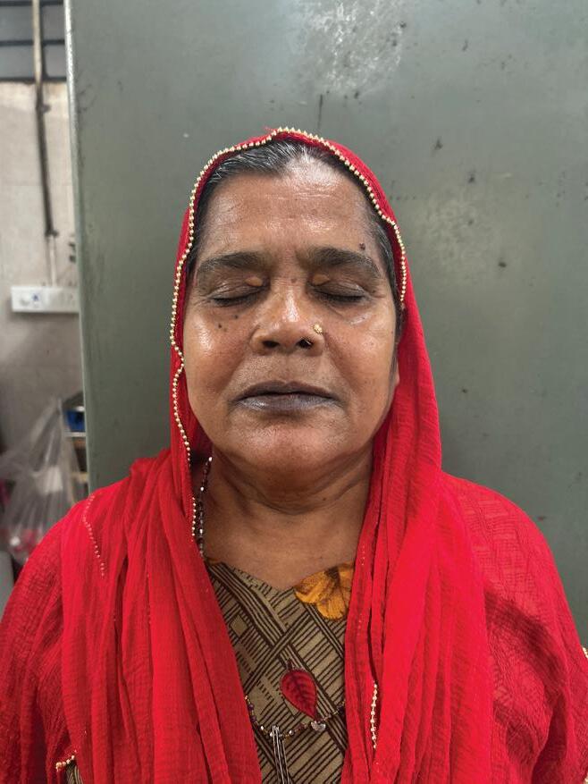
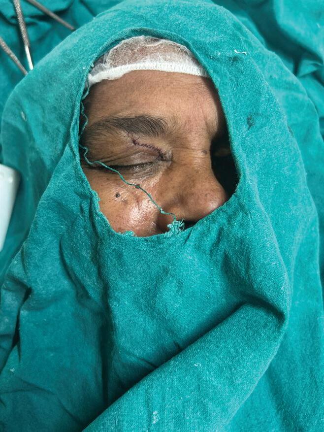
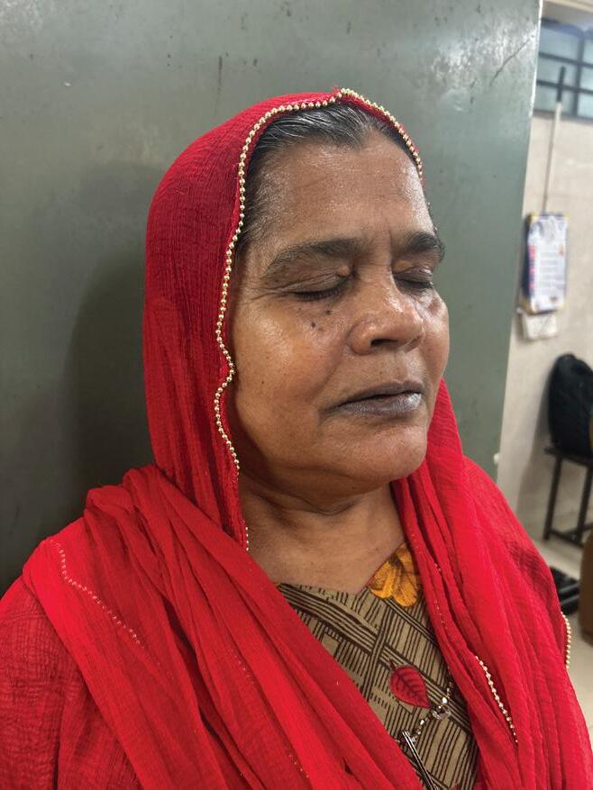
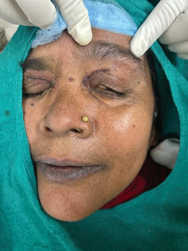
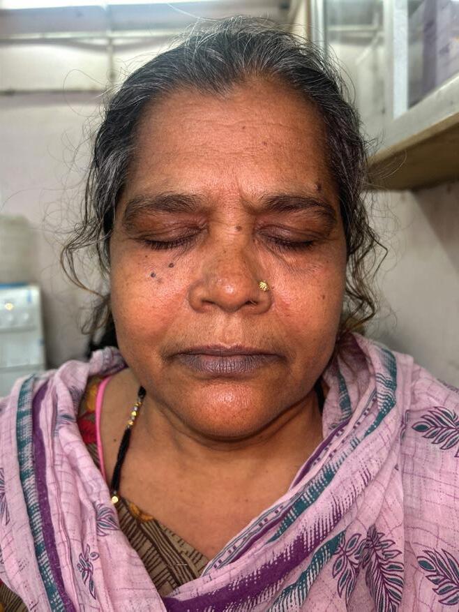
A 60-year-old female presented with soft, yellowish papular lesions on the medial aspects of the upper eyelids, clinically consistent with xanthelasma palpebrarum. The lesions were slowly progressive, asymptomatic, and caused no visual impairment; a systemic evaluation for dyslipidemia was advised. Surgical excision was chosen to provide a definitive, cosmetically favourable outcome. Precise incisions were placed along the natural eyelid creases, followed by meticulous dissection and removal of the lipid-laden plaques while preserving the orbicularis oculi and eyelid margin. The wound edges were approximated with fine sutures to promote optimal healing. Postoperative review revealed complete lesion resolution, a smooth eyelid contour without notching
or deformity, and excellent cosmetic results with scars nearly imperceptible within natural folds. The patient reported high satisfaction, and no early recurrence was noted.
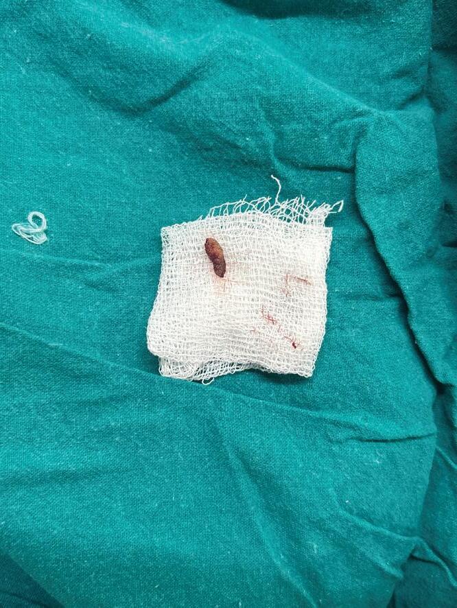
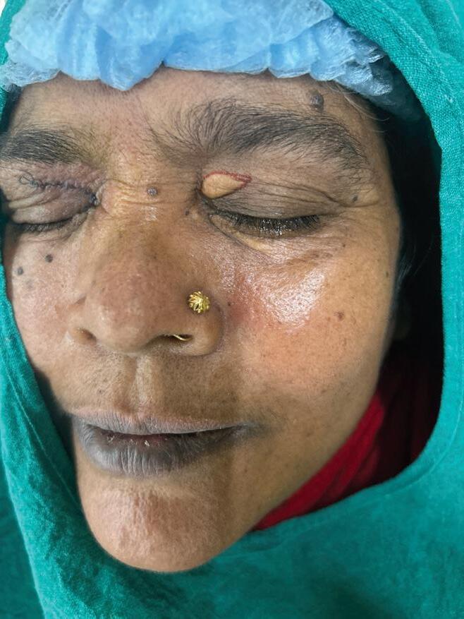
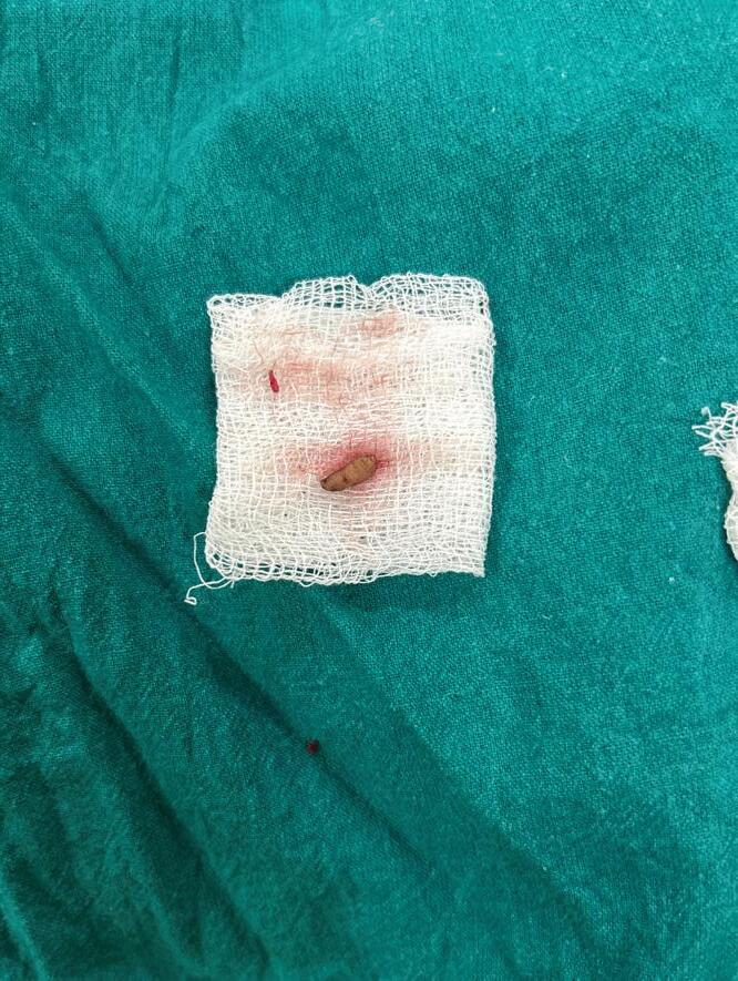
2 (a): Pre-treatment clinical photograph showing xanthelasma palpebrarum involving the right upper eyelid. (b): Intraoperative view of the right upper eyelid following surgical excision of xanthelasma palpebrarum. (c): Excised xanthelasma palpebrarum tissue specimen after surgical removal.
Figure
Figure 1: Soft, yellowish papular lesions on the medial aspects of the upper eyelids consistent with xanthelasma palpebrarum
Figure 2 (a)
Figure 2 (c)
Figure 2 (b)
Surgical Excision of Bilateral Xanthelasma Palpebrarum: Clinical Insights and Definitive Surgical Management
3 (a): Pre-treatment
clinical photograph showing xanthelasma palpebrarum involving the left upper eyelid. (b): Intraoperative view of the left upper eyelid showing marked xanthelasma palpebrarum prior to surgical excision.
(c): Excised xanthelasma palpebrarum tissue specimen from the left upper eyelid.
Figure 4: Postoperative follow-up showing complete excision of xanthelasma palpebrarum
Diagnosis
The diagnosis of xanthelasma palpebrarum is primarily clinical, based on recognition of its characteristic yellowish, soft plaques on the eyelids and their typical distribution. Careful clinical examination is usually
sufficient for confirmation. Lipid profile testing serves as an important adjunct, as xanthelasma may be associated with underlying dyslipidemia. A fasting lipid panel—including total cholesterol, LDL-C, HDL-C, and triglycerides— is recommended to identify abnormalities such as hypercholesterolemia or hypertriglyceridemia, while acknowledging that a significant proportion of patients may have normal lipid levels. Detection of dyslipidemia aids in systemic and metabolic risk assessment and helps guide long-term management.3
Histopathologically, ........ xanthelasma palpebrarum is characterized by aggregates of lipidladen histiocytes, known as foam cells, located predominantly in the upper reticular dermis, often in perivascular and periadnexal distributions. These cells contain intracytoplasmic vacuoles filled with esterified cholesterol, forming the hallmark of the lesion.2 Dermoscopy typically demonstrates homogeneous or lobulated yellow to yellow-white structureless areas corresponding to dermal
Figure
Figure 3 (a)
Figure 3 (b)
Figure 3 (c)
foamy histiocytes, along with whitish septa representing fibrous strands.4
Accurate diagnosis relies on integrated clinical assessment to ensure appropriate management.
Treatment
Surgical excision of xanthelasma palpebrarum (XP) represents a definitive intervention aimed at complete removal of cholesterol-laden deposits within the superficial dermis, with meticulous attention to preserving eyelid structure and optimizing cosmetic outcomes. The procedure is individualized based on lesion size, depth, location, and the patient eyelid anatomy. Preoperatively, the lesion margins and natural eyelid crease are precisely delineated, followed by infiltration of a local anesthetic solution combined with a vasoconstrictor to provide effective analgesia and hemostasis.5, 6
A general surgical procedure for xanthelasma palpebrarum excision begins with sublesional infiltration of local anesthesia, followed by an adequate waiting period for complete anesthetic effect.
A precise incision is made along the superior and inferior margins of the lesion to expose the underlying orbicularis oculi muscle. The overlying skin is elevated and excised in an uncapping manner, allowing clear visualization of the lesion. Careful dissection is performed to separate the xanthelasma from adjacent dermal structures, after which the lipid-rich core is removed from the surface of the orbicularis muscle. Under magnification, residual foam cells adherent to the skin flap are meticulously excised to ensure complete clearance, and any localized infiltration of the orbicularis muscle is removed to restore normal tissue integrity. Because of decompression achieved during un-capping, the wound generally closes without tension. Layered closure is then performed using fine sutures, with a light pressure dressing applied to maintain flap adherence and support optimal postoperative healing. The process of surgical excision involves the physical removal of lipid-laden macrophages and associated dermal deposits, thereby
eliminating the pathological substrate responsible for lesion formation. Complete excision interrupts the cycle of lipid deposition and tissue inflammation, significantly lowering recurrence rates.5, 6
In cases of larger lesions or when aesthetic correction is necessary, advancement or rotation of musculocutaneous flaps is employed, sometimes in combination with upper eyelid blepharoplasty techniques, to restore eyelid contour, maintain natural fold height, and prevent excessive tension or distortion. Hemostasis is achieved using light cautery, and layered closure with fine sutures ensures precise skin-edge approximation, minimizing scar formation and preserving functional eyelid dynamics.5, 6
Postoperatively, patients were advised to keep the surgical site clean and dry, ensuring proper hygiene to prevent infection and support optimal wound healing. Regular monitoring for signs of inflammation or complications was recommended. Overall, surgical excision remains
Surgical Excision of Bilateral Xanthelasma Palpebrarum: Clinical Insights and Definitive Surgical Management
the treatment of choice for extensive, recurrent, or deep-seated xanthelasma lesions, offering a combination of functional restoration and durable aesthetic outcomes when performed with precision.5, 6
Other treatments for xanthelasma palpebrarum (XP) include systemic, topical, and energy-based modalities, each targeting the lipid-laden dermal deposits through distinct mechanisms. Systemic therapy comprises agents such as probucol, an antioxidant that may inhibit atherogenesis by limiting oxidative modification of LDL cholesterol and thereby reducing foam-cell formation, and alirocumab, a monoclonal antibody against Proprotein Convertase Subtilisin/ Kexin Type 9 (PCSK9), primarily indicated for hypercholesterolemia........ management and potentially decreasing the substrate for lesion development. Topical therapies involve controlled destructive or cytotoxic approaches, including chemical peels such as trichloroacetic acid (TCA), which induce coagulation
of epidermal and superficial dermal layers, liquid nitrogen cryotherapy, which promotes vasoconstriction and microthrombi formation leading to tissue ischemia and selective cell necrosis, and intralesional pingyangmycin, a broadspectrum antitumor antibiotic utilized to target foam cells directly and promote lesion regression. Energy-based devices offer precise, minimally invasive treatment options, including radio-frequency ablation, which delivers controlled thermal energy to disrupt foam cells and remodel tissue, and various laser modalities. CO2 lasers, considered the goldstandard ablative device, vaporize cellular water layer by layer to achieve precise lesion removal, whereas Er:YAG lasers provide purely ablative effects with smaller thermal coagulation zones, faster healing, and reduced postinflammatory hypoor hyperpigmentation. Additional laser options include Q-switched Nd:YAG lasers, argon lasers for lesion coagulation, potassium titanyl phosphate (KTP) lasers employing selective photothermolysis,
pulsed dye lasers, and diode lasers, historically used for photothermal destruction of mid-dermal sebaceous structures.7
These approaches allow tailored XP treatment, optimizing efficacy while minimizing complications and preserving aesthetics.
Discussion
Xanthelasma palpebrarum (XP) is the most common cutaneous xanthoma, characterized by soft, yellowish plaques located predominantly on the medial aspects of the upper and lower eyelids. It occurs in approximately 1.1% of women and 0.3% of men, most frequently between 35 and 55 years of age, and often presents symmetrically. The differential diagnosis of XP is broad and requires careful clinical assessment due to overlap with various dermatologic and systemic conditions that may mimic its appearance. Sebaceous hyperplasia may resemble XP but typically presents as small, umbilicated papules rather than flat plaques. Juvenile xanthogranuloma may appear as yellowish lesions but is usually solitary, dome-shaped,
and primarily affects children. Nodular basal cell carcinoma, though occasionally yellowish, often demonstrates telangiectasia, pearly borders, or ulceration, warranting exclusion due to its malignant potential. Adult-onset asthma and periocular xanthogranuloma (AAPOX) presents with indurated, infiltrated yellow-brown plaques accompanied by systemic features, while palpebral sarcoidosis may show firm, violaceous papules or plaques with granulomatous inflammation. Lipoid proteinosis produces waxy yellow eyelid lesions but is associated with hoarseness and mucocutaneous infiltration. Necrobiotic xanthogranuloma presents as indurated, often ulcerated plaques associated with paraproteinemia and systemic involvement. Distinguishing these conditions is essential, as several carry significant systemic implications requiring targeted evaluation and management.2
Xanthelasma palpebrarum is generally benign and does not directly cause medical complications;
however, the lesions may be cosmetically concerning for patients. Treatment-related adverse effects can include pain, erythema, scarring, and pigmentary alterations, with similar incidence across different therapeutic modalities. Cryotherapy and chemical cauterization may result in significant scarring and pigmentary changes. Given the frequent association of xanthomas with dyslipidemia, affected individuals have an increased risk of cardiovascular events, including myocardial infarction, ischemic heart disease, advanced atherosclerosis, and other potentially life-threatening outcomes.2
Among available therapeutic modalities, surgical excision remains the most definitive treatment for XP, particularly for welldemarcated, large, deep, or recurrent lesions. Surgical management allows direct removal of lipidladen macrophages and dermal deposits, offering immediate correction. Incisions placed along natural eyelid creases yield superior cosmetic
outcomes, and the excision can be combined with aesthetic procedures such as blepharoplasty when indicated. Unlike chemical or energybased treatments, surgery provides controlled removal of the entire lesion burden, reducing the likelihood of incomplete clearance. Patient education for XP should emphasize its benign nature and association with dyslipidemia and cardiovascular risk, highlighting the importance of lipid evaluation, lifestyle modification, and medical management when indicated. Counseling on the likelihood of recurrence and expected outcomes supports informed decision-making. Prognosis is generally excellent, with significant cosmetic improvement achievable, and long-term results are optimized when systemic risk factors, particularly lipid abnormalities, are addressed.2
Conclusion
Xanthelasma palpebrarum, while benign, poses cosmetic concerns and signals increased cardiovascular risk due to lipid abnormalities. Surgical excision is the preferred
approach, offering precise removal, immediate aesthetic improvement, and durable results. It allows controlled clearance with preservation of eyelid anatomy and can be combined with adjunctive procedures for extensive lesions. Patient education, lipid assessment, and lifestyle modification are essential to optimize both cosmetic and systemic outcomes.
Reference
1. Zak A, Zeman M, Slaby A, Vecka M. Xanthomas: clinical and pathophysiological relations. Biomed Pap Med Fac Univ Palacky Olomouc Czech Repub. 2014; 158(2):181-188. doi:10.5507/bp.2014.016.
2. Al Aboud AM, Shah SS, Blair K, Al Aboud DM. Xanthelasma Palpebrarum. In: StatPearls. Treasure Island (FL): StatPearls Publishing; March 1, 2024
3. Kavoussi H, Ebrahimi A, Rezaei M, Ramezani M, Najafi B, Kavoussi R. Serum lipid profile and clinical characteristics of patients with xanthelasma palpebrarum. An Bras Dermatol. 2016; 91(4):468471. doi:10.1590/abd18064841.20164607.
4. Bhogar K, Ritu K. Role of ultrasound biomicroscopy assessment in xanthelasma
Surgical
palpebrarum. Oman J Ophthalmol. 2025; 18(2):257258. Published 2025 Jun 24. doi:10.4103/ojo.ojo_94_24.
5. Lin Y, Wu L. Aesthetic Surgical Treatment of Large Xanthelasma palpebrarum. Aesthetic Plast Surg. 2024; 48(23):4828-4832. doi:10.1007/s00266-02404435-x.
6. Lee HY, Jin US, Minn KW, Park YO. Outcomes of surgical management of xanthelasma palpebrarum. Arch Plast Surg. 2013; 40(4):380-386. doi:10.5999/aps.2013.40.4.380
7. Laftah Z, Al-Niaimi F. Xanthelasma: An Update on Treatment Modalities. J Cutan Aesthet Surg. 2018; 11(1):1-6. doi:10.4103/JCAS. JCAS_56_17.
Intralesional immunotherapy by Vitamin D3 for treatment of Periungual Warts: A Case Report
Intralesional immunotherapy by Vitamin D3 for treatment of Periungual Warts: A Case Report
Dr. Amit Murkute
MBBS, DDVL, DNB
Consultant Dermatologist,
Cosmetologist and Hair Transplant Surgeon
Founder of Elite Skin and Hair Clinic, Pune
Introduction
Viral warts, commonly known as verrucae or papillomas, are benign lesions caused by infection with human papillomavirus (HPV). They can be categorized as cutaneous or mucosal depending on the HPV types involved.1 The clinical presentation of viral warts encompasses various characteristic features depending on their location and type. Warts commonly appear as raised, rough lesions on the skin, ranging from small papules to larger, cauliflower-like growths. They can be skin-colored, white, pink, or tan, often with a rough, scaly surface.
Common sites for warts include the hands, fingers, feet (especially on pressure points like soles), and around nails. Warts can also occur on other body areas such as the face, knees, and elbows. Warts may present single or in clusters, with growth rates that can be slow or rapid. The clinical spectrum of cutaneous warts includes, verruca vulgaris (common warts) with hyperkeratotic, exophytic, dome-shaped papules or nodules found on fingers, hands, knees, elbows, or areas prone to trauma. Flat warts (plane warts) flat-topped papules with minimal scaling and slight elevation, typically
2-4 mm in diameter. Intermediate warts that exhibit features of both common warts and flat warts. Plantar warts with painful, callused, endophytic papules on the soles of the feet with deep sloping sides and a central depression. Mosaic warts refer to coalesced warts forming a tile-like pattern on the plantar surface. Filiform warts with frondlike projections, often on the face and characterized by rapid growth. Periungual warts occurring along nail margins, including the proximal nail fold and hyponychium, potentially causing nail matrix damage and onycholysis. Anogenital warts (condyloma acuminata) found in perineal and genital areas, commonly transmitted sexually and vertically to neonates from infected mothers. Oral warts with small pink or white papules on the oral mucosa. Individuals with compromised skin barriers, such as those with dermatitis, or those undergoing immunosuppressive ....... therapy or living with HIV
Intralesional immunotherapy by Vitamin D3 for treatment of Periungual Warts: A Case Report
infection, are particularly susceptible to developing warts.2
The pathophysiology of viral warts, caused by various human papillomavirus (HPV) types, involves key steps in their development and persistence. HPV, a doublestranded DNA virus with over 150 known types, infects the skin through microabrasions, targeting keratinocytes in the basal epidermal layer. Once inside, it replicates its circular DNA using host cell machinery and produces viral proteins essential for replication and pathogenesis. By residing within keratinocytes, HPV evades immune surveillance and alters host immune responses. Infected cells undergo hyperproliferation, forming the hyperkeratotic tissue characteristic of warts. Common causative types include HPV 1, 2, 3, 4, 10, 27, 29, and 57. Diagnosis is typically straightforward for clinicians due to their distinctive appearance, and management is tailored to lesion site and characteristics to achieve complete resolution and prevent recurrence. Intralesional administration of vitamin D3 has recently emerged as a novel and effective immunotherapeutic strategy for the treatment of verrucae.1,3
Case report
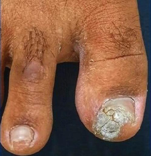
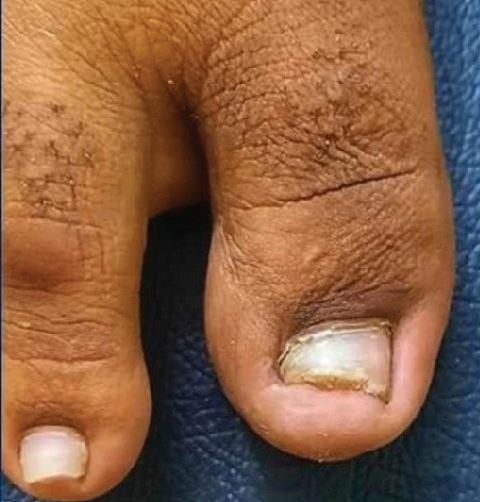
A 34-year-old male presented with persistent periungual viral warts—rough, cauliflower-like plaques encircling the toes—present for several months prior to consultation. Clinical diagnosis was established based on the characteristic morphology and location of the lesions. The treatment plan comprised intralesional Vitamin D3 immunotherapy (60 lakh IU), with 3–4 units injected directly into each wart every two weeks, for a total of four sessions. The lesions resolved completely, with no recurrence observed during follow-up. The patient reported high satisfaction with the results, noting both the effectiveness and convenience of the therapy. This case highlights the efficacy of intralesional Vitamin D3 as a safe, effective, and well-tolerated option for the prompt clearance of periungual viral warts.
Before treatment
After treatment
Diagnosis
Diagnosing viral warts primarily involves visual inspection of their characteristic appearance—raised, ..... rough-textured growths ranging from small papules to larger nodules, often flesh-colored, pink or darker than surrounding skin. They commonly affect fingers, hands, knees, elbows, face, and plantar surfaces of the feet. Dermatoscopy aids diagnosis by revealing specific vascular patterns and surface textures, while biopsy is reserved for atypical cases. Differential diagnosis is important to distinguish warts from other lesions. Histopathology confirms HPV infection through features such as hyperkeratosis, acanthosis, papillomatosis, and koilocytes. Swabbing, scraping, PCR and Southern blot hybridization can detect HPV DNA and guide management. Accurate diagnosis relies on clinical evaluation supported by supplementary tools to ensure effective treatment and better outcomes.3,4,5,6
Treatment
The therapeutic approach
Figure 1: Periungual viral warts characterized by rough, cauliflower-like plaques around the toe
to viral warts is tailored to wart type, location, and patient needs, aiming for symptom relief, clearance, and prevention of HPV transmission. Topical and intralesional options include imiquimod 5% cream (stimulates cytokine production for immune clearance), salicylic acid (keratolytic that softens tissue and enhances clearance), and bleomycin (induces DNA strand breaks in recalcitrant lesions). Interferon alfa inhibits viral replication and promotes immune recognition; 5-fluorouracil (5-FU) disrupts DNA synthesis in resistant or widespread warts; cidofovir inhibits viral DNA polymerase in HPV-resistant cases; and retinoids reduce hyperkeratosis and impair viral replication. Procedural options include electrodessication, cryotherapy (destroys tissue, triggers local immunity) and laser therapy. Photodynamic therapy (PDT) with 5-aminolevulinic acid (ALA) is also effective. Selection is based on lesion characteristics and patient preference to maximize efficacy and reduce recurrence.7,8
Intralesional immunotherapy by Vitamin D3 for treatment of Periungual Warts: A Case Report
Intralesional immunotherapy with Vitamin D3 is a targeted approach for managing challenging viral warts, such as periungual warts, mosaic plantar warts, or multiple difficultto-treat lesions. This method involves injecting therapeutic agents directly into the wart lesions to enhance the local immune response and promote wart clearance. Vitamin D3 (60 lakh IU) is a promising agent in this therapy, although other substances like interferon and various immunomodulators are also utilized. The procedure begins with a thorough patient evaluation, including history and physical examination to confirm the diagnosis and assess wart characteristics, followed by treatment planning to determine the suitability of the therapy. During the injection procedure, local anesthesia may be utilized to minimize discomfort, employing either a topical anesthetic or local nerve block as required. A maximum of six injections can be administered into each wart at two-week intervals. The therapeutic agent is
injected directly into the wart using a fine-gauge needle, ensuring thorough distribution throughout the lesion and adjusting the volume according to the size and number of warts. Intralesional therapy, including the use of Vitamin D3, has demonstrated efficacy in clearing warts and reducing recurrence rates due to its ability to deliver high local concentrations of the therapeutic agent, thereby enhancing treatment efficacy while minimizing systemic effects. The therapy works through direct cytotoxic effects on wart tissue, with agents like Vitamin D3 targeting wart cells and interferon inhibiting HPV replication. Additionally, it stimulates a localized immune response by enhancing T-cell and macrophage function and activating dendritic cells for effective antigen presentation and T-cell activation. The localized inflammation induced by the injection helps draw immune cells to the site, aiding in the recognition and destruction of HPVinfected cells. By increasing the expression
of viral antigens on infected cells, the therapy enhances antigen presentation, strengthens immune memory against HPV, and reduces the likelihood of recurrence. Overall, intralesional therapy effectively combines direct cytotoxic effects and localized immune stimulation to eradicate viral warts, offering a focused treatment option, especially for cases resistant to standard therapies.9
Discussion
Human papillomaviruses (HPVs) cause chronic, often asymptomatic infections in humans and some animals. Beta and Gamma types usually remain subclinical, shedding virions without disease, while certain Alpha types cause visible papillomas through immune evasion. Viral proteins E6, E7, and E5 disrupt host cell cycle control, with high-risk types like HPV 16 and 18 promoting neoplastic transformation. HPV-induced warts are frequently persistent or recurrent, with 51.7% of patients reporting moderate to severe discomfort and 38.8% experiencing
social or leisure limitations. Warts affect both sexes, especially children aged 2–12 years, and rank among the top three dermatoses treated. Spontaneous regression occurs in ~23% within 2 months, 30% within 3 months, and 65–78% within 2 years. Recurrence risk depends on subtype, immune status, and lesion size/duration. Risk factors include occupational meat handling, communal showers or pools, and close contact with affected individuals, while immunocompromised and atopic patients are more susceptible. Preventive measures— with limited evidence— include covering warts while swimming, avoiding shared items, keeping feet dry, and maintaining good hygiene. HPV vaccination (quadrivalent or nanovalent) protects against common wart-causing types and is advised for adolescents before sexual activity and unvaccinated adults to reduce infection, transmission, and overall burden.8,10
Numerous treatment
modalities exist for cutaneous warts, but many have notable limitations. Traditional destructive procedures—such as electrocoagulation, .......... cryotherapy, laser surgery, 5-fluorouracil, and salicylic acid—are widely used but often cause scarring, high recurrence, and discomfort. They can be cumbersome, costly, and time-consuming, with effects largely confined to the treated site, limiting their use for multiple lesions. Immunotherapy offers a promising alternative, effective for solitary, recalcitrant, and recurrent warts. It works by enhancing immune recognition of viral antigens in wart tissue and triggering a delayed-type hypersensitivity response to eradicate human papillomaviruses (HPVs). Intralesional administration of vitamin D3 has recently emerged as a promising and effective immunotherapeutic option for the treatment of warts. Its mechanism of action includes the regulation of epidermal cell proliferation and differentiation, as well as the modulation
of cytokine production. Furthermore, vitamin D3 activates Toll-like receptors, resulting in the upregulation of human macrophages and the expression of vitamin D receptors (VDR) and vitamin D1-hydroxylase genes, which contribute to the synthesis of antimicrobial peptides. A growing body of evidence supports the efficacy of intralesional vitamin D3 in the treatment of cutaneous warts, underscoring its potential as a viable therapeutic option in clinical practice.7
Conclusion
Intralesional vitamin D3 injection immunotherapy offers notable advantages in managing warts, especially those resistant to conventional treatments. By enhancing local immune responses and stimulating immune effector cells, this therapy promotes effective clearance of wart lesions and induces antiviral peptides targeting human papillomavirus (HPV). Patients experience reduced pain and quicker recovery, leading to improved satisfaction and compliance. For clinicians,
Intralesional immunotherapy by Vitamin D3 for treatment of Periungual Warts: A Case Report
this approach broadens treatment options and allows for personalized care, particularly in complex cases. Ongoing research into the immunomodulatory effects and optimal protocols for vitamin D3 will further enhance its efficacy, positioning it as a key strategy in the management of viral warts and the overall improvement of patient quality of life.
References
1. Kuriyama Y, Kosaka M, Kaneko A, et al. Skin surface material for detecting human papillomavirus infection of skin warts. J Dermatol. 2023;50(11):14501458. doi:10.1111/13468138.16920.
2. Hewavisenti RV, Arena J, Ahlenstiel CL, Sasson SC. Human papillomavirus in the setting of immunodeficiency: Pathogenesis and the emergence of next-generation therapies to reduce the high associated cancer risk. Front Immunol. 2023; 14:1112513. Published 2023 Mar 7. doi:10.3389/ fimmu.2023.1112513.
3. Lipke MM. An armamentarium of wart treatments. Clin Med Res. 2006;4(4):273-293. doi:10.3121/ cmr.4.4.273.
4. Agarwal M, Khunger N, Sharma S. A Dermoscopic Study of Cutaneous Warts and Its Utility in Monitoring Real-Time Wart Destruction by Radiofrequency Ablation. J Cutan Aesthet Surg. 2021;14(2):166171. doi:10.4103/JCAS. JCAS_47_20.
5. Kuriyama Y, Kosaka M, Kaneko A, et al. Skin surface material for detecting human papillomavirus infection of skin warts. J Dermatol. 2023;50(11):14501458. doi:10.1111/13468138.16920.
6. García-Oreja S, Álvaro-Afonso FJ, Sevillano-Fernández D, Tardáguila-García A, LópezMoral M, Lázaro-Martínez JL. A non-invasive method for diagnosing plantar warts caused by human papillomavirus (HPV). J Med Virol. 2022;94(6):28972901. doi:10.1002/jmv.27514.
7. Zhu P, Qi RQ, Yang Y, et al. Clinical guideline for the diagnosis and treatment of cutaneous warts (2022). J Evid Based Med. 2022;15(3):284301. doi:10.1111/jebm.12494.
8. Al Aboud AM, Nigam PK. Wart. In: StatPearls. Treasure Island (FL): StatPearls Publishing; August 14, 2023.
9. Latif I, Sultan J, Aslam A, Hassan I, Devi R. Role of Intralesional Vitamin D3 in the Treatment
Intralesional immunotherapy by Vitamin D3 for treatment of Periungual Warts: A Case Report
of Cutaneous Warts. J Cutan Aesthet Surg. 2021;14(4):404408. doi:10.4103/JCAS.
JCAS_170_20.
10. Doorbar J, Egawa N, Griffin H, Kranjec C, Murakami I. Human papillomavirus molecular biology and disease association. Rev Med Virol. 2015;25 Suppl 1(Suppl Suppl 1):2-23. doi:10.1002/ rmv.1822
MUMBAI 2025
MUMBAI 2025
MUMBAI 202
1 Day Conference, Hands on Workshop and Exhibition
AESTHETICCON, a 1 Day Conference, Hands on Workshop and Exhibition focused on practical learning experiences in Aesthetic Dermatology.
AESTHETICCON, a 1 Day Conference, Hands on Workshop and Exhibition focused on practical learning experiences in Aesthetic Dermatology.
Brought to you by “The Aestheticians Journal” serving you since 2010 with 12 years in print and digital publications and over a 100 educational workshops and conferences.
Brought to you by “The Aestheticians Journal”
To the point: A unique knowledge sharing platform
To the point: A unique knowledge sharing platform
Skill up:
Skill up:
Hands on training by Masters in Aesthetic Dermatology with International Certificate Expo: Update yourself of the New Products and Latest Devices
Hands on training by Masters in Aesthetic Dermatology with International Certificate Expo: Update yourself of the New Products and Latest Devices
AEESTHETICCON Mumbai 202 is just the event for you with practical insights shared in the Conference, tips while training in the Hands on workshop and interaction with product and device manufacturers.
Spend the day catching up and meeting with your fellow Dermatologists colleagues.
Aestheticcon Mumbai 2025 a must attend event. For furt detaills and to register for the conference and hands on workshop call us at +91 8928866175
OBSERVERSHIP IN AESTHETICS
3-Nights, 4-Days Program Highlights*
2 Half-days
Package to be announced shortly
Inclusions:
2 Half-days
Observership in Aesthetic Procedures
Lectures by DHA Certified Faculty with upto 6 DHA@CME Credits# CME Credits# with
* 3-Nights 4-Days Stay, in a 4-STAR Hotel with breakfast and dinner, Lunch at Academy/Aesthetic Centre, To and fro - Hotel to Academy/Aesthetic Centre SIC◊
Exclusions:
Air fare to Dubai and back , airport transfer, Sight seeing etc.
@ DHA- Dubai Health Authority
# Proposed s/t confirmation
◊ SIC- Seat in Coach
Interested Drs kindly call for further details: +91