22, Shreeji Bhavan, 275-279, Samuel Street, Masjid Bunder (W), Mumbai-4000 03, INDIA.
EMAIL: info@residerm.com
TEL: + 91 22 2345 1404
Printed, Published, Edited and Owned by Dom Daniel Printed at Swastik Printer, Gala No.9 & 10, Vishal Industrial Estate, Bhandup (West), Mumbai- 400078. Published at 22 Shreeji Bhavan, 275/279, Samuel Street, Masjid Bunder (West), Mumbai - 400003. India.
“Residerm ” takes no responsibility for unsolicited photographs or material
ALL PHOTOGRAPHS, UNLESS OTHERWISE INDICATED, ARE USED FOR ILLUSTRATIVE PURPOSE ONLY.
Views expressed in this Journal are those of the contributors and not of the publisher. Reproduction in whole or in parts of texts or photography is prohibited. Manuscripts, Photographs and art are selected at the discretion of the publisher free of charge (advertising excluded). Whether published or not, no material will be returned and remains the property of the publishing house, which may make use of it as seen fit. This may include the withdrawal of publication rights to other publishing houses.
All rights reserved. Reproducing in any manner without prior written permission prohibited.
Published for the period of December -2025
DEEP DIVES INTO RARE AND CHALLENGING CASES: INSIGHTS FOR THE RESIDENT DERMATOLOGIST
Welcome to the December issue of The RESIDERM. As we move towards the close of another academic year, this edition brings you a thoughtfully curated blend of rare presentations, practical insights, and evidence-based approaches to strengthen your clinical understanding.
Our first feature article, “Acquired Perforating Dermatosis in a Patient with Chronic Renal Failure: A Case Report,” highlights a rare yet clinically significant condition often overlooked in routine practice. It walks readers through the diagnostic complexities, characteristic histopathological findings, and essential clues for early recognition. The case underscores the importance of tailored management in patients with systemic comorbidities, offering practical insights for residents.
The second article, “Comprehensive Management of Postauricular Keloid: A Case Report and Review of Current Therapeutic Strategies,” presents a detailed overview of a challenging and often recurrent aesthetic and functional problem. This piece bring together clinical decisionmaking, multimodal treatment approaches, and an update on evolving strategies that can help residents refine their management plans.
We encourage all residents to continue sharing unique cases, observations and perspectives for future editions. Your contributions are essential in building a rich repository of dermatology learning.
Wishing you an engaging and insightful read.
Thanks & Cheers!
- Dom Daniel Executive Editor & Publisher
Acquired Perforating Dermatosis in a Patient with Chronic Renal Failure: A Case Report
Dr. Vaishnavi S
3rd Year Resident (2023)
Department of Dermatology
Jagadguru Jayadeva Murugarajendra Medical College (JJMMC), Davangere, Karnataka
Fellow in Dermatosurgery
St John's Medical College Hospital, Bangalore, Karnataka
Dr. Pavithra J R
Assistant professor
Vydehi Institute of Medical Sciences and Research Centre, Bangalore, Karnataka
Comprehensive Management of Postauricular Keloid: A Case Report and Review of Current Therapeutic Strategies
Dr. Bala Narasimhulu
MBBS, MD
Consultant Dermatologist
Mamilla Skin and VD Clinic
Vijayawada, Andhra Pradesh
Acquired Perforating Dermatosis in a Patient with Chronic Renal Failure: A Case Report
Acquired Perforating Dermatosis in a Patient with Chronic Renal Failure: A Case Report
Dr. Vaishnavi S
3rd Year Resident (2023) Department of Dermatology
Jagadguru Jayadeva Murugarajendra Medical College (JJMMC), Davangere, Karnataka Fellow in Dermatosurgery
St John's Medical College Hospital, Bangalore, Karnataka
Dr. Pavithra J R
Assistant professor
Vydehi Institute of Medical Sciences and Research Centre Bangalore, Karnataka
Introduction
Perforating dermatoses are disorders characterized by elimination of dermal material transepidermally. The acquired perforating dermatosis (APD) affects adult patients with renal failure (CRF), diabetes mellitus (DM) and rarely other systemic diseases. About 11% of patients with acquired perforating dermatosis are on dialysis.
Perforating dermatoses are a group of skin conditions that are distinguished by a papulonodular rash with transepidermal elimination of dermal components such as collagen, elastic tissue or necrotic connective tissue. Acquired perforating dermatosis is a chronic disease that presents with severely pruritic follicular hyperkeratotic papules on the hair-bearing limbs of
adults. There are four classic forms of primary perforating dermatosis, including Kyrle disease, reactive perforating collagenosis, elastosis perforans serpiginosum and perforating folliculitis. Acquired perforating dermatosis is a secondary form of this condition that affects adults with diabetes mellitus, chronic renal failure and in rare cases, other systemic diseases. This condition is chronic in nature, although it can also be associated with other conditions such as liver disease, malignant disease, hypothyroidism and HIV and is sometimes seen in patients receiving dialysis. In patients receiving dialysis, acquired perforating dermatosis is
Acquired Perforating Dermatosis in a Patient with Chronic Renal Failure: A Case Report
known to occur in about 10% of cases. The differential diagnosis includes prurigo nodularis, folliculitis, arthropod bites, multiple keratoacanthomas, psoriasis and lichen planus. Treatment options include topical and systemic agents, such as retinoids, corticosteroids and antibiotics, as well as phototherapy and excision of lesions. The choice of treatment depends on the severity of the condition and the underlying cause.1,2,3,4,5,6,7,8
Case Report
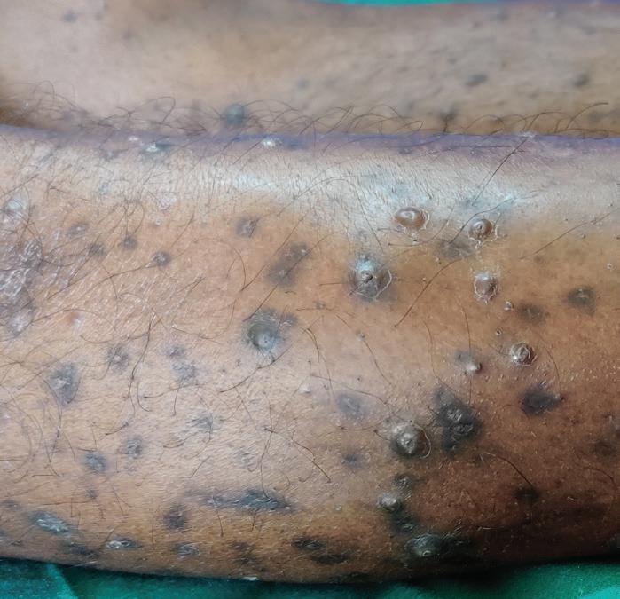
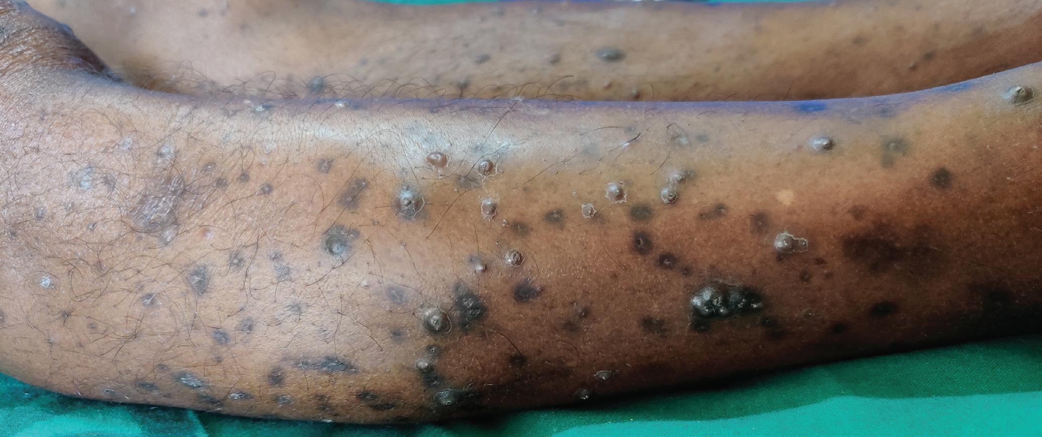
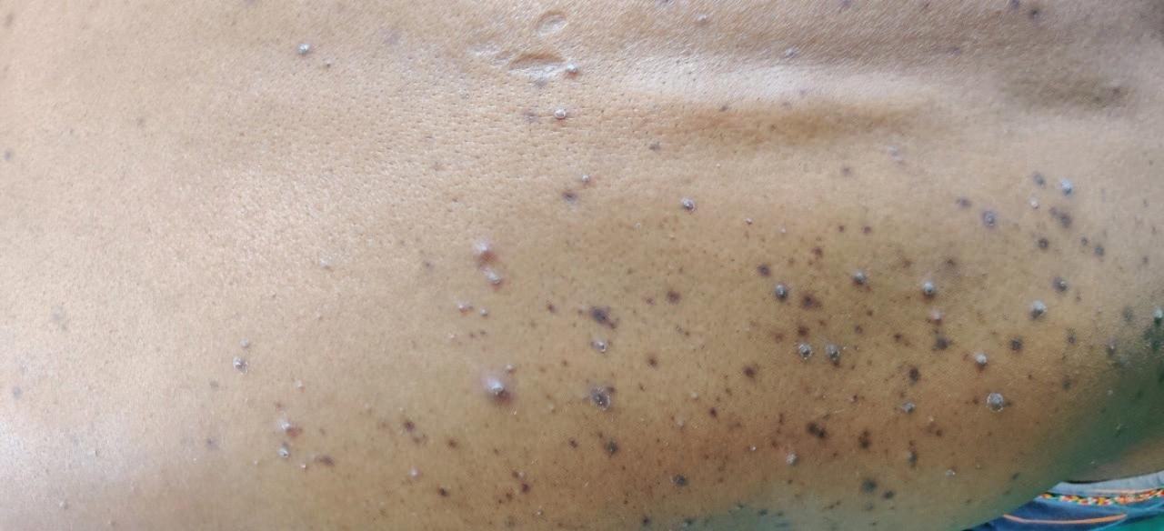
We report a 50 year old male patient who presented with generalized pruritis for about two months, resulting in skin lesions over the lower limbs, upper arms, trunk and face. Pathological history revealed hypertension, diabetes mellitus and chronic renal failure and the patient was on haemodialysis twice weekly for two and half years. Dermatological examination revealed dark brown to blackish papules and nodules with a keratotic center on the lower limbs, upper limbs, trunk and face. Differential diagnoses after examination were acquired perforating dermatoses and prurigo nodularis and diagnosis confirmed on histopathology. Laboratory tests showed blood urea of 111 and serum creatinine of 11.4 mg/ dL, hemoglobin was 7.8 g/dL. Patient was treated with topical potent corticosteroid, urea containing moisturizer, doxycycline 100mg BD and oral antihistamines.
Acquired Perforating Dermatosis in a Patient with Chronic Renal Failure: A Case Report
After Treatment
Figure 1: Dark brown to blackish papules and nodules with a keratotic center on the lower limbs
Diagnosis 9,10,11,12,13
Acquired perforating dermatosis (APD) is a rare skin disorder characterized by transepidermal elimination of dermal components, typically presenting with pruritic papules or nodules that develop central keratin plugs. It's often associated with systemic diseases, most commonly chronic renal failure (CRF). Here's how a diagnosis of APD in a patient with CRF might be approached:
1. Clinical Examination: A thorough examination of the skin is essential. Look for papules or nodules, often with a central keratotic plug. These lesions may be pruritic.
2. Patient History: Inquire about the patient's medical history, specifically focusing on any underlying systemic conditions. CRF, diabetes mellitus and other metabolic disorders are common associations with APD.
3. Laboratory Investigations:
• Renal Function Tests: Assess renal function through serum creatinine levels, blood urea nitrogen (BUN) and estimated glomerular filtration rate (eGFR).
• Serum Calciumand Phosphorus Levels: Abnormalities in calcium and phosphorus metabolism are often seen in patients with CRF, which may contribute to the development of APD.
4. Skin Biopsy: A skin biopsy may be performed to confirm the diagnosis. Histopathological ............. examination typically reveals a hyperkeratotic epidermis with transepidermal elimination of dermal material, such as collagen or elastic fibers.
5. Differential Diagnosis:
Consider other conditions that may present with similar skin manifestations, such as prurigo nodularis, perforating folliculitis or Kyrle's disease. Distinguishing features may include histopathological findings and associated systemic conditions.
Treatment 1, 5, 9, 13, 14, 15
Acquired perforating dermatoses (APD) is a group
of skin conditions that involve the formation of small bumps or papules on the skin that become crusted or scaly and eventually develop a central opening or perforation. The exact cause of APD is not known, but it is associated with other medical conditions such as diabetes, kidney disease and liver disease.
The evaluation and management of patients with acquired perforating dermatosis typically involves a multidisciplinary approach, with input from dermatologists, endocrinologists, ......... nephrologists and other specialists as needed.
Treatment is aimed at relieving pruritus and addressing any underlying medical conditions that may be contributing to the condition. The choice of treatment depends on the severity of the condition and the underlying cause and may include the use of topical or systemic medications, phototherapy or surgical excision of lesions. Close collaboration between healthcare providers can help ensure that patients with acquired perforating dermatosis receive optimal care and management. Treatment
Acquired Perforating Dermatosis in a Patient with Chronic Renal Failure: A Case Report
is usually directed at relieving pruritus, which can exacerbate the condition by triggering koebnerization. Treatment strategies for perforating dermatosis are often based on anecdotal evidence and may include the use of oral or topical retinoids, topical or intralesional corticosteroids, phototherapy (narrowband or broad-band UVB, or psoralen plus UVA), antibiotics (e.g., doxycycline) or destructive methods such as cryotherapy, surgical debridement and laser therapy. It is important to closely monitor patients receiving retinoid therapy, as these medications can have potential side effects, including hepatotoxicity, hyperlipidemia and teratogenicity. Patients with pre-existing cardiac disease may be at increased risk for adverse events associated with retinoid therapy.
Treatment of APD involves managing the underlying medical condition if present and treating the skin lesions. Here are some of the treatment options for APD:
1. Topical steroids: Topical steroids can be used to reduce inflammation and itching associated with APD lesions.
2. Topical retinoids:........
Topical retinoids are a type of medication that helps to normalize the growth and shedding of skin cells and may be useful in treating APD.
3. Emollients and......... moisturizers: Regular use of emollients and moisturizers can help to soften the skin and reduce the risk of skin injury and subsequent APD lesions.
4. Oral medications:......
Certain oral medications, such as antihistamines, can help to alleviate itching associated with APD.
5. Laser therapy: Laser therapy has been shown to be effective in treating APD lesions by reducing inflammation and promoting healing.
6. Phototherapy:..............
Phototherapy involves exposing the skin to ultraviolet light and can be useful in treating APD.
Discussion
Acquired perforating dermatosis typically presents with pruritic papules, which are often hyperkeratotic and umbilicated and located on the hair-bearing limbs of adults. Generalized papules may also be present in some cases. The manifestation of acquired perforating
dermatosis mainly as ulcerations on the lower limbs is a rare presentation. However, it is important to consider this condition in the differential diagnosis of any ulceration of the lower limbs, especially in patients with renal disease and diabetes mellitus. The clinical presentation of APD can vary widely and ulceration is a rare manifestation of this condition. APD most commonly presents as discrete, hyperkeratotic papules or nodules, which may be pruritic or painful. The lesions are typically located on the trunk and extremities, but can occur anywhere on the body.16,17,18
Kyrle’s disease (KD) lesions are firm, hyperkeratotic, usually occurring on the extremities. They are often pruritic, Koebner’s phenomenon may occur. Histopathologically, Kyrle’s disease shows keratotic, partly parakeratotic plug containing basophilic debris, vessel wall thickening and lymphocytic infiltrates in the dermis. APD represents a cutaneous response to superficial trauma caused by scratching. Diabetic vasculopathy and elevated concentrations of fibronectin may be causative. It is treated with keratolytics, electrocautery,
cryotherapy, CO2 laser, UV irradiation, surgical excision, isotretinoin, highdose vitamin A, tretinoin cream, emollients, oral antihistamines and oral clindamycin.19, 20,21,22
Perforating folliculitis is another rare condition that primarily affects young adults. It is characterized by the appearance of papules and pustules that surround hair follicles. Histopathologically, it is characterized by the elimination of keratinous material and bacteria through the follicular infundibulum. Reactive perforating collagenosis is a condition that primarily affects young adults and is characterized by the appearance of dome-shaped papules on the extremities. Histopathologically, it is characterized by the elimination of altered collagen fibers through the epidermis. Elastosis perforans serpiginosa is a rare disorder that primarily affects young adults and is characterized by the appearance of serpiginous plaques on the neck, face and extremities. Histopathologically, it is characterized by the elimination of abnormal elastic fibers through
the epidermis. Prurigo nodularis is a chronic dermatological condition characterized by intensely itchy (pruritic) nodules or papules on the skin. The clinical presentation of prurigo nodularis typically includes the following features: Prurigo nodules are firm, raised lesions that can vary in size from a few millimeters to several centimeters in diameter. These nodules may be dome-shaped or flat-topped and are often grouped or clustered together. Pruritus is the hallmark symptom of prurigo nodularis and is often severe and persistent. The itching can be so intense that it significantly impacts the patient's quality of life, leading to scratching and excoriation of the skin. Due to the intense itching, patients with prurigo nodularis often scratch or pick at the nodules, leading to excoriations, crusts and secondary changes such as erosions or ulcerations. These secondary changes can further exacerbate the itching and discomfort. Prurigo nodularis lesions can occur anywhere on the body but are most commonly found on extensor surfaces of the limbs, particularly the elbows and knees. However, they can also
affect the trunk, scalp and neck. Prurigo nodularis is a chronic condition characterized by persistent or recurrent episodes of itching and the formation of new nodules over time. The lesions may wax and wane in severity but often persist for months to years without spontaneous resolution. Chronic scratching and inflammation can lead to hyperpigmentation of the skin surrounding the nodules. This can result in darkening of the skin, particularly in individuals with darker skin tones. In addition to excoriations and crusts, prurigo nodularis lesions may exhibit other secondary changes such as lichenification (thickening of the skin), scaling and scarring, especially in longstanding cases. Effective management typically involves a combination of symptomatic relief, addressing underlying triggers or comorbidities and behavioral interventions to minimize scratching and further exacerbation of the condition.1,14,23,24,25,26,27
Conclusion
In conclusion, perforating disorders of the skin are a group of uncommon dermatological conditions that can be challenging to diagnose and manage.
Acquired Perforating Dermatosis in a Patient with Chronic Renal Failure: A Case Report
Accurate diagnosis relies on histopathological examination, which helps to classify these disorders according to the type of epidermal disruption and the nature of the eliminated material. However, acquired perforating dermatosis can also present as ulcerations on the lower limbs. This presentation can be easily misdiagnosed as a venous ulcer or a diabetic foot ulcer. Therefore, it is essential to consider acquired perforating dermatosis in the differential diagnosis of any ulcerations on the lower limbs, especially in patients with underlying renal disease and diabetes mellitus. Patients with APD and CRF require regular follow-up to monitor renal function, assess skin lesions for progression or recurrence and adjust treatment as necessary. Educate the patient about their condition, including the association between APD and CRF, the importance of adherence to treatment and follow-up appointments and measures for symptom management and prevention of complications. The prognosis for APD in patients with CRF varies depending on the underlying renal disease, response to treatment and presence of any complications.
Early recognition and management of both the skin disorder and renal dysfunction are crucial for optimizing outcomes. Collaboration with dermatologists, nephrologists and other specialists may be necessary for a comprehensive evaluation and management plan.
References
1. Harbaoui S, Litaiem N. Acquired Perforating Dermatosis. [Updated 2023 Feb 13]. In: StatPearls [Internet]. Treasure Island (FL): StatPearls Publishing; 2024 Jan-. Available from: https://www.ncbi. nlm.nih.gov/books/NBK539715/
2. Fernandes KA, Lima LA, Guedes JC, Lima RB, D'Acri AM, Martins CJ. Acquired perforating dermatosis in a patient with chronic renal failure. An Bras Dermatol. 2016;91(5 suppl 1):10-13. doi:10.1590/abd18064841.20164619
3. Al-Bader, Shaima1; ElReshaid, Kamel2; Madda, John3. Acquired Perforating Dermatosis: A Disorder Treatable with Mycophenolate Mofetil. Saudi Journal of Kidney Diseases and Transplantation 34(2):p 142-146, Mar–Apr 2023. | DOI: 10.4103/1319-2442.39189
4. Graña JM, Lorente L, Ortega C, et al. Acquired perforating dermatosis in patients with chronic renal failure. A report of two cases and a review of the
5. Imam T H, Patail H, Khan N et al. Acquired Perforating Dermatosis in a Patient on Peritoneal Dialysis: A Case Report and Review of the Literature. Case Reports in Nephrology Volume 2018, Article ID 5953069, 3 pages https://doi. org/10.1155/2018/5953069
6. Fernandes, Karen & Lima, Lourenço & Guedes, Juliana & Barbosa Lima, Ricardo & D'Acri, Antonio & Martins, Carlos. (2016). Acquired perforating dermatosis in a patient with chronic renal failure. Anais Brasileiros de Dermatologia. 91. 10-13. 10.1590/abd18064841.20164619.
7. Ramirez-Fort, Marigdalia & Khan, Farhan & Rosendahl, Cliff & Mercer, Stephen & Shim-Chang, Helen & Levitt, Jacob. (2013). Acquired perforating dermatosis: A clinical and dermatoscopic correlation. Dermatology online journal. 19. 18958. 10.5070/ D3197018958.
8. Kim SW, Kim MS, Lee JH, Son SJ, Park KY, Li K, Seo SJ, Han TY. A Clinicopathologic Study of Thirty Cases of Acquired Perforating Dermatosis in Korea. Ann Dermatol. 2014 Apr;26(2):162-171. https://doi. org/10.5021/ad.2014.26.2.162
9. Hong SB, Park JH, Ihm CG, Kim NI. Acquired perforating dermatosis in patients with
chronic renal failure and diabetes mellitus. J Korean Med Sci. 2004;19(2):283-288. doi:10.3346/jkms.2004.19.2.283
10. L. González-Lara, S. GómezBernal, F. Vázquez-López, B. Vivanco-Allende, Acquired Perforating Dermatosis: A Report of 8 Cases, Actas DermoSifiliográficas (English Edition), Volume 105, Issue 6, 2014, Pages e39-e43, ISSN 15782190, https://doi.org/10.1016/j. adengl.2014.05.007.
11. Wang, Mei-Fang & Mei, Xue-Ling & Wang, Li & Li, Lin-Feng. (2020). Clinical characteristics and prognosis of acquired perforating dermatosis: A case report. Experimental and Therapeutic Medicine. 19. 10.3892/etm.2020.8651.
12. Lynde CB, Pratt MD. Clinical Images: Acquired perforating dermatosis: association with diabetes and renal failure. CMAJ. 2009;181(9):615. doi:10.1503/ cmaj.082013
13. Hideyuki Kosumi, Hiroaki Iwata, Masumi Tsujiwaki, Hiroshi Shimizu; Diagnosis at a Glance: Acquired Perforating Dermatosis. Diabetes Care 1 April 2018; 41 (4): 911–912. https://doi. org/10.2337/dc17-2572
14. Wang W, Liao Y, Fu L, Kan B, Peng X and Lu Y (2021) Dermoscopy Features of Acquired Perforating Dermatosis Among 39 Patients. Front. Med. 8:631642. doi: 10.3389/fmed.2021.631642
15. Al-Bader, Shaima; ElReshaid, Kamel; Madda, John. Acquired Perforating Dermatosis: A Disorder Treatable with Mycophenolate Mofetil. Saudi Journal of Kidney Diseases and Transplantation 34(2):p 142146, Mar–Apr 2023. | DOI: 10.4103/1319-2442.391892
16. Lynde, Carrie & Pratt, Melanie. (2009). Clinical Images: Acquired perforating dermatosis: association with diabetes and renal failure. CMAJ : Canadian Medical Association journal = journal de l'Association medicale canadienne. 181. 615. 10.1503/ cmaj.082013.
17. Maiberger M, Nunley JR, Jeffrey Callen J. Ofori AO. Perforating dermatoses. Jan 2024.
18. García-Malinis, Ana & Sánchez, Elena & Sánchez-Salas, María & Prado, Elena & Coscojuela, Carmen & Gilaberte, Yolanda. (2017). Acquired perforating dermatosis: clinicopathological study of 31 cases, emphasizing pathogenesis and treatment. Journal of the European Academy of Dermatology and Venereology : JEADV. 31. 10.1111/jdv.14220.
19. Dharmadji, Hartati & Firdaus, Chaerani & Sugiri, Unwati & Sutedja, Eva & Achdiat, Pati & Tsaqilah, Laila & Gunawan, Hendra. (2022). Generalized Lesions of Kyrle’s Disease: A Rare Case. International Medical Case Reports Journal. Volume 15. 187-191. 10.2147/IMCRJ.
S358523.
20. Rice AS, Zedek D. Kyrle Disease. [Updated 2023 Jun 15]. In: StatPearls [Internet]. Treasure Island (FL): StatPearls Publishing; 2024 Jan-. Available from: https:// www.ncbi.nlm.nih.gov/books/ NBK532886/
21. Forouzandeh, Mahtab & Stratman, Scott & Yosipovitch, Gil. (2020). The Treatment of Kyrle’s Disease: A Systematic Review. Journal of the European Academy of Dermatology and Venereology. 34. 10.1111/ jdv.16182.
22. L. Macca, F. Vaccaro, F. Li Pomi, F. Borgia, N. Irrera, M. Vaccaro. Kyrle disease: a case report and literature review. European Review for Medical and Pharmacological Sciences 2023; 27: 10705-10715
23. Sawant S, Gaikwad N, Hajirnis K, Vasani R. Acquired perforating collagenosis: A clinicopathological study of ten. Indian J Pathol Oncol 2019;6(4):677-681.
24. Mullins TB, Sickinger M, Zito PM. Reactive Perforating Collagenosis. [Updated 2022 Aug 25]. In: StatPearls [Internet]. Treasure Island (FL): StatPearls Publishing; 2024 Jan-. Available from: https://www.ncbi.nlm.nih. gov/books/NBK459214/
25. Kestner RI, Ständer S, Osada N, Ziegler D, Metze D. Acquired Reactive Perforating Dermatosis is a Variant of Prurigo Nodularis. Acta Derm
Acquired Perforating Dermatosis in a Patient with Chronic Renal Failure: A Case Report
26. Agnes Rosarina Prita Sari, Monika Puspitasari, Hardyanto Soebono, Niken Trisnowati; Acquired Perforating Disorder: A Case with Multiple Underlying Diseases. Case Rep Dermatol 9 September 2022; 14 (2): 107–111. https://doi. org/10.1159/000524466
27. Biswas A. Folliculitis/ Perifolliculitis. In: Pearls and Pitfalls in Inflammatory Dermatopathology. Cambridge University Press; 2016:218-238.
Comprehensive Management of Postauricular Keloid: A Case Report and Review of Current Therapeutic Strategies
Dr. Bala Narasimhulu
MBBS, MD
Consultant Dermatologist
Mamilla Skin and VD Clinic
Vijayawada, Andhra Pradesh
Introduction
Keloids are pathological fibroproliferative lesions of the skin that arise from aberrant wound healing, leading to excessive and disorganized accumulation of extracellular matrix (ECM) components, primarily type I and III collagen. They are characterized by persistent fibroblast hyperactivity, reduced apoptosis, and chronic inflammation. Clinically, keloids present as firm, rubbery, raised nodules or plaques extending beyond the boundaries of the initial dermal injury. They may appear as smooth, clawlike, or bosselated lesions
with variable pigmentation, ranging from erythematous to violaceous or brown. These scars are often accompanied by itching, tenderness, or pain, and can lead to functional limitations when located near joints or mobile areas. Commonly affected regions include the earlobes, shoulders, and anterior chest—sites subject to mechanical tension and repetitive trauma—while palms, soles, and genitalia are rarely involved. Keloids are more prevalent in individuals with darker skin phototypes (Fitzpatrick IV–VI), with incidence rates reported between 4.5% and 16%. They typically develop
between the ages of 10 and 30 years, with a slight female preponderance attributed to increased risk from ear piercing and hormonal factors.1
Genetic predisposition plays a major role, supported by familial clustering and associations with rare syndromes such as Rubinstein–Taybi and Goeminne syndromes, though no specific causative gene has been definitively identified. Environmental and local triggers, including surgical trauma, burns, acneiform eruptions, insect bites, tattoos, or piercings, often precipitate keloid formation. Mechanical tension during wound healing and hormonal fluctuations during puberty and pregnancy further aggravate fibroblast activation and ECM deposition.2,3
Pathogenetically, keloids result from an intricate network of cellular and molecular dysregulation. Angiogenic imbalance plays a central role, with upregulated vascular endothelial growth factor (VEGF) signaling through VEGFR1 and VEGFR2 promoting
abnormal endothelial proliferation and immature neovascularization. Concurrent reduction in antiangiogenic endostatin sustains chronic inflammation and nutrient supply. The hypoxic microenvironment stabilizes hypoxia-inducible factor-1 alpha (HIF-1α), which enhances glycolysis, fibroblast survival, and collagen synthesis via TGFβ-mediated pathways. The Janus kinase/signal transducer and activator of transcription (JAK/STAT) pathway—particularly ...... JAK1, JAK2, and STAT3—is hyperactivated by interleukin-6 (IL-6), leading to enhanced fibroblast proliferation, ECM production, and upregulation of TGF-β expression. The Notch signaling cascade, involving NOTCH1–NOTCH3 and the JAG1 ligand, acts synergistically with TGF-β, NF-κB, and Wnt/β-catenin pathways to promote fibrosis. Crosstalk between PI3K/Akt and MAPK/ERK signaling further reinforces fibroblast activation and survival. Epigenetic modifications play an emerging role in keloid persistence. Global DNA
hypomethylation, altered histone acetylation, and dysregulation of microRNAs (miR-21, miR-29) and long noncoding RNAs (lncRNA H19) contribute to sustained fibroblast activation and excessive collagen synthesis. Aberrant paracrine communication between keratinocytes, fibroblasts, and endothelial cells perpetuates inflammation and fibrosis. Collectively, these cellular, molecular, and epigenetic mechanisms establish a self-sustaining cycle of fibroproliferation, excessive ECM deposition, and progressive keloid growth—defining keloids as a complex, chronic, and multifactorial dermal fibrosis disorder.2,3
Case Report
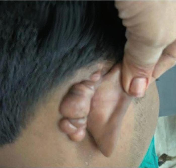
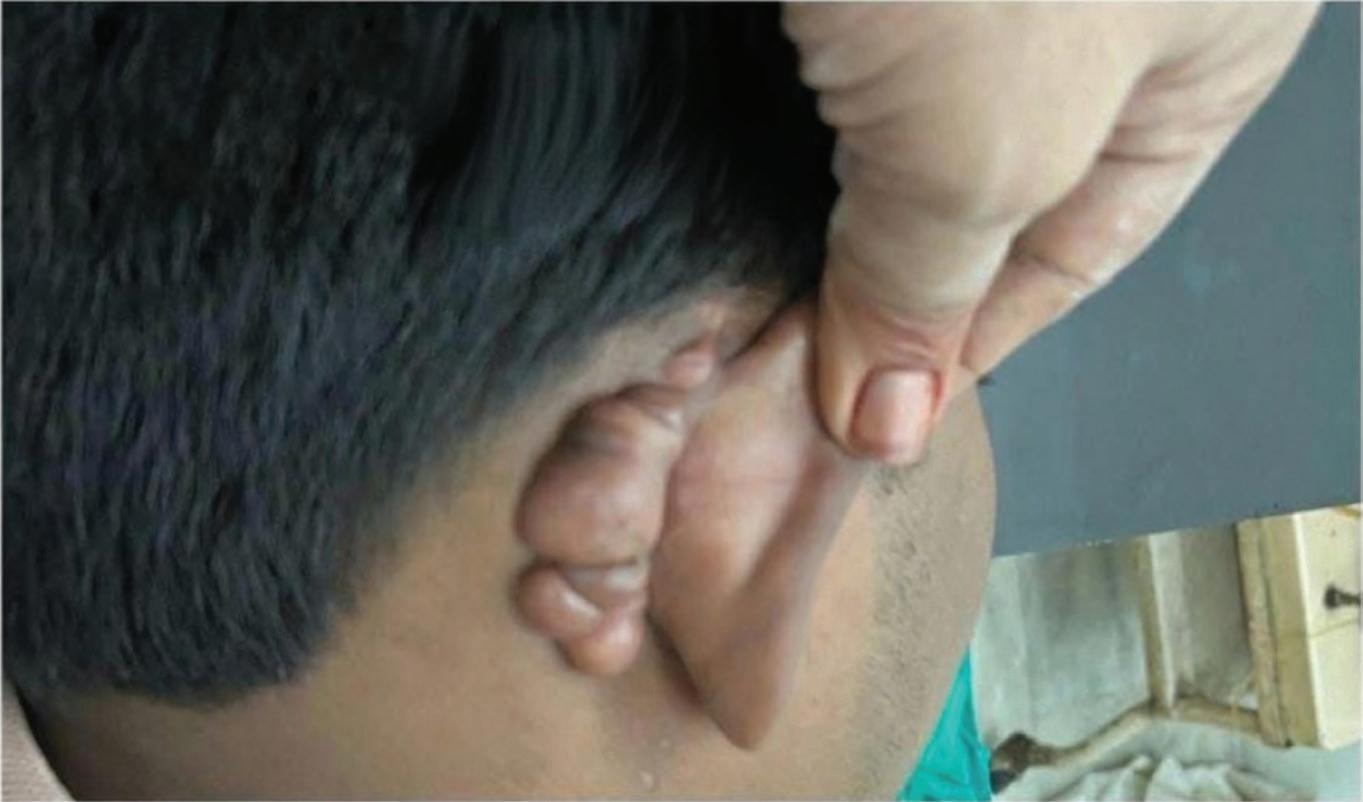
A 25-year-old male presented to the dermatology outpatient department with a firm, nontender, hyperpigmented lesion located in the postauricular region, progressively increasing in size over the past year. The lesion initially developed following minor trauma associated with ear piercing. On clinical examination, the keloid was found to involve
Comprehensive
the postauricular sulcus and extended onto the adjacent mastoid area. It appeared as a lobulated, elevated, rubbery mass with welldefined margins extending beyond the original wound boundary — characteristic of keloidal morphology. The overlying skin was smooth, hyperpigmented, and taut, without ulceration or discharge. The patient reported mild pruritus and occasional discomfort when lying on the affected side, but no history of infection, bleeding, or restriction of auricular movement was noted. Based on clinical presentation and morphology, a diagnosis of postauricular keloid was established. After thorough counselling regarding treatment options and recurrence risk, surgical excision was planned. Under local anesthesia, complete excision of the fibrotic mass was performed with careful undermining to preserve surrounding normal skin and minimize tension at the closure site. Meticulous hemostasis was achieved, and layered primary closure was done using fine non-absorbable sutures to ensure minimal scarring. The excised
tissue was sent for histopathological examination, which confirmed dense collagen deposition arranged in whorled bundles with fibroblast proliferation, consistent with keloid. Postoperatively, the patient was advised routine wound care with topical antibiotic application and avoidance of pressure or trauma to the area. Prophylactic intralesional triamcinolone acetonide (10 mg/mL) was administered around the suture line two weeks post-surgery to reduce the risk of recurrence. At the 27-day follow-up, the surgical site exhibited satisfactory epithelialization and healthy scar formation. There was significant reduction in lesion bulk, minimal hypertrophic response, and no immediate evidence of recurrence or wound dehiscence. The aesthetic and functional outcomes were satisfactory, and the patient was advised continued follow-up for long-term surveillance and potential adjuvant therapy if recurrence occurred. This case highlights the importance of combining precise surgical excision with adjunctive corticosteroid therapy and diligent postoperative care in managing auricular keloids. Early follow-up demonstrated promising short-term results, underscoring the efficacy of a meticulous, tensionminimized surgical approach in preventing recurrence and optimizing cosmetic outcomes.
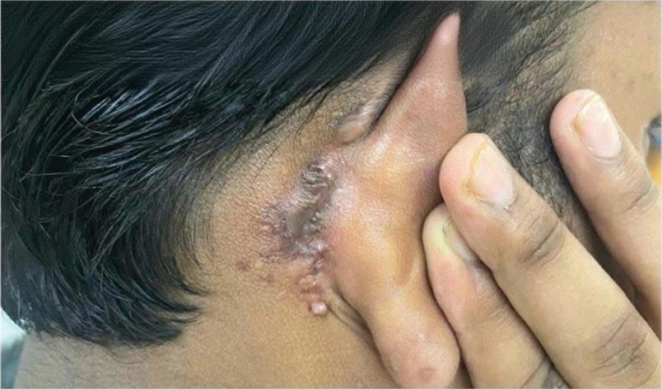
Figure 1: Firm, non-tender, hyperpigmented lesion located in the postauricular region (Before treatment)
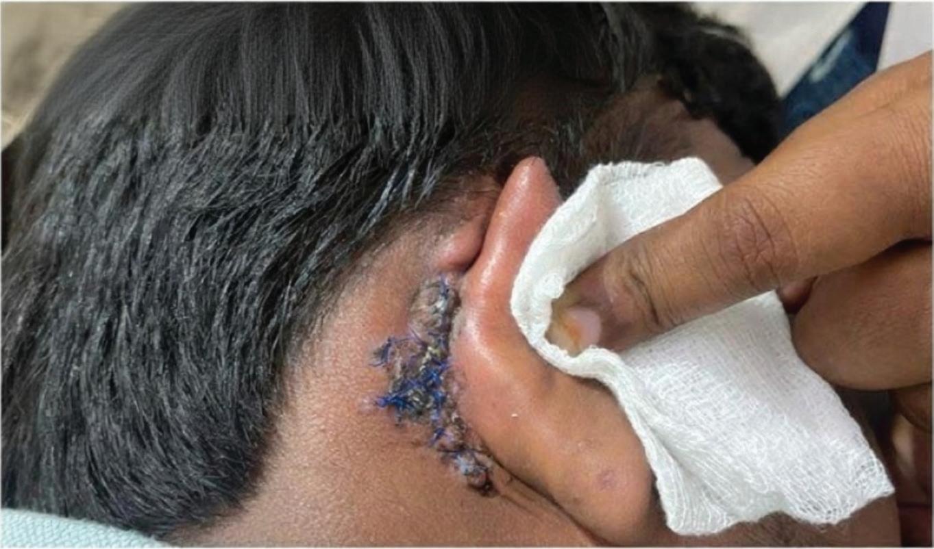
Figure 2: Immediate postoperative appearance following surgical excision of the keloid
Figure 3: 27 days post-excision, the surgical site exhibited satisfactory epithelialization and healthy scar formation and no signs of recurrence or wound dehiscence
Diagnosis
whitish background, linear or polymorphous vessels, and white streaks reflecting dense collagen. These features help differentiate keloids from hypertrophic scars and other dermal lesions.4 High-frequency ultrasound is a useful, noninvasive tool for evaluating keloids. It allows accurate assessment of lesion depth, internal structure, vascularity, and involvement of nearby tissues. Keloids typically appear as welldefined, hypoechoic or heterogeneous dermal to subdermal masses with posterior acoustic enhancement. Color Doppler may show increased vascularity, indicating fibroproliferation and neovascularization. Ultrasound also helps differentiate keloids from hypertrophic scars.5
Biopsy is essential for diagnosing keloids in atypical cases or to exclude other dermal pathologies. Histology shows dense, disorganized hyalinized collagen bundles with sparse fibroblasts and minimal inflammation. MRI is used to assess keloid size, depth, and involvement of surrounding tissues. Keloids appear iso- to hypointense on T1 and variably hyperintense
Diagnosis of keloids primarily depends on clinical examination, the cornerstone of assessment. It involves visual and tactile evaluation of scar size, shape, texture, and symptoms like pruritus or pain. This evaluation is essential to differentiate keloids from hypertrophic scars and other dermal lesions. Dermoscopy is a useful non-invasive tool in the assessment of keloids and serves to support diagnosis. It typically reveals a homogeneous pink or Comprehensive Management of Postauricular Keloid: A Case
on T2 due to collagen and edema. MRI helps differentiate keloids from other lesions. Photographic documentation is a key noninvasive tool for objectively assessing keloid size, shape, and color. It aids in monitoring progression and treatment response, supports clinical evaluation, and enhances patient communication.6
A comprehensive diagnostic strategy combining clinical and imaging modalities is critical for accurate diagnosis, guiding effective management and improving patient outcomes.
Treatment
Surgical Excision of Keloids
Surgical excision remains a widely utilized and effective treatment modality for keloids, particularly in cases where functional impairment or disabling scar contracture is present. When executed with precision and accompanied by adjunctive therapies, surgical excision offers favourable aesthetic and symptomatic outcomes while reducing recurrence risk.6, 7, 8
Preoperative Planning and Anaesthesia
A meticulous preoperative assessment is essential, with careful marking of keloid
margins to ensure complete excision while preserving adjacent viable skin. Local anaesthesia, typically 1% lidocaine with epinephrine, is administered to provide analgesia and minimize intraoperative bleeding via vasoconstriction.2, 7, 8
Surgical Technique
The procedure commences with incision along the previously demarcated keloid margins. The overlying marginal skin is elevated to expose the underlying fibrous core. Total excision of the keloid is performed, ensuring removal of the entire fibrotic mass. In cases involving large or anatomically sensitive lesions, such as auricular keloids, partial or intralesional debulking may be employed to reduce volume while maintaining structural integrity and minimizing cosmetic distortion. Excision is conducted using a scalpel or electrocautery, with attention to preserving the subdermal vascular plexus to promote optimal wound healing. Incisions should ideally be beveled or aligned parallel to Langer’s lines to enhance cosmesis and reduce scar tension. Preservation of surrounding healthy dermis and subcutaneous tissue
is prioritized where feasible.2, 7, 8
Wound Closure and Tension Management
Wound closure is achieved using fine, interrupted sutures, with emphasis on achieving a tensionfree closure. Techniques such as wound edge eversion and the use of deep dermal sutures can be employed to counteract the natural contracture forces and reduce scar elevation. Excessive tension during closure is a known contributor to keloid recurrence and must be rigorously avoided.2, 7, 8
Postoperative Follow-Up
Patients require close postoperative surveillance for early detection of recurrence, which may manifest within weeks to months following excision. Recurrences are typically managed with additional intralesional corticosteroid injections or alternate adjunctive therapies as indicated. Patient counseling should include guidance on minimizing mechanical stress to the surgical site and adhering to prescribed topical or systemic therapies.2, 7, 8
Debulking of keloids, through partial excision or volume-reducing
techniques, is considered a near-permanent treatment modality when combined with appropriate adjunctive therapies, offering substantial reduction in lesion size and long-term symptomatic relief.
Discussion
Keloids are benign fibroproliferative lesions that arise from an exaggerated and dysregulated wound healing response, characterized by excessive collagen deposition and fibrosis extending beyond the original wound margins. These lesions can develop in any anatomical region, often without an identifiable predisposing trauma, making their pathogenesis complex and multifactorial. Approximately 10 to 15% of keloid cases occur without a clear precipitating factor. Common triggers include surgical trauma, burns, acne, and infections. The underlying pathogenesis involves persistent fibroblast activation, impaired apoptosis, and disrupted extracellular matrix remodeling, with transforming growth factorbeta playing a central role. Epidemiologically, keloids show a higher prevalence in females (20.4%) compared to males (12.9%) in the general population,
potentially due to hormonal influences such as estrogen, which enhances collagen synthesis in fibroblasts. This hormonal effect is supported by the frequent onset of keloids during puberty and pregnancy, periods marked by elevated estrogen levels. Additionally, factors like ear lobe piercings contribute to the increased incidence in females, with an estimated keloid formation rate of approximately 2.5%. Age is also a significant factor, with the highest prevalence occurring between 10 and 30 years of age.9
The differential diagnosis of keloids requires meticulous clinical and histopathological evaluation. Hypertrophic scars remain confined to the original injury and may regress spontaneously, unlike keloids that extend beyond wound margins. Dermatofibroma presents as a firm, hyperpigmented nodule showing a “dimple sign” on compression. Dermatofibrosarcoma ...... protuberans is a locally aggressive spindle cell tumor with indurated, irregular nodules and no trauma history. Keloidal morphea or scleroderma manifests as indurated plaques with possible systemic connective tissue features. Xanthoma disseminatum
mimics keloids but appears as symmetric xanthomatous lesions with potential systemic involvement. Lobomycosis, a chronic fungal infection, produces keloid-like nodules on extremities after environmental exposure. Biopsy is essential for atypical, rapidly growing, or ulcerated lesions to confirm diagnosis and exclude neoplastic or infectious causes.2
Keloids are benign dermal fibroproliferative lesions that develop following cutaneous trauma, surgery, burns, or inflammation. They may appear within one to three months after injury, though delayed onset— sometimes years later—is not uncommon. Rarely, keloids may develop spontaneously, often following unrecognized minor trauma. Clinically, they may present as pedunculated nodules or broad plaque-like growths that extend beyond the original wound margins, distinguishing them from hypertrophic scars, which remain confined and typically less than 4 mm in height. Keloids vary in color—erythematous, fleshtoned, or hyperpigmented— and may change over time. Symptomatic presentations
are common; studies report pruritus in up to 86% and pain in about 46% of patients, along with tenderness or burning. Although histologically benign, their progressive growth and disfiguring nature can result in significant cosmetic and psychosocial distress. Effective keloid management requires a multimodal and individualized approach tailored to lesion characteristics, chronicity, and prior treatment history. First-line therapy combines silicone gel or sheeting with intralesional corticosteroids, typically triamcinolone acetonide, which suppresses fibroblast activity and collagen synthesis. Refractory cases may benefit from non-corticosteroid ......... intralesional agents such as 5-fluorouracil, bleomycin, verapamil, botulinum toxin A, hyaluronidase, or platelet-rich plasma (PRP), used alone or synergistically with corticosteroids. Cryotherapy, including intralesional cryoneedles, induces localized necrosis and flattening and is often used in combination regimens. Laser and lightbased therapies—notably pulsed dye laser, CO₂, and erbium:YAG lasers—target
vascularity, erythema, and dermal remodeling. Postoperative radiotherapy, such as external beam radiation or brachytherapy, effectively reduces recurrence when initiated immediately after excision by inhibiting fibroblast proliferation. Pressure therapy using garments or silicone tapes minimizes tensile forces, enhancing scar remodeling. Adjunctive and emerging treatments include topical imiquimod, extracorporeal shockwave therapy, systemic agents like pentoxifylline, and biologics targeting fibrotic pathways. Surgical excision remains essential for bulky or symptomatic lesions but should always be followed by adjuvant therapy to prevent recurrence and ensure durable, cosmetically acceptable outcomes.1,2
Conclusion
Keloid scars remain a persistent therapeutic challenge due to their multifactorial pathogenesis, unpredictable behaviour, and high rates of recurrence. Although various treatment modalities have been explored, no single approach guarantees complete resolution. Surgical excision continues to serve as a cornerstone in keloid management,
offering satisfactory outcomes particularly when combined with appropriate postoperative adjuvant therapies aimed at reducing recurrence. Longterm success depends on individualized treatment planning, careful surgical technique, and ongoing monitoring. Continued clinical research and evidence-based refinement of existing strategies are essential to optimize outcomes and establish standardized, effective management protocols.
References
1. Merlino L, Dominoni M, Pano MR, Pasquali MF, Senatori R, Zino G, Gardella B. Recent Progress in Keloid Mechanism and Treatment: A Comprehensive Review. Biomedicines. 2025; 13(9):2276. https://doi.org/10.3390/ biomedicines13092276
2. McGinty S, Siddiqui WJ. Keloid. In: StatPearls. Treasure Island (FL): StatPearls Publishing; July 17, 2023.
3. Latoni DI, McDaniel DC, Tsao H, Tsao SS. Update on the Pathogenesis of Keloid Formation. JID Innov. 2024; 4(6):100299. Published 2024 Jul 25. doi:10.1016/j.xjidi.2024.100299.
4. Yoo MG, Kim IH. Keloids and hypertrophic scars: characteristic vascular structures visualized by using dermoscopy. Ann
Comprehensive Management of Postauricular Keloid: A Case Report and Review of
5. Liu Y, Xiong X, Cao N, Zhao Y. Diagnosis and Treatment of Keloid: Method Summary and Effect Evaluation. Clin Cosmet Investig Dermatol. 2023; 16:37753783. Published 2023 Dec 29. doi:10.2147/CCID.S446018
6. Walsh LA, Wu E, Pontes D, et al. Keloid treatments: an evidence-based systematic review of recent advances. Syst Rev. 2023; 12(1):42. Published 2023 Mar 14. Doi: 10.1186/ s13643-023-02192-7
7. Kim SW. Management of keloid scars: noninvasive and invasive treatments. Arch Plast Surg. 2021; 48(2):149-157. doi:10.5999/aps.2020.01914.
8. Wilson AM. Eradication of keloids: Surgical excision followed by a single injection of intralesional 5-fluorouracil and botulinum toxin. Can J Plast Surg. 2013; 21(2):87-91. Doi: 10.1177/229255031302100208.
9. Merlino L, Dominoni M, Pano MR, et al. Recent Progress in Keloid Mechanism and Treatment: A Comprehensive Review. Biomedicines. 2025; 13(9):2276. Published 2025 Sep 16. doi:10.3390/ biomedicines13092276
Bio Remodeller & Skin
Dr. Pallavi Sule MBBS, DDV Dermatologist and Cosmetologist Dr. Pallavi Sule’s Clinic, Mumbai Faculty
Dr. Dattatray Sonawane MD (Skin) Founder and Consultant Dermatologist La Peau Laser World, Thane
for Botulinum Toxin Type A
Dr. Abhay Talathi MD, DNB, FCPS, DVD Dermatologist and Cosmetologist SkinSpace Clinic, Mumbai
Dr. Pallavi Sule welocome message for Aestheticconf Workshop