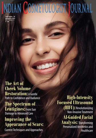
Indian Cosmetologist Journal Digital Sept'25 Issue
EXECUTIVE EDITOR & PUBLISHER DOM DANIEL
CORPORATE OFFICE
22, SHREEJI BHAVAN, 275-279, SAMUEL STREET, MASJID BUNDER (W), MUMBAI-4000 03, INDIA.
EMAIL: info@indiancosmetologistjournal.com
WEBSITE: indiancosmetologistjournal.online
PRINTED, PUBLISHED, EDITED AND OWNED BY DOM DANIEL
PRINTED AT SWASTIK PRINTER, GALA NO.9 & 10, VISHAL INDUSTRIAL ESTATE, BHANDUP (WEST), MUMBAI400078.
PUBLISHED AT 22 SHREEJI BHAVAN, 275/279, SAMUEL STREET,
MASJID BUNDER (WEST), MUMBAI - 400003. INDIA.
“INDIAN COSMETOLOGIST JOURNAL” TAKES NO RESPONSIBILITY FOR UNSOLICITED PHOTOGRAPHS OR MATERIAL
ALL PHOTOGRAPHS, UNLESS OTHERWISE INDICATED, ARE USED FOR ILLUSTRATIVE PURPOSE ONLY.
VIEWS EXPRESSED IN THIS JOURNAL ARE THOSE OF THE CONTRIBUTORS AND NOT OF THE PUBLISHER. REPRODUCTION IN WHOLE OR IN PARTS OF TEXTS OR PHOTOGRAPHY IS PROHIBITED. MANUSCRIPTS, PHOTOGRAPHS AND ART ARE SELECTED AT THE DISCRETION OF THE PUBLISHER FREE OF CHARGE (ADVERTISING EXCLUDED). WHETHER PUBLISHED OR NOT, NO MATERIAL WILL BE RETURNED AND REMAINS THE PROPERTY OF THE PUBLISHING HOUSE, WHICH MAY MAKE USE OF IT AS SEEN FIT. THIS MAY INCLUDE THE WITHDRAWAL OF PUBLICATION RIGHTS TO OTHER PUBLISHING HOUSES.
ALL RIGHTS RESERVED. REPRODUCING IN ANY MANNER WITHOUT PRIOR WRITTEN PERMISSION PROHIBITED.
PUBLISHED FOR THE PERIOD OF SEPTEMBER -2025
Advancing Aesthetic Excellence: Innovations, Insights, and the Future of Cosmetology
Welcome to this issue of the Indian Cosmetologist Journal, featuring a rich collection of insights and innovations shaping the future of aesthetic medicine. In this edition, we explore diverse yet interconnected themes that reflect the precision, science, and artistry defining modern cosmetology.
IN THIS EDITION, WE EXPLORE DIVERSE YET INTERCONNECTED THEMES THAT REFLECT THE PRECISION, SCIENCE, AND ARTISTRY
DEFINING MODERN COSMETOLOGY.
We open with “The Art of Cheek Volume Restoration: A Gentle Path to Confidence and Radiance”, illustrating how subtle enhancements can restore natural balance and self-assurance. “The Spectrum of Lentigines: From Sun Damage to Advanced Care” delves into the complexities of pigmentation disorders, underscoring our commitment to evidence-based approaches in managing these common yet challenging conditions.
Advances in scar treatment take center stage in “Improving the Appearance of Scars: Current Techniques and Approaches”, while “Focused Ultrasound (HIFU): Revolutionizing Non-Invasive Treatment” highlights a breakthrough technology transforming skin tightening and rejuvenation. Finally, “AI-Guided Facial Analysis: Transforming Personalized Aesthetics and Healthcare” offers a forward-looking perspective on data-driven, individualized patient care—where technology and human expertise intersect to create better outcomes.
This issue embodies our mission: to equip practitioners with trusted knowledge, inspire innovation, and celebrate the synergy of art and science in cosmetology. We hope these articles enrich your practice and spark new ideas to elevate patient care.
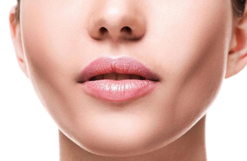
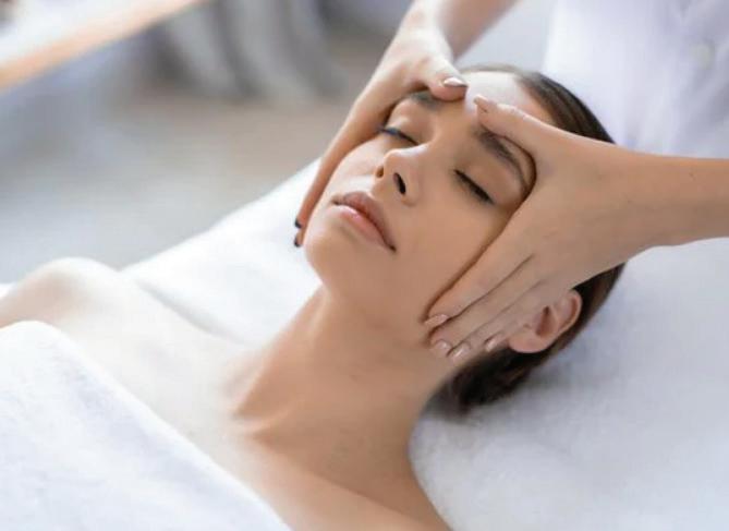
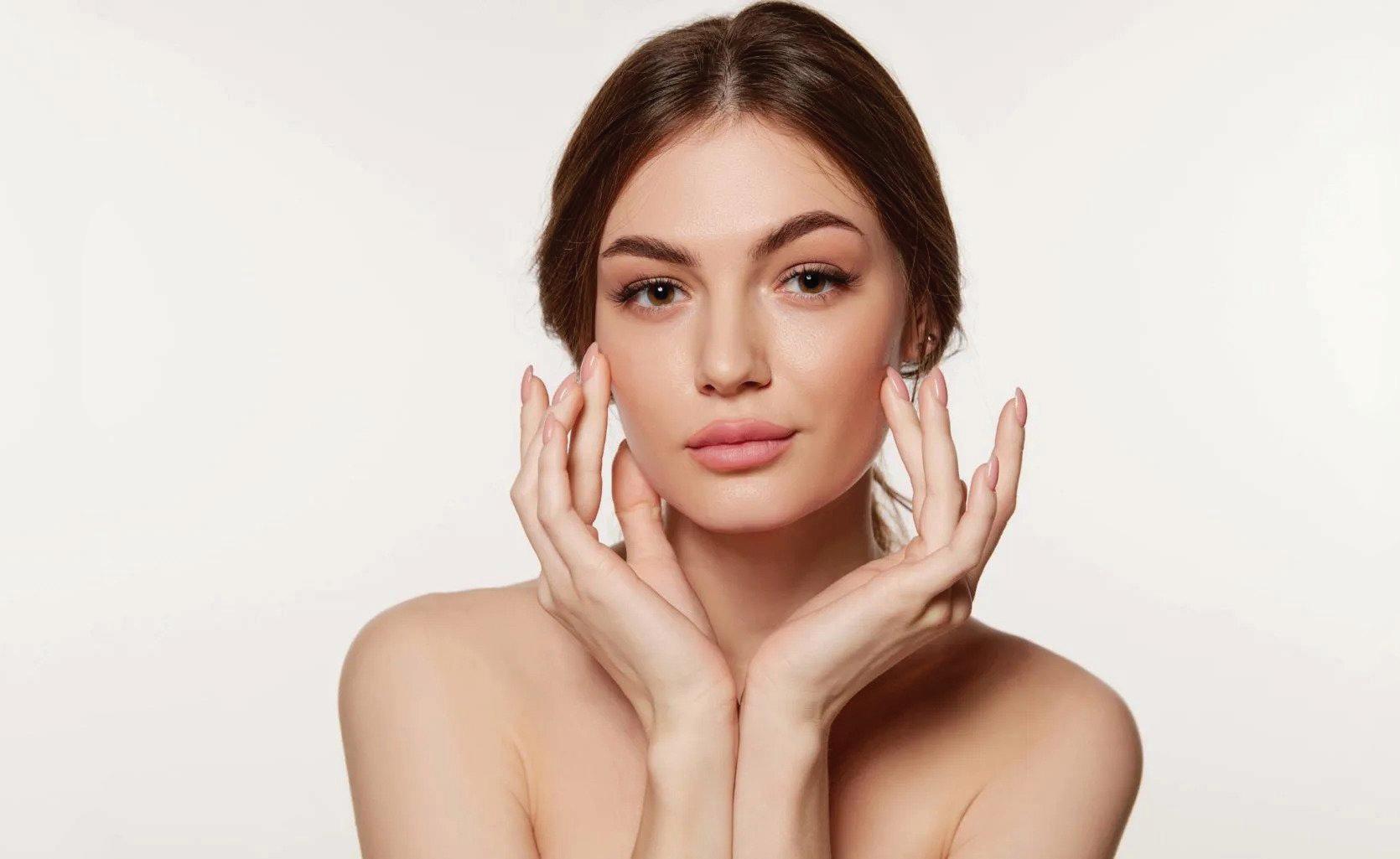
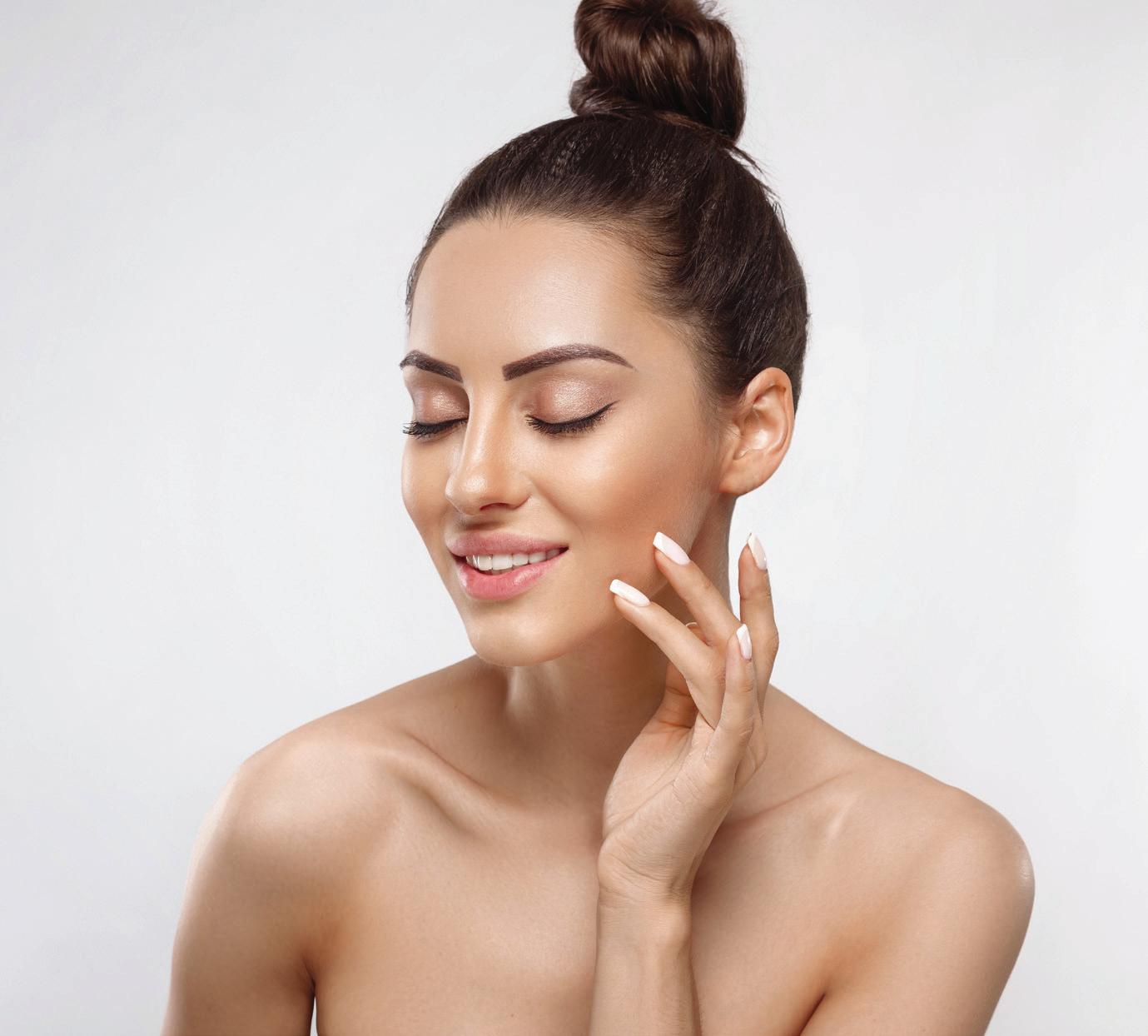
THE ART OF CHEEK VOLUME RESTORATION: A GENTLE PATH TO CONFIDENCE AND RADIANCE
INTRODUCTION
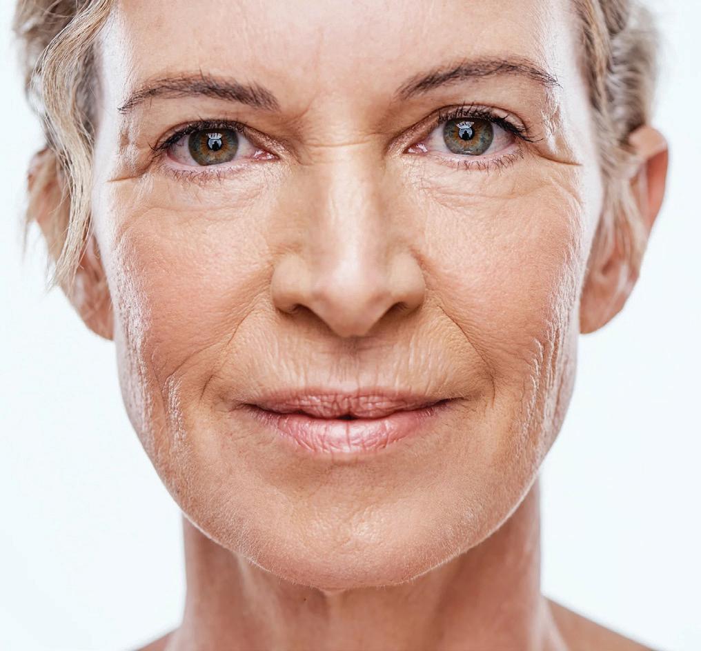
The cheeks are one of the most important features that give the face its shape and youthful glow. When cheeks are full and firm, they support the skin and create a balanced, attractive look. In youth, the face typically exhibits a V-shaped contour characterized by full cheeks and a well-defined jawline. However, as we age, the body produces less collagen and elastin proteins that keep skin firm and elastic. As a result, the skin
may sag, fine lines and wrinkles can appear, and the cheeks lose their fullness. Over time, the fat pads that give cheeks their fullness shrink and move downward, causing hollow areas in the midface and sagging along the jawline, often called jowls. While aging is the main cause of these changes, lifestyle factors such as excessive sun exposure, poor diet, smoking, and genetics can speed up the process. When cheeks lose their natural fullness, the face can appear
tired, sunken, or gaunt, which can affect a person confidence and how they feel about their appearance. As a result, restoring cheek volume has become a key goal in facial beauty and skin care treatments today. Fortunately, advances in aesthetic medicine have introduced a variety of non-surgical treatments that effectively restore volume and enhance facial contours without the need for invasive procedures.1
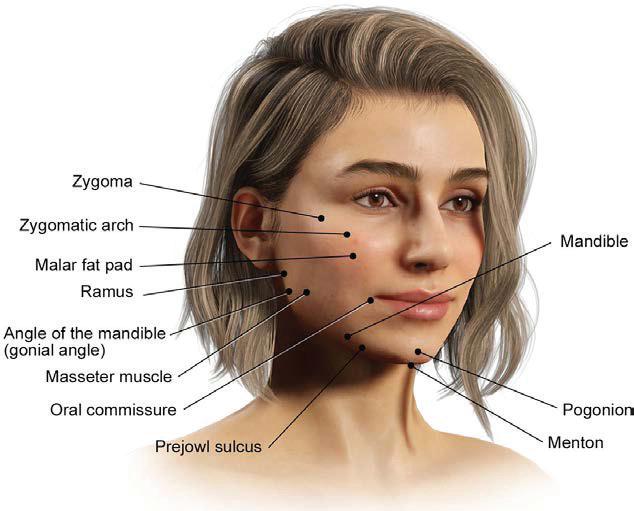
ANATOMY OF THE CHEEK: THE FOUNDATION OF FACIAL BEAUTY AND EXPRESSION
The anatomy of the cheek reveals the structural elements that define facial harmony, contour, and a youthful appearance. For professionals in medical aesthetics, understanding these layers is essential to delivering subtle, precise, and natural-looking enhancements.2
Bony Framework
• Zygoma and Zygomatic Arch form the high cheekbone structure that gives the face its elegant upper contour and lateral projection.2
• Mandible, particularly the Ramus, Angle of the Mandible (gonial angle), Pogonion, and Menton, shapes the jawline and chin, contributing to facial balance and definition in the lower face.2
These bones serve as the essential foundation for the overlying soft tissues. Their structure directly influences how facial contours develop and how agerelated changes become visible over time.
Fat Compartments
• The Malar Fat Pad is one of the most significant fat pads in the midface. It provides volume and a lifted appearance to the cheeks. As it shifts or diminishes with age, it can lead to hollowing, nasolabial folds, or sagging.2
• The Prejowl Sulcus is a common area where volume loss presents early, contributing to visible jowling, which is often a focus in aesthetic rejuvenation.2
Muscular Layer
• The Masseter Muscle lies at the side of the jaw and is
crucial for mastication. From an aesthetic standpoint, it influences facial width. In cosmetic practice, it is often addressed to create a more refined, tapered lower face.2
Functional and Expressive Zones
• The Oral Commissure, or corners of the mouth, along with the surrounding muscles, are involved in various expressions such as smiling and frowning. With age, these areas may become downturned and are commonly treated to restore a more uplifted, youthful appearance.2
Together, these components form a multi-layered system that supports both facial aesthetics and essential functions such as expression, chewing, and structural support.2
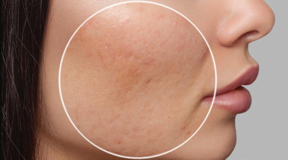
WHAT ARE CHEEK HOLLOWS AND VOLUME LOSS?
Cheek hollows are indentations that develop as the natural volume in the midface starts to diminish. This often affects the area beneath the eyes and across the upper cheeks, giving the face a more angular or shadowed appearance. It can result from factors like weight fluctuations, stress, or reduced facial support over time. These changes may lead to softer cheek contours and more noticeable folds around the mouth. Even mild volume loss can subtly affect facial symmetry and overall expression.3
Causes of Cheek Hollows and Volume Loss
1. Natural aging process:
As we age, the skin produces less collagen and elastin proteins responsible for firmness and elasticity causing the skin to become thinner and less supportive. Additionally, the bones in the midface slowly resorb or reduce in density, further decreasing the underlying structure that gives the cheeks their shape. Together, these changes cause the cheeks to lose volume and sag, creating hollow areas and a less defined facial contour.3
2. Genetic factors: Genetics significantly influence the timing and extent of facial volume loss. Some people naturally have less facial fat or thinner skin, which can make signs of volume loss appear earlier or more pronounced. Genetic predispositions also influence the way fat is distributed and how resilient skin is to aging.3
3. Lifestyle Factor: External habits and environmental conditions can accelerate facial volume loss. Significant or rapid weight loss reduces fat in the body as well as in the face, often resulting in sunken cheeks. Prolonged sun exposure leads to ultraviolet radiation damage, which compromises collagen integrity and promotes premature skin aging. Smoking also degrades collagen and restricts blood flow, diminishing overall skin health and facial volume.3
4. Hormonal Changes and Health Conditions: Hormonal fluctuations, especially during menopause, can lead to decreased collagen production and changes in fat distribution, accelerating facial volume loss. Additionally, certain health conditions such as chronic
inflammation, autoimmune disorders, or nutritional deficiencies can negatively impact skin quality and facial fat, contributing to hollow cheeks and loss of youthful fullness.4
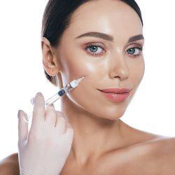
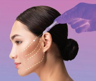
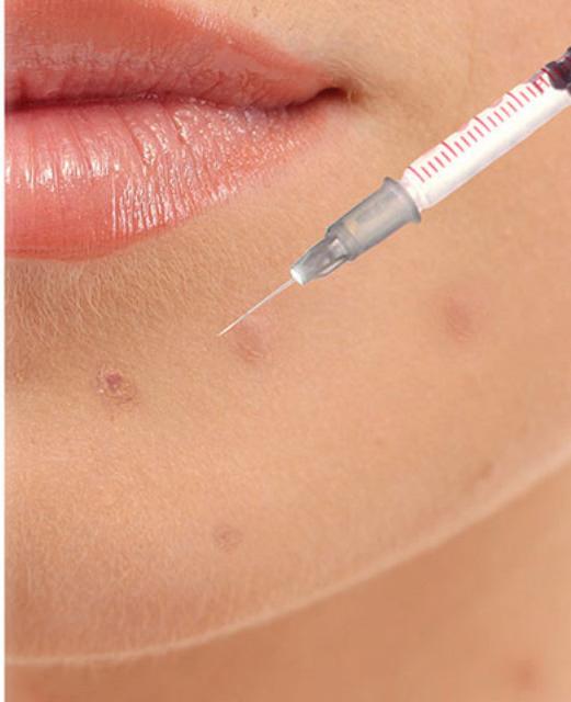
TREATMENT OPTIONS
•
Injectable fillers
➢ Hyaluronic acid (HA) filler is a quick, non-surgical way to restore lost midface volume, enhance facial contours, and create a more youthful appearance. It addresses one of the key signs of aging cheek volume loss without surgery or downtime. Hyaluronic acid fillers work by attracting and retaining moisture in the injected area, instantly restoring volume to hollow cheeks. They
provide structural support and lift, enhancing facial contours and creating a more youthful appearance.5
➢ Collagen-based fillers are used for hollow cheeks to restore volume and improve facial contour by integrating into the skin and stimulating natural collagen production. They provide immediate, natural-looking results and have a long history of safe use.
However, non-human collagen fillers may require skin testing due to allergy risks, and most need refrigerated storage. Their effects last a shorter time than some newer filler, so repeat treatments are necessary to maintain results. Overall, collagen fillers offer a biologically compatible option for subtle and effective cheek rejuvenation.5
• Thread lifts
It offers a minimally invasive alternative to surgery by lifting and tightening sagging midface tissues to restore volume and improve facial contours. The procedure works by inserting dissolvable threads under the skin, which physically lift the cheeks while simultaneously stimulating the body’s natural collagen production, leading to firmer, more youthful skin over time. Thread lifts provide immediate improvement with minimal downtime and can be combined with fillers or other treatments for enhanced results, making them a popular choice for patients seeking subtle facial rejuvenation without surgery.5
• Surgical Method
➢ Fat grafting offers a natural and long-lasting way to restore volume in hollow cheeks using the patient own fats. It works by harvesting fat from another area of the body, processing it, and then carefully injecting it into the cheek region, where the fat cells integrate with existing tissues are enhanced to offer better contour,
support, and a rejuvenated, more youthful look. Unlike temporary fillers, fat grafting can stimulate skin quality improvement through stem cells and often provides more durable results, making it an efficient option for those seeking long-lasting midface rejuvenation.6
ESSENTIAL LIFESTYLE FACTORS TO PRESERVE
CHEEK VOLUME AND PREVENT
FACIAL VOLUME LOSS
➢ Anti-Inflammatory, Collagen-Boosting Diet
Eating a balanced diet rich in vitamin C (citrus fruits, bell peppers), omega-3 fatty acids (salmon, flaxseeds, chia seeds), and adequate protein (lean meats, collagen peptides) supports collagen synthesis and skin strength. Limit sugar intake to prevent collagen damage caused by glycation.7
➢ Stress Management and Quality Sleep
Chronic stress elevates cortisol levels, which can accelerate collagen breakdown. Prioritize 7–9 hours of restful sleep nightly to boost growth hormone production, essential for skin repair and collagen renewal. Practices such as meditation, yoga, or deep breathing can also reduce stress.7
➢ Facial Exercises
Targeted facial exercises focusing on muscles like the zygomaticus major and minor improve muscle tone and provide natural lift. Simple routines
such as cheek puffing, smiling widely, and facial yoga can be performed regularly for best results.7
➢ Sun Protection
Ultraviolet (UV) radiation accelerates collagen degradation and skin aging. Use broad-spectrum sunscreen daily, wear protective clothing, and avoid excessive sun exposure to preserve skin integrity and cheek volume.7
➢ Hydration
Proper hydration maintains skin elasticity and volume. Drink adequate water throughout the day to keep skin plump and support metabolic processes that maintain tissue health.7
➢ Regular Physical Activity
Exercise improves blood circulation, delivering oxygen and nutrients to the skin and supporting healthy tissue maintenance. It also helps manage stress and promote better sleep quality, indirectly benefiting facial skin and volume.7
CONCLUSION
The gradual reduction of midfacial volume is a multifactorial process influenced by biological aging and environmental factors, significantly impacting facial aesthetics. Modern interventions, both minimally invasive and surgical, provide effective means to re-establish facial volume and contour, thereby enhancing structural support and improving appearance. Complementary lifestyle strategies aimed at promoting skin integrity and tissue health are essential adjuncts to these treatments. Overall, a comprehensive approach that integrates medical therapies with preventive care optimizes outcomes and supports long-term facial rejuvenation.
REFERENCES
1. Muhn C, Rosen N, Solish N, et al. The evolving role of hyaluronic acid fillers for facial volume restoration and contouring: a Canadian overview. Clin Cosmet Investig Dermatol. 2012;5:147-158. doi:10.2147/CCID.S30794
2. Nguyen JD, Duong H. Anatomy, Head and Neck, Cheeks. In: StatPearls. Treasure Island (FL): StatPearls Publishing; August 14, 2023.
4. Moghadam S, Roca Y, LaGuardia JS, et al. Effect of Duration of Feminizing Hormone Therapy on Facial Fat Volumes. Plast Reconstr Surg. 2024;154(5):1081-1088. doi:10.1097/PRS.0000000000011200
5. Clark NW, Pan DR, Barrett DM. Facial fillers: Relevant anatomy, injection techniques, and complications. World J Otorhinolaryngol Head Neck Surg. 2023;9(3):227-235. Published 2023 Aug 2. doi:10.1002/wjo2.126
6. Vasavada A, Raggio BS. Autologous Fat Grafting for Facial Rejuvenation. In: StatPearls. Treasure Island (FL): StatPearls Publishing; July 31, 2023.
7. Hong GW, Kim SB, Park Y, et al. Anatomical Considerations for Thread Lifting Procedure. J Cosmet Dermatol. 2025;24(1):e16618. doi:10.1111/jocd.16618
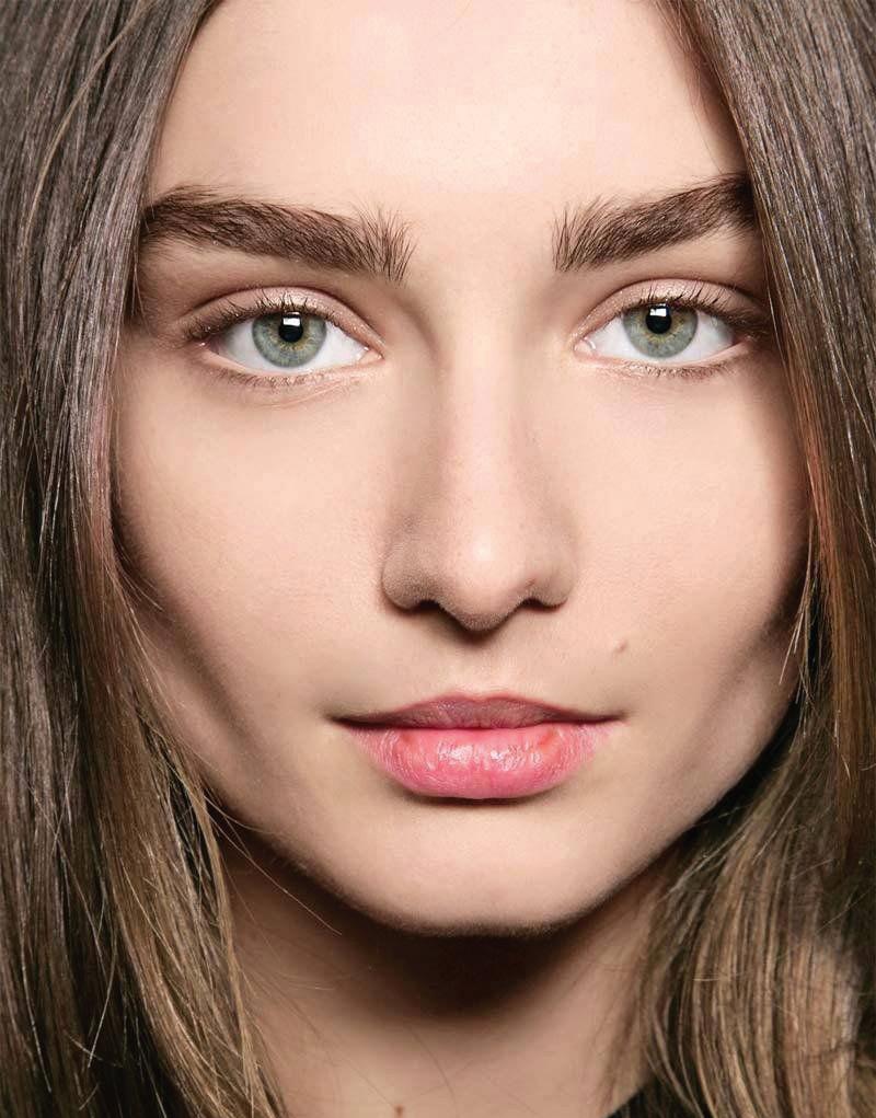
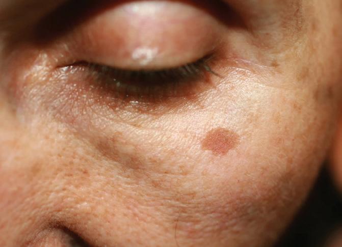
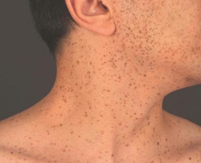
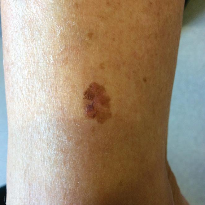
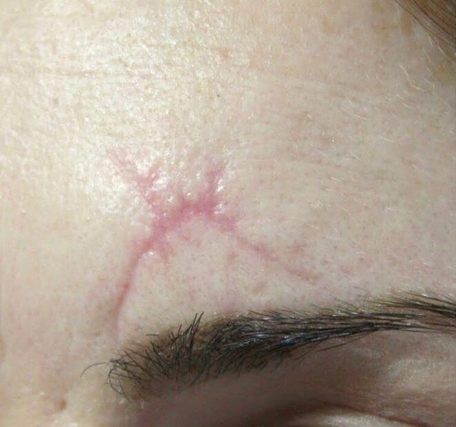
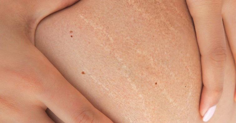
THE SPECTRUM OF LENTIGINES: FROM SUN DAMAGE TO ADVANCED CARE
INTRODUCTION
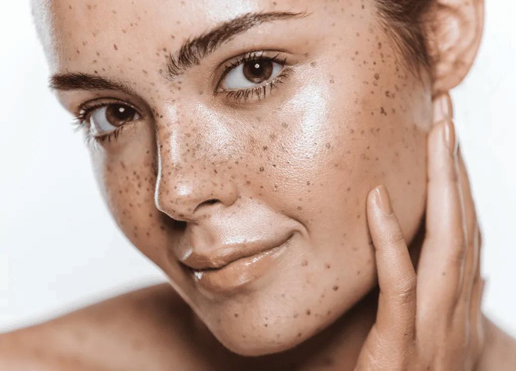
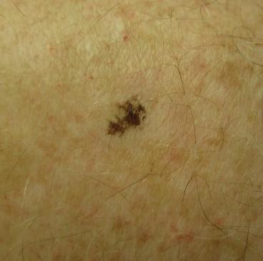
Lentigines are clearly defined, pigmented spots that appear on the skin as a result of increased melanin production or a higher concentration of melanocytes in the epidermis. Often referred to as age spots, sun spots, or liver spots, they are most commonly seen in
women during midlife and beyond, especially in those with a history of frequent sun exposure. Though medically harmless, these marks are visible signs of sun-induced skin ageing and can sometimes resemble more serious conditions such as melanoma, making regular skin checks important. Their presence
not only affects the complexion but also serves as a reminder of the long-term effects of UV exposure. As awareness of sun safety and skincare deepens, many women are now seeking both preventative care and gentle treatment solutions to maintain radiant, even-toned skin throughout the years.1
CONTRIBUTING FACTORS
➢ Chronic Ultraviolet (UV) Radiation Exposure: Prolonged sun exposure leads to DNA damage in skin cells, stimulating melanocyte proliferation and increased melanin production.
➢ Aging: Reduced ability of the skin to repair UV-induced damage over time results in the accumulation of pigmented lesions.
➢ Genetic Predisposition: Some individuals have a higher susceptibility to developing lentigines due to hereditary factors.
➢ Hormonal Changes: Hormonal shifts during pregnancy or while taking birth control pills can impact the activity of pigment-producing cells in the skin.
➢ Environmental Pollutants: Exposure to pollutants may exacerbate skin damage and contribute to lentigine formation.
➢ Photosensitizing Medications: Certain drugs increase skin sensitivity to UV radiation, promoting lentigine development.
TYPES OF LENTIGENS
➢ Solar lentigines, also known as actinic lentigines, are benign pigmented lesions that result from chronic sun exposure. They do not fade with reduced UV exposure and may darken over time. These spots typically appear after the fourth decade of life but can develop earlier in individuals with fair skin or a history of significant sun exposure.2
➢ LEOPARD syndrome (Multiple Lentigines Syndrome) is a rare autosomal dominant disorder, most often caused by mutations in the PTPN11gene. LEOPARD is an acronym used to describe the main signs of the condition: spots on the skin (Lentigines), heart rhythm problems, wide-set
➢ Ink-spot lentigo, also called reticulated black solar lentigo, is a benign, darkly pigmented macule with irregular, splatter-like borders, often found on sun-exposed areas such as the shoulders and upper back. Caused by prolonged UV exposure, it may mimic melanoma due to its color and shape.3
eyes, narrowing of the pulmonary valve, genital abnormalities, slow growth, and hearing loss. Lentigines usually develop in early childhood, especially on the face and upper torso.4
➢ Lentigomaligna is a type of melanoma in situ found primarily in older adults on chronically sun-exposed areas like the face and arms. It usually shows up as a flat spot that grows slowly and has uneven colouring, often in shades of brown, black, or gray. While it remains confined to the epidermis initially, it can become invasive over time, developing into lentigomalignamelanoma.5
TREATMENT OPTIONS
➢ Topical treatments
These are commonly used for managing lentigines, particularly when the goal is cosmetic improvement. Hydroquinone is one of the most effective skin-lightening agents, working by inhibiting melanin production and gradually fading pigmented spots. Tretinoin, a retinoid, promotes skin cell turnover and helps to lighten hyperpigmentation over time. Other topical agents such as azelaic acid, kojic acid, and vitamin C are also used either alone or in combination to reduce pigmentation and brighten the skin. These treatments are often considered first-line options for mild cases.2
➢ Procedural Techniques
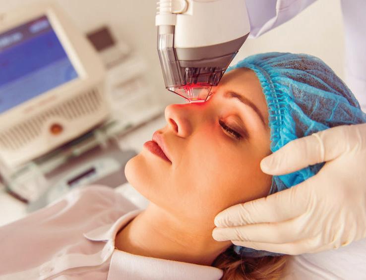
Intense Pulsed Light (IPL) therapy is a highly effective and non-invasive treatment option for lentigines, targeting the pigmented cells in the skin using broadspectrum light. The light energy is absorbed by melanin, which heats and breaks down the pigment, allowing the body to naturally clear it over time. IPL is valued for its ability to treat multiple pigmented spots in a single session with minimal downtime, as patients typically experience only mild redness or swelling that resolves quickly. In addition to reducing pigmentation, IPL can improve overall skin texture and stimulate collagen production, offering added benefits for skin rejuvenation. While IPL works well for many skin types, it is important to take precautions with darker skin to minimize the chance of pigment changes or irritation.6
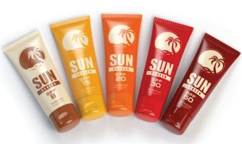
➢ Preventive Measures
• Consistent application of a broad-spectrum sunscreen with an SPF of 30 or higher is essential and should be performed daily, regardless of weather conditions, including overcast or cloudy days. This practice helps protect the skin from both UVA and UVB rays, significantly reducing the risk of suninduced pigmentation and preventing the formation of lentigines.
• Wearing protective clothing, such as long-sleeved garments, wide-brimmed hats, and UV-blocking sunglasses, is a vital measure to shield the skin from harmful ultraviolet radiation when outdoors. These
• Fractional laser treatment is an effective way to reduce lentigines by targeting and breaking down pigmented cells with tiny laser beams. It helps your skin heal and look smoother, with few side effects and minimal recovery time. Multiple sessions may be needed for best results, and sun protection afterward is important to prevent spots from returning. Overall, fractional lasers offer a safe and controlled method to clear lentigines and rejuvenate the skin.7
Integrating multiple approaches allows for tailored management of pigmented lesions, promoting clearer, healthier skin. Ongoing care and protection are vital to maintain treatment benefits and prevent recurrence.
physical barriers provide an additional layer of defence, especially during extended periods of sun exposure, helping to minimize the risk of developing lentigines and other sun-related skin damage.
• Limiting sun exposure during these times significantly reduces the risk of developing lentigines and other forms of photoaging.
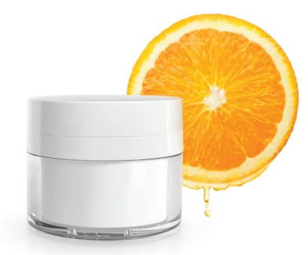
• The application of antioxidants, such as topical vitamin C, helps to mitigate oxidative stress by neutralizing free radicals, thereby protecting the skin from photodamage and reducing the likelihood of lentigine formation.
CONCLUSION
Understanding lentigines and their underlying triggers is an important step toward achieving healthier, more resilient skin. While these spots are benign, they reflect the lasting impact of both environmental exposure and genetic predisposition. Modern skincare innovations have made it possible to treat lentigines with confidence while also achieving noticeable improvements in skin tone and radiance. Early attention to irregular changes remains essential, particularly to rule out more serious concerns. A continuous awareness around sun protection and self-monitoring empowers women to take charge of their skincare journey. As innovation progresses, we can look forward to more refined and effective solutions making it possible to care for our skin with both confidence and clarity. A personalized, proactive routine remains the key to maintaining a luminous, even-toned complexion at every age.
REFERENCES
1. Puckett Y, Geist R, Chen CSJ. LentigoMaligna Melanoma. [Updated 2025 Jun 25]. In: StatPearls [Internet]. Treasure Island (FL): StatPearls Publishing; 2025 Jan-.
2. Saki N, Modabber V, Kasraei H, Kasraee B. Successful treatment of solar lentigines by topical application of stabilized cysteamine: A vehicle-controlled, double-blind randomized study. Health Sci Rep. 2024; 7(2):e1930. Published 2024 Feb 25. doi:10.1002/hsr2.1930
3. Salvi, M., Cappilli, S., Ricci, C., Palmisano, G., Di Stefani, A. and Peris, K. (2024), Digital imaging of ink spots lentigo: Confocal microscopy and line-field confocal optical coherence tomography assessment. JEADV ClinPract, 3: 1308-1311
4. Wang, T., Lin, Y., Sun, L., Mao, L., Gao, X., Liu, X. and Liu, H. (2024), An incomplete LEOPARD syndrome presented with generalized lentigines. J CosmetDermatol, 23: 711-713
5. Naik PP. Diagnosis and Management of LentigoMaligna: Clinical Presentation and Comprehensive Review. J Skin Cancer. 2021; 2021:7178305. Published 2021 Jul 24. doi:10.1155/2021/7178305
6. Fischer DL, Han H, Gade A, Nestor MS. Intense pulsed light for the treatment of pigmented and vascular disorders and lesions: A review. Dermatological Reviews. 2021; 2: 69-81.
7. Lueangarun S, Tempark T. Efficacy of 532-nm Nd: YAG Fractional Picosecond Laser with 9 mm High Coverage Handpiece Micro Lens Array in the Treatment of Facial Partial Unilateral Lentiginosis: A Case Report. J Lasers Med Sci. 2024; 15:e24. Published 2024 Jul 27. doi:10.34172/jlms.2024.24
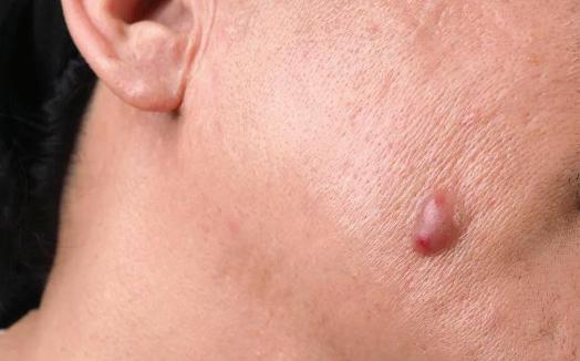
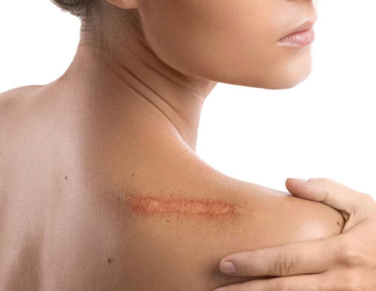
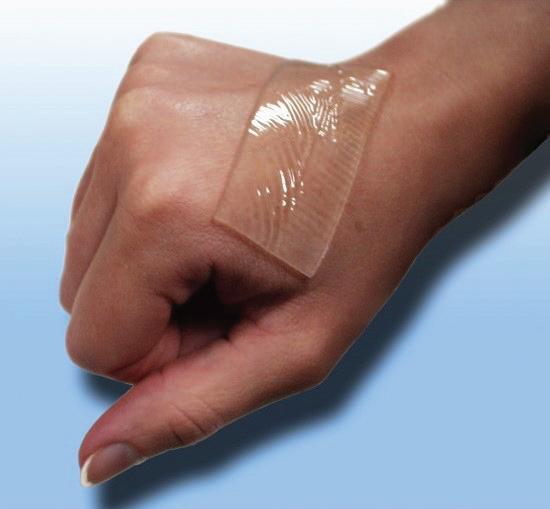
IMPROVING THE APPEARANCE OF SCARS: CURRENT TECHNIQUES AND APPROACHES
INTRODUCTION
Scars are the skin natural way of reflecting healing, recovery, and resilience. Whenever the skin experiences an injury—whether from a cut, burn, surgery, acne, or a chronic skin condition—the body instinctively begins building new tissue to close the wound and restore its protective barrier. The result is a scar, unique
in shape, texture, and tone, formed through the body intricate healing process. While most scars gradually fade over time, their appearance can influence how individuals, especially women, feel about their skin and overall confidence. Although scars lack the natural features of healthy skin such as pigmentation, sweat
glands, or hair follicles, they remain visible reminders of the body extraordinary ability to repair itself. For many, understanding the science behind scar formation is the first step in embracing their skin journey with compassion and confidence.1
UNDERSTANDING SCARS AND ITS TYPES
When the skin is injured, the body repairs it through inflammation, tissue formation, and remodeling. Immune cells clean the wound, and then fibroblasts produce collagen to rebuild the skin structure. Over time, collagen reorganizes to strengthen the healed area, but scar tissue differs from normal skin because it has fewer glands and a different collagen arrangement, which makes scars visible. Scars form because the body replaces damaged skin with fibrous tissue, and factors like wound size and healing affect their appearance. Facial scars go beyond surface-level marks. They are the visible result of the skin extraordinary ability to heal. When the skin undergoes injury, the body initiates a complex repair process to restore its barrier.
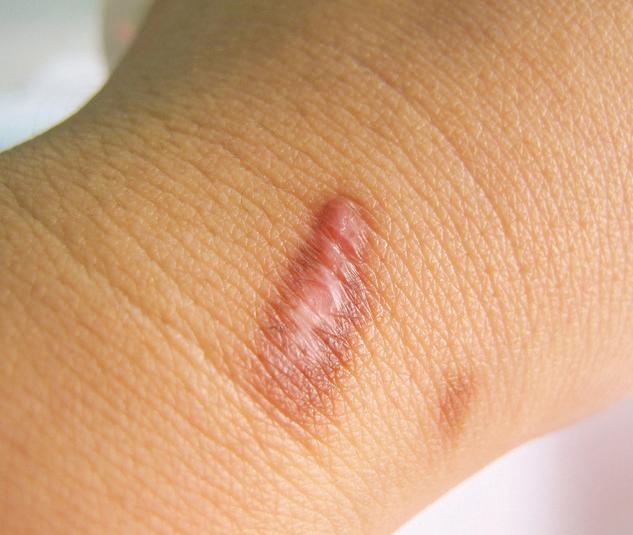
1. Hypertrophic scar - A hypertrophic scar is a raised, thickened scar that stays within the original wound edges. It forms due to excess collagen after skin injury, such as surgery, trauma, or burns. These scars are usually red or pink, may itch or hurt, and typically appear within weeks of healing. Over time, they often improve and become less noticeable.2
2. Keloid scar - Keloid scars are raised, thickened scars that extend beyond the original wound, often growing larger over time. They can be itchy, painful, or tender to the
touch. Keloids are more common in people with darker skin tones and may occur after injuries, surgeries, or even minor skin trauma. Unlike hypertrophic scars, they grow past the wound edges and are often longlasting.3
3. Stretch marks (striae) on the face are rare but can occur with rapid skin growth or the use of steroids. They appear as thin, linear marks that may be pink, red, or purple initially before fading to a lighter color over time. While they are more common on areas like the abdomen or thighs, they can occasionally develop on the face, especially during hormonal changes
or rapid weight fluctuations.4
4. Traumatic scars result from cuts, lacerations, or injuries to the skin. They can vary greatly in size and shape and often have jagged or irregular edges. These scars reflect the nature and severity of the original wound and may be more noticeable depending on how the injury healed.5
METHODS FOR IMPROVING SCAR APPEARANCE
Non-invasive techniques
➢ Silicone Gels and Sheets
Silicone gels and sheets are popular, non-invasive treatments for reducing scar thickness, redness, and discomfort. They create a moist environment that helps regulate collagen production and improve scar texture. Effective for both new and old scars especially hypertrophic and keloid types they are safe, easy to use, and
backed by strong clinical evidence. Consistent use over several weeks is key to achieving the best results.6
➢ Pressure therapy and massage
These are hands-on techniques used to improve the appearance of scars, especially hypertrophic and burn-related scars. Pressure therapy involves applying constant pressure using garments or dressings, which helps flatten and soften the scar by
reducing blood flow and collagen build-up. Massage, on the other hand, increases circulation, breaks down excess scar tissue, and helps improve flexibility and skin texture.7
help reduce thick scars by calming inflammation and slowing excess collagen production. They work to flatten and soften the scar over time. Regular sessions are needed for optimal results. These injections are a highly effective treatment for stubborn scars, offering noticeable improvements with consistent use.8
➢ Laser therapies improve scars by resurfacing skin and boosting healthy tissue growth. They help reduce redness, flatten raised scars, and smooth texture. Different lasers target various scar types, and multiple sessions are usually needed for best results. These treatments are minimally invasive and have a relatively quick recovery time compared
to surgical options.9
Emerging technologies
➢ Plasma radiofrequency (RF)
treatment uses ionized gas combined with radiofrequency energy to precisely target and remodel scar tissue. It stimulates collagen production and skin regeneration, improving the texture and appearance of scars such as acne, hypertrophic, and surgical scars. This minimally invasive method delivers controlled thermal energy, reducing damage to surrounding skin while promoting skin tightening and even tone. Multiple sessions are usually needed for the best results. Plasma RF offers minimal downtime and fewer side effects compared to traditional laser treatments and are safe for most skin types when performed by trained professionals.10
➢ Ultrasound therapy for scar treatment uses sound waves to penetrate deep into the scar tissue, helping to break down dense collagen fibers and promote tissue remodeling. This process increases blood flow and stimulates healing in the affected area, which can reduce scar thickness, improve flexibility, and soften the scar. The treatment is non-invasive, painless, and has minimal side effects. Ultrasound is often used in combination with other therapies to
enhance overall results and is suitable for treating various types of scars, including hypertrophic and burn scars.11
PSYCHOLOGICAL
IMPACT
Scars can have a significant psychological impact, affecting self-esteem, confidence, and emotional well-being. Visible or severe scars may lead to feelings of embarrassment, anxiety, or social withdrawal. Providing emotional support and counselling is important to help individuals cope with these challenges. Encouraging open communication and offering reassurance can improve mental health and promote a positive outlook during scar treatment and recovery.
CONCLUSION
Scars are more than just physical marks they often carry deep emotional and personal significance. Many individuals, especially when scars are visible or prominent, they can affect self-image and confidence. Understanding the nature and types of scars allows for greater awareness and compassion, both personally and socially. By acknowledging the emotional and psychological impact of scars, we can promote a more empathetic approach to care and support. Embracing skin, with or without scars, is a vital step toward self-acceptance and emotional well-being.
REFERENCES
1. Lin X, Lai Y. Scarring Skin: Mechanisms and Therapies. Int J Mol Sci. 2024;25(3):1458. Published 2024 Jan 25. doi:10.3390/ijms25031458
2. Mony MP, Harmon KA, Hess R, Dorafshar AH, Shafikhani SH. An Updated Review of Hypertrophic Scarring. Cells. 2023;12(5):678. Published 2023 Feb 21. doi:10.3390/cells12050678
3. Zhao SY, Wu D, Cheng C, Xie JH. Advances and future directions in keloid research: Pathogenesis, diagnosis and personalized treatment strategies. World J Clin Cases. 2023;11(34):8094-8098. doi:10.12998/wjcc.v11.i34.8094
4. Mendes N, Alves PJ, Barros M, Rodrigues JM, Machado J. A Narrative Review of Current Striae Treatments. Healthcare (Basel). 2022;10(12):2565. Published 2022 Dec 17. doi:10.3390/healthcare10122565
5. Tian Q, Zhang YX, Wang JJ, Huang GB. Advances in Photoelectric Therapy for the Early Intervention and Treatment of Traumatic Scars. Clin Cosmet Investig Dermatol. 2023; 16:869-877. Published 2023 Apr 4. doi:10.2147/CCID. S407361
6. Nguyen A, Huynh C, Goh A, Co A, Hassan O, Phan S. A systematic review of the management of postoperative scars with silicone gel-based products in randomized controlled trials. Dermatol Online J. 2023;29(4):10.5070/ D329461860. Published 2023 Aug 15. doi:10.5070/D329461860
7. De Decker I, Beeckman A, Hoeksema H, et al. Pressure therapy for scars: Myth or reality? A systematic review. Burns. 2023;49(4):741-756. doi:10.1016/j.burns.2023.03.007
8. Yuan B, Upton Z, Leavesley D, Fan C, Wang XQ. Vascular and Collagen Target: A Rational Approach to Hypertrophic Scar Management. Adv Wound Care (New Rochelle). 2023;12(1):38-55. doi:10.1089/wound.2020.1348
9. Campolmi P, Quintarelli L, Fusco I. A Multimodal Approach to Laser Treatment of Extensive Hypertrophic Burn Scar: A Case Report. Am J Case Rep. 2023; 24:e939022. Published 2023 Jun 5. doi:10.12659/AJCR.939022
10. Baroni A, Verolino P. Plasma Radiofrequency Ablation for Scar Treatment. Journal of Clinical Medicine. 2022; 11(1):140. https://doi.org/10.3390/jcm11010140
11. Riis Porsborg S, Krzyslak H, Pierchala MK, et al. Exploring the Potential of Ultrasound Therapy to Reduce Skin Scars: An In Vitro Study Using a Multi-Well Device Based on Printable Piezoelectric Transducers. Bioengineering (Basel). 2023;10(5):566. Published 2023 May 9. doi:10.3390/bioengineering10050566
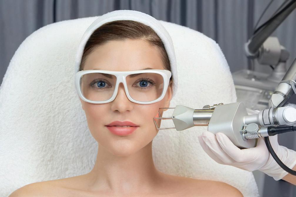
The skin is our body largest organ, more than just a protective shield it is a dynamic interface between us and the world, reflecting not only our health but also our age, lifestyle, and well-being. Maintaining its vitality has become a priority for many, driving the evolution of treatments that nurture and enhance its natural resilience. In this landscape of
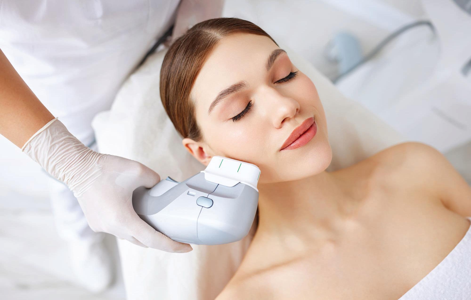
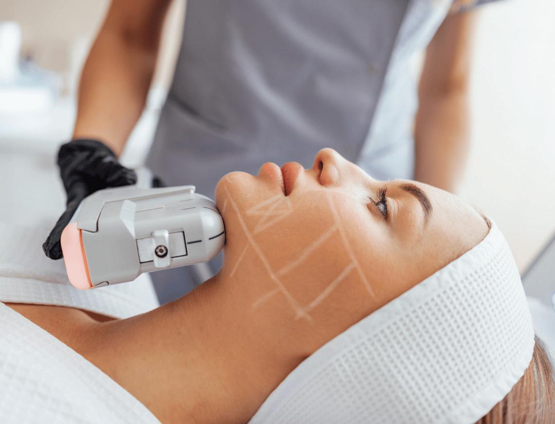
advanced skincare, High-Intensity Focused Ultrasound (HIFU) has emerged as a groundbreaking technology in the realm of noninvasive treatments. By harnessing the power of focused ultrasound waves, HIFU offers a precise and effective way to rejuvenate the skin and treat medical conditions without the need for surgery. With a rising preference for minimally invasive
procedures, both in aesthetics and healthcare, the demand for treatments that combine safety, efficacy, and minimal downtime has never been greater. HIFU perfectly aligns with this modern sensibility, delivering remarkable results while preserving the natural integrity of the body.1
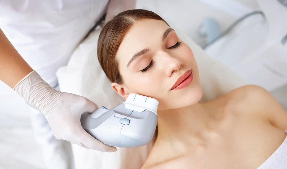
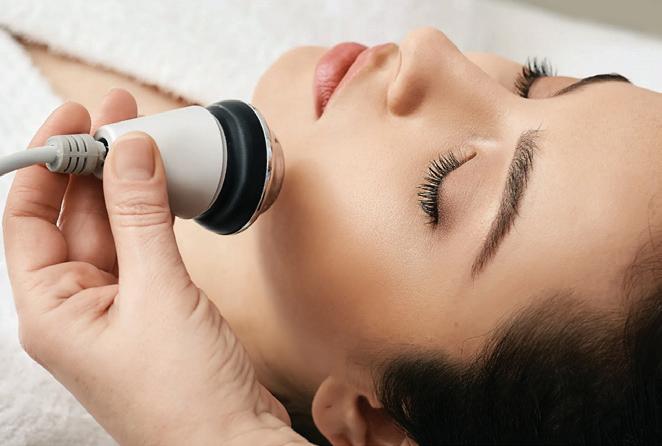
WHAT IS HIFU?
High-Intensity Focused Ultrasound (HIFU) is a non-invasive therapeutic technology that uses focused ultrasound energy to precisely target tissue beneath the skin surface. By concentrating high-frequency sound waves on a specific area, HIFU generates thermal energy that stimulates collagen production and, in some cases, causes targeted tissue destruction without harming the surrounding skin. This controlled energy delivery makes HIFU effective for skin tightening and lifting, and medical applications, such as tumour ablation, with its ability to treat deep layers without surgery or downtime.2
HOW DOES HIFU WORK?
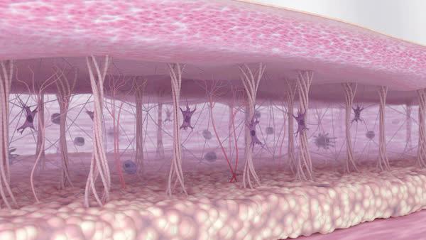
HIFU delivers precisely targeted ultrasound energy to specific skin depths typically 1.5 mm, 3.0 mm, and 4.5 mm stimulating tissue beneath the surface. These waves converge at a precise point, generating thermal energy that raises the temperature in targeted tissues to around 60–70°C. This controlled heating causes thermal coagulation a process that triggers immediate tissue tightening and initiates the body natural woundhealing response.3 As the tissue responds to this heat, fibroblasts the cells responsible for producing collagen are activated. This results in a gradual regeneration of
collagen and elastin fibers, which are essential for maintaining skin structure, elasticity, and firmness.3
HIFU targets and affects only the tissues at the focal point, while leaving the skin surface and surrounding areas completely untouched. This allows for effective skin lifting and tightening without the risks or downtime associated with invasive procedures. Results typically develop over 2 to 3 months and can continue to improve for up to 6 months, offering a natural, long-lasting enhancement.3
APPLICATIONS
➢ Role in Wrinkle Reduction and Skin
Tightening
HIFU works by delivering focused ultrasound energy deep beneath the skin surface to precisely target areas prone to wrinkles and sagging. The heat generated stimulates an immediate tightening effect and activates the body natural healing process, encouraging the production of collagen and elastin essential proteins that restore skin firmness and smoothness. This dual action not only helps reduce fine lines and wrinkles but also strengthens the deeper layers that support skin structure, resulting in improved skin tone, lifted contours, and a more youthful appearance. Over several weeks to months, these natural rebuilding processes continue, providing lasting and gradual enhancement without the need for surgery or downtime.4
➢ Role in Facial Uplift
High-Intensity Focused Ultrasound initiates a controlled thermal response within the foundational layers of the face, particularly in regions responsible for facial support. By concentrating energy at a specific depth beneath the skin, HIFU triggers localized heating that promotes cellular activity and encourages structural renewal. This process enhances the density and resilience of the tissue, resulting in a visible lifting of the facial profile. Over time, the skin adapts to this internal stimulation, leading to firmer features, improved definition, and a naturally elevated appearance all achieved without invasive techniques or disruption to the surface.5, 6
➢ Role in Medical Treatment
High-Intensity Focused Ultrasound (HIFU) is a non-invasive medical technology that precisely targets and destroys abnormal tissues such as tumours, fibroids and breast cancer. It is also used to treat neurological disorders as well as to manage chronic pain by focusing ultrasound energy on specific areas. HIFU offers an effective alternative to surgery, with minimal risks and faster recovery.5, 6
ADVANTAGES OF HIFU
➢ HIFU is a non-invasive procedure that does not require surgical incisions, making it a preferred option for patients seeking alternatives to traditional surgery. It utilizes focused ultrasound waves to target and treat specific tissues without affecting surrounding healthy areas.4
➢ Patients undergoing HIFU treatments often experience minimal downtime. Most can resume their daily activities shortly after the procedure, as it typically does not involve significant recovery periods associated with invasive surgeries.4
➢ HIFU has a favourable safety profile, with a low incidence of complications. Most side effects are transient and mild, and such as temporary redness or swelling at the treatment site. Serious adverse events are rare, making it a safer alternative to more invasive treatments.7
➢ HIFU has been shown to deliver long-lasting results with a naturallooking appearance. Treated areas often exhibit noticeable improvements in skin firmness and elasticity, contributing to a more youthful and revitalized look.4
LIMITATIONS
• Like all ultrasound-based technologies, HIFU can be affected by imaging issues such as shadowing and distortion, which can make it harder to accurately target the treatment area.8
• Treating tumours located deep in the body or near bones is challenging, because bones can block or interfere with the ultrasound waves.8
• HIFU cannot pass through gas, so areas behind gas-filled organs like the bowel may not be reachable for treatment.8
• Reflected ultrasound waves in HIFU carry a lot of energy, which can sometimes cause burns in the tissue between the skin and the treatment target.8
• If the ultrasound beam is not precisely focused, it may deliver energy to the wrong area, causing unintended damage to nearby or surface tissues.8
• To ensure safety and effectiveness, HIFU should only be used on carefully selected patients, with proper planning and skilled operators to avoid complications.8
REFERENCES
CONCLUSION
High-Intensity Focused Ultrasound (HIFU) is a leading non-invasive treatment offering precise, safe, and effective solutions in both medical and aesthetic fields. Its ability to target deep tissues without harming the surface makes it ideal for those seeking subtle, non-surgical improvements. As technology advances, HIFU is expanding into oncology, neurology, and cosmetic applications, with AI integration and real-time imaging enhancing precision. Emerging uses like tissue regeneration, combination therapies, and non-thermal methods such as histotripsy, along with portable devices and growing clinical validation, are set to make HIFU a key tool in personalized minimally invasive care.
1. Turner B, Cranston D. A Review of High-Intensity Focused Ultrasound. International Journal of Translational Medicine. 2024; 4(1):197-207. https://doi.org/10.3390/ijtm4010011
2. Yao R, Hu J, Zhao W, Cheng Y, Feng C. A review of high-intensity focused ultrasound as a novel and non-invasive interventional radiology technique. J Interv Med. 2022;5(3):127-132. Published 2022 Jun 22. doi:10.1016/j.jimed.2022.06.004
3. Biskanaki F, Tertipi N, Sfyri E, Kefala V, Rallis E. Complications and Risks of High-Intensity Focused Ultrasound (HIFU) in Esthetic Procedures: A Review. Applied Sciences. 2025; 15(9):4958. https://doi.org/10.3390/ app15094958
4. Haykal D, Sattler S, Verner I, Madhumita M, Cartier H. A Systematic Review of High-Intensity Focused Ultrasound in Skin Tightening and Body Contouring. Aesthet Surg J. 2025;45(7):690-698. doi:10.1093/asj/ sjaf053
5. Lari, S., Kohandel, M. & Kwon, H.J. Model based deep learning method for focused ultrasound pathway scanning. Sci Rep 14, 20042 (2024). https://doi.org/10.1038/s41598-024-70689-9
6. di Biase L, Falato E, Caminiti ML, Pecoraro PM, Narducci F, Di Lazzaro V. Focused Ultrasound (FUS) for Chronic Pain Management: Approved and Potential Applications. Neurol Res Int. 2021;2021:8438498. Published 2021 Jun 29. doi:10.1155/2021/8438498
7. Ali MM, Raphael Mpehle C, Olusola E, et al. A systematic review of the side effects of high-intensity focused ultrasound ablation of uterine fibroids. Proc (Bayl Univ Med Cent). 2024;37(6):947-956. Published 2024 Aug 21. doi:10.1080/08998280.2024.2387497
8. Tempany CM, McDannold NJ, Hynynen K, Jolesz FA. Focused ultrasound surgery in oncology: overview and principles. AJR Am J Roentgenol. 2008;190(1):191-199. doi:10.2214/AJR.07.2671
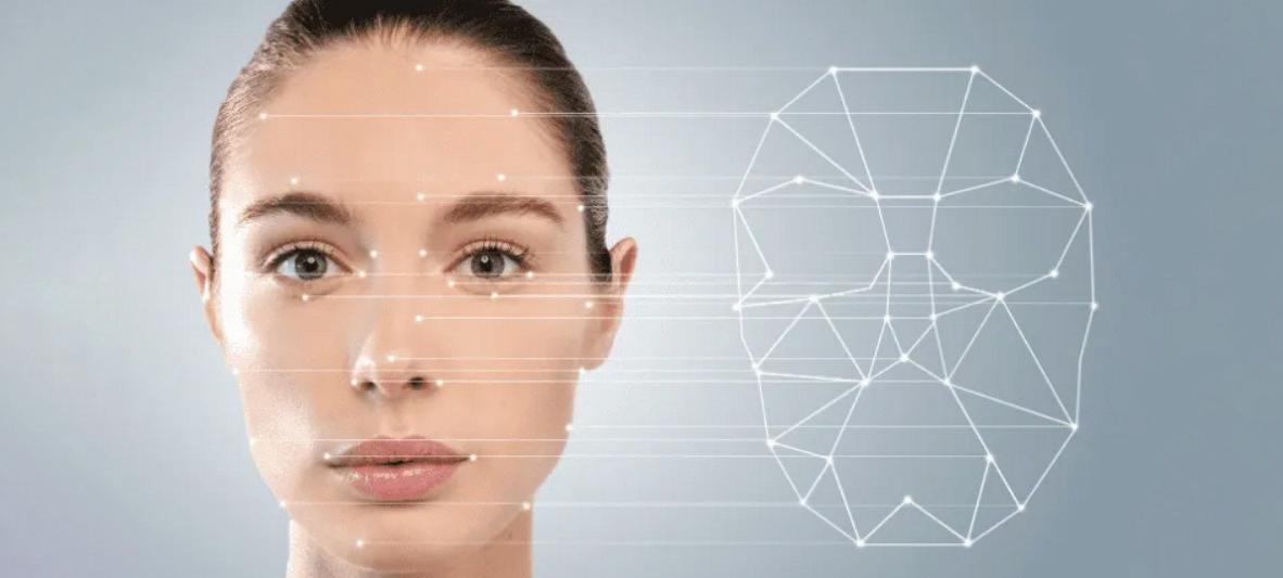
AI-GUIDED FACIAL
ANALYSIS: TRANSFORMING PERSONALIZED AESTHETICS AND HEALTHCARE
INTRODUCTION
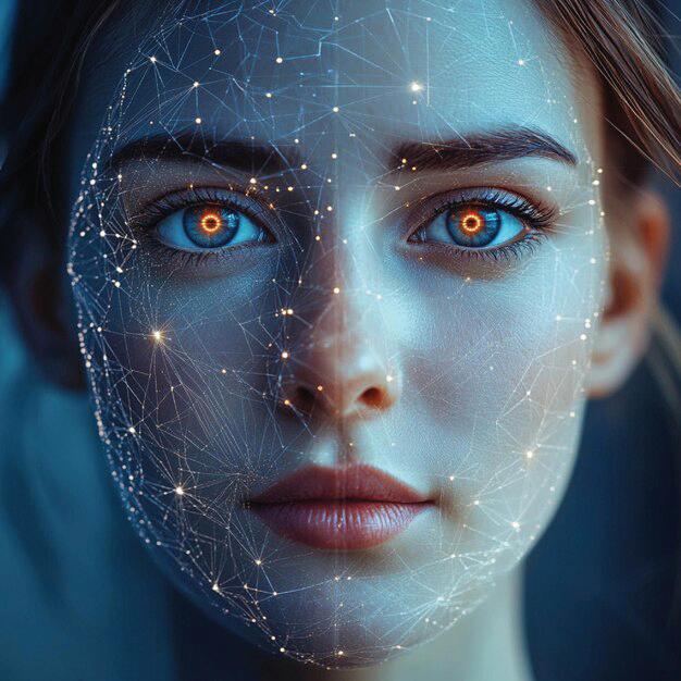
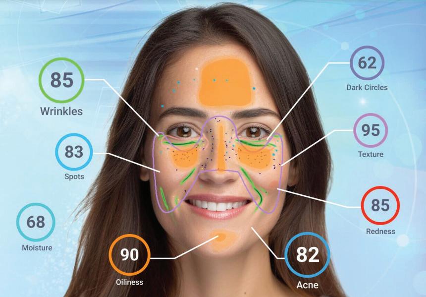
AIguided facial analysis is a cutting-edge technology that harnesses the power of artificial intelligence to examine, interpret, and understand facial features with remarkable precision. Rapidly gaining momentum in skincare, medical aesthetics, and holistic wellness, this non-invasive and data-driven approach is transforming the way professionals assess and manage skin health and facial aesthetics. By analysing key facial characteristics such as
skin texture, tone, elasticity, and symmetry, AI systems deliver highly personalized insights that surpass traditional methods. From identifying early signs of aging to monitoring changes in skin condition and supporting customized treatment planning, this technology offers real-time, evidence-based data to guide clinical decisions. Its ability to process vast volumes of data within seconds ensures efficiency, consistency, and objectivity, achieving a level
of analytical precision that extends beyond the capabilities of human evaluation alone. AI facial analysis represents a new benchmark for care that is scientifically grounded and outcome-driven. As the technology evolves, its role in clinical and aesthetic practice is expected to expand further by enhancing diagnostic accuracy, optimizing treatment protocols, and contributing to more effective, personalized care across the field of medical aesthetics.1
AI and ML are the core technologies behind facial analysis systems, enabling them to learn from large image datasets and recognize features like skin texture, wrinkles, tone, and symmetry. Deep learning models, especially CNN, allow for accurate and consistent evaluation, often matching the
performance of trained professionals in tasks like skin condition detection or facial aging analysis. These systems continuously improve with use, adapting to diverse facial types and clinical needs. Their ability to provide objective, standardized, and non-fatigued assessments makes them highly valuable in both medical and aesthetic fields, cosmetic planning, and telehealth applications.2
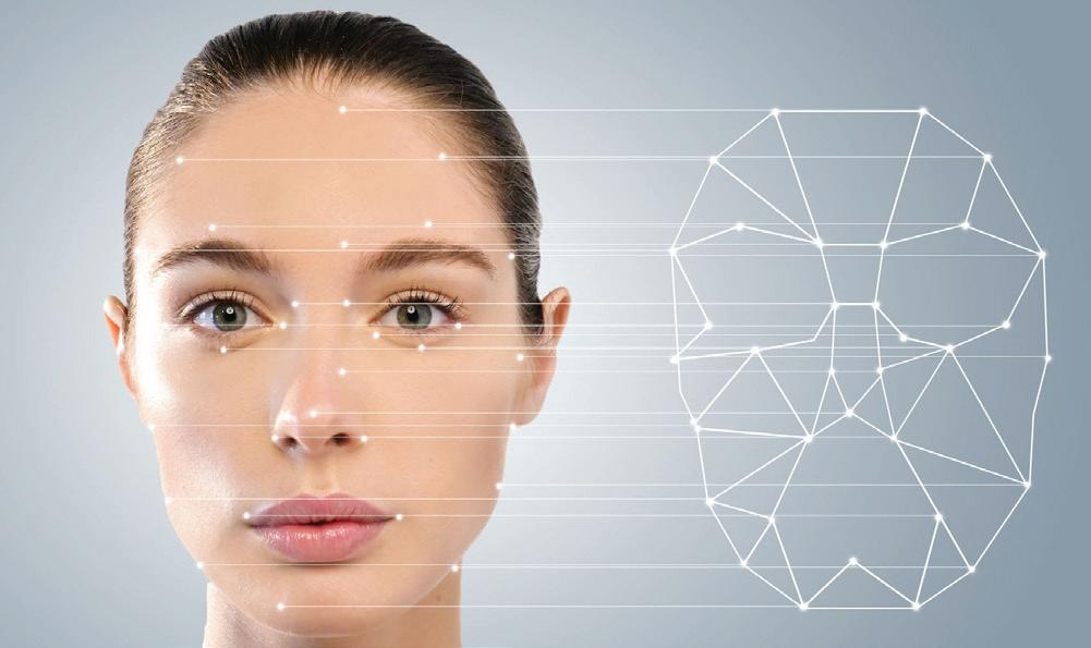
➢ Facial Recognition & Mapping
Facial recognition and mapping are foundational technologies in AI-guided facial analysis systems. These systems analyze human faces using advanced computer vision and machine learning techniques to extract, interpret, and respond to facial features.3
• 3D Mapping
Artificial intelligence (AI) excels at analyzing facial features, particularly in identifying key landmarks and assessing facial symmetry with high precision. Using 3D contour data and deep learning models like Convolutional Neural Networks (CNNs), AI can automatically assess facial symmetry before and after facial correction procedures. These systems provide accurate and human-like evaluations, helping improve treatment planning. AI methods can also create a facial midline, which supports more precise 3D symmetry analysis. Overall, AI reduces the need for manual checks and makes facial analysis faster and more efficient.4
• Thermal Facial Analysis
Thermal facial analysis leverages infrared imaging to capture facial temperature patterns, which are then interpreted by AI models to recognize faces, detect emotions, and even assess physiological or health-related states. Deep learning algorithms trained on thermal infrared images can recognize unique facial heat patterns and vascular structures, ensuring accurate identification in lowlight environments and offering biometric data that extends beyond conventional visual characteristics. Thermal approaches also support expression recognition when fused with visible-light data; they enhance accuracy under varying conditions.5
WORKFLOW OF AN AI-GUIDED FACIAL ANALYSIS SYSTEM
➢ Face detection serves as the foundational step in facial analysis, tasked with accurately identifying and isolating human faces within images or video frames. Modern AI models like CNN, R-CNN, SSD, and anchor-free methods make this process fast, accurate, and suitable for real-time use. This step is crucial because all further analysis like identifying facial features, verifying identity, or detecting health conditions depends on accurately locating the face.1
➢ Face Alignment, or Facial Landmark Detection, identifies key facial points like the eyes, nose, and mouth to standardize face positioning after detection. This improves face recognition, expression analysis, and AR effects. Two main methods are Heatmap
Regression, which estimates likely landmark areas, and Coordinate Regression, which predicts exact point locations using deep learning. Advanced models like Deep Adaptive Graph Networks enhance accuracy, even with tilted faces, poor lighting, or occlusions.1
➢ Face reconstruction builds a 3D model of the face from one or more 2D images, helping us understand its shape and depth more clearly. It is especially helpful in medical and surgical fields. Some methods use a single photo and AI to predict the 3D form, while others combine multiple images for a more detailed and accurate model that works well even with different angles or hidden features.1
➢ Facial recognition identifies or verifies a person identity by analyzing their unique facial features. It is commonly used in security, health checks, and spotting facial traits. Advanced AI models make recognition more accurate. Beyond identification, analyzing facial features can also help detect signs of genetic disorders or other health conditions.1
➢ Attribute-based health detection uses AI to spot facial features linked to diseases, like unevenness or unusual jaw and eye shapes. Advanced models can analyze many traits at once, helping with early screening and more accurate diagnoses.1
APPLICATIONS
➢ Facial analysis and simulation: AI enables realistic virtual simulations of surgical or cosmetic procedures, helping patients and doctors visualize expected outcomes before treatment.6
➢ Personalized treatment planning: By analyzing unique facial features, AI assists clinicians in designing customized treatment plans that suit each patient needs.6
➢ Robotic-assisted procedures: AI-driven facial data guides robotic systems during surgery, increasing precision and reducing risks.6
➢ Patient outcome prediction: AI models predict treatment results and progress by monitoring facial changes and related health indicators.6
➢ Enhanced patient experience: With more accurate analysis and tailored care, AI improves patient satisfaction and overall treatment effectiveness.6
LIMITATIONS
• AI models may perform poorly for underrepresented ethnic groups due to lack of diverse training data.6
• Accurate AI analysis often depends on large amounts of detailed patient data, which can be time-consuming to collect.6
• Implementing AI systems can be expensive and may only apply to specific facial procedures or conditions.6
• The AI reasoning process is often unclear, making it difficult for clinicians to fully understand or trust the results.6
• Reliance on AI might reduce human contact, potentially affecting the patient emotional comfort and trust in care.6
CONCLUSION
AI-driven facial analysis marks a major advancement in integrating intelligent systems across healthcare, aesthetics, and other fields. This technology introduces innovative diagnostic possibilities while enhancing precision and operational efficiency in ways previously unattainable. By enabling deeper structural insight and multi-layered pattern recognition, it expands the potential of digital evaluation in both clinical and non-clinical environments. Despite its impressive capabilities, responsible use and continued refinement are essential to ensure inclusivity, interpretability, and user-centred outcomes. As research and development progress, this field is poised to redefine how facial data is utilized across diverse professional domains.
REFERENCES
1. Lei C, Dang K, Song S, et al. AI-assisted facial analysis in healthcare: From disease detection to comprehensive management. Patterns (N Y). 2025;6(2):101175. Published 2025 Feb 4. doi:10.1016/j.patter.2025.101175
2. Theodosiou AA, Read RC. Artificial intelligence, machine learning and deep learning: Potential resources for the infection clinician. J Infect. 2023;87(4):287-294. doi:10.1016/j.jinf.2023.07.006
3. Qiang J, Wu D, Du H, Zhu H, Chen S, Pan H. Review on Facial-Recognition-Based Applications in Disease Diagnosis. Bioengineering (Basel). 2022;9(7):273. Published 2022 Jun 23. doi:10.3390/bioengineering9070273
4. Zhu Y, Zhao Y, Wang Y. A Review of Three-Dimensional Facial Asymmetry Analysis Methods. Symmetry. 2022; 14(7):1414. https://doi.org/10.3390/sym14071414
5. Weidlich VA. Thermal Infrared Face Recognition. Cureus. 2021;13(3):e13736. Published 2021 Mar 6. doi:10.7759/cureus.13736
6. Al-Dhubaibi MS, Mohammed GF, Atef LM, Bahaj SS, Al-Dhubaibi AM, Bukhari AM. Artificial Intelligence in Aesthetic Medicine: Applications, Challenges, and Future Directions. J CosmetDermatol. 2025;24(6):e70241. doi:10.1111/jocd.70241