SURGICAL ONCOLOGIST

SURGICAL ONCOLOGIST

Hysteroscopy
The first step in diagnosing uterine cancer is often a physical exam. During a physical exam for uterine cancer, your healthcare provider will check your reproductive organs, including the uterus, ovaries, and cervix, for any abnormalities. Your doctor will perform a pelvic exam to check for any abnormalitiesinyouruterus,ovaries,orcervix.
A pelvic exam for uterine cancer is a type of physical exam that is specifically focused on checking for signs of uterine cancer. This exam includes a gentle press on your lower abdomen to check the size, position, and shape of your uterus and ovaries. Your healthcare provider will also insert fingers to check for any unusual lumps or growths in your vagina, cervix, uterus, fallopian tubes, ovaries, and rectum. They will also assess the uterus, cervix, ovaries, and fallopian tubes for any signs of cancerous growth or tumors.

A Pap test, also known as a Pap smear, is a c screening test used to detect abnormal cells or cha the cervix that may indicate the presence of uterine

The test involves collecting a sample of cells from th and examining them under a microscope for any ab changes.
During a Pap test, a healthcare provider will use a sp to hold open the walls of the vagina and examine the They will then use a small brush or spatula to c sample of cells from the cervix. The sample is then s laboratory where it is examined for any abnormal cha
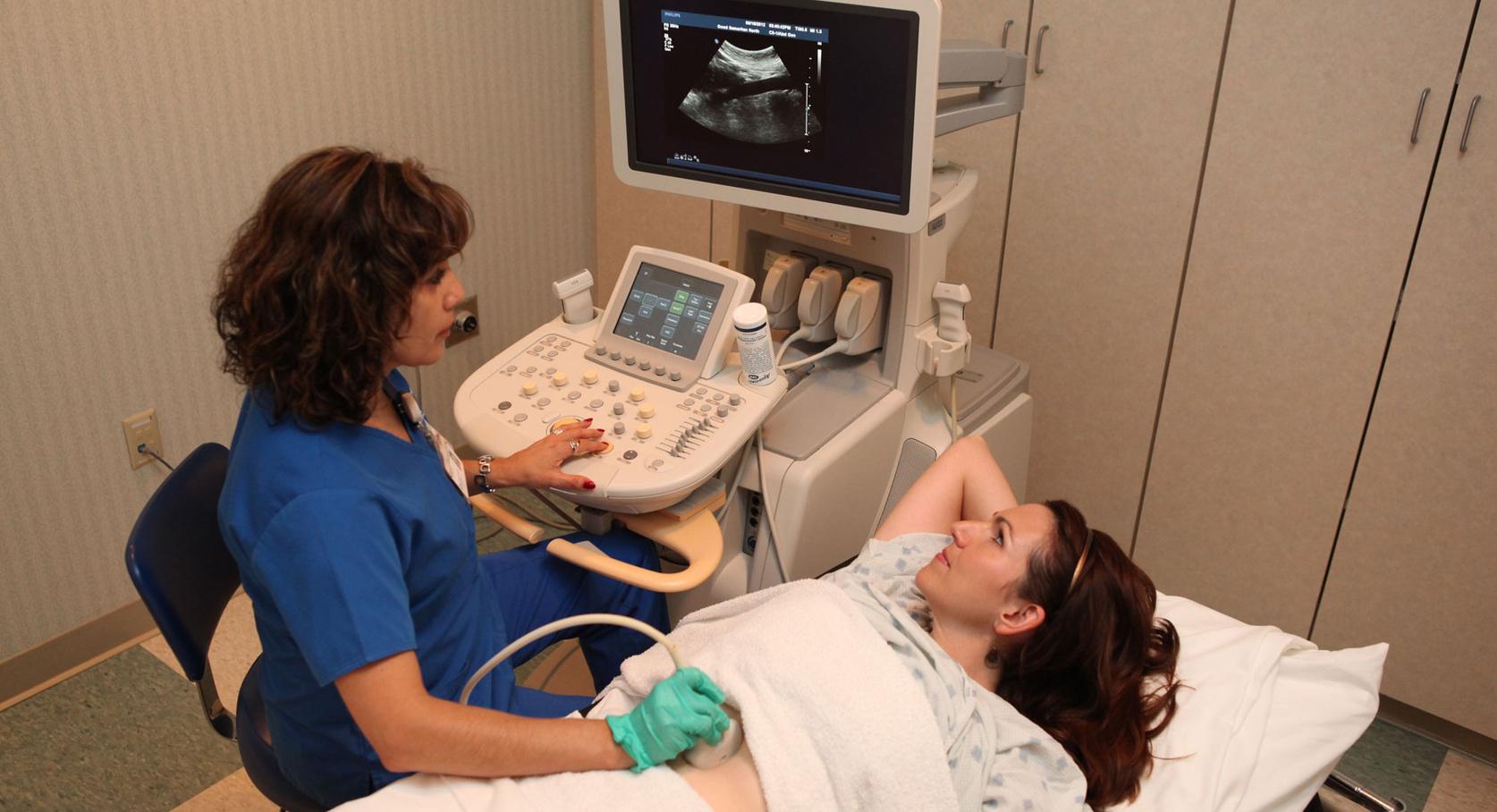
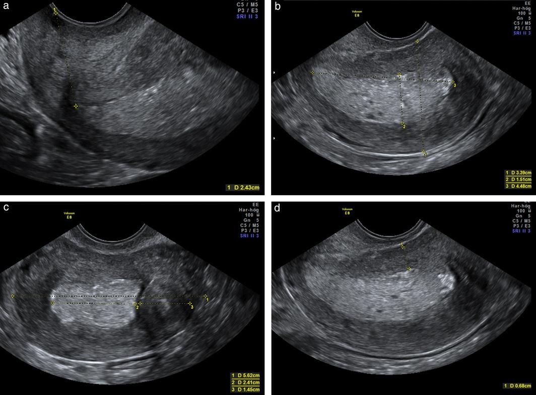
During an ultrasound for uterine cancer, a healthcare provider will use a handheld device called a transducer to send sound waves through the body. The sound waves bounce off the organs and create an image on a computer screen. The healthcare provider may also use a special wand called a transvaginal ultrasound probe, which is inserted into the vagina to obtain more detailed imagesoftheuterusandovaries.
Ultrasound can help detect abnormalities in the size, shape, or texture of the uterus or other reproductive organs, which may indicate the presenceofcancer.Itcanalsohelpdeterminethe stage of the cancer and whether it has spread to nearbyorgansortissues.
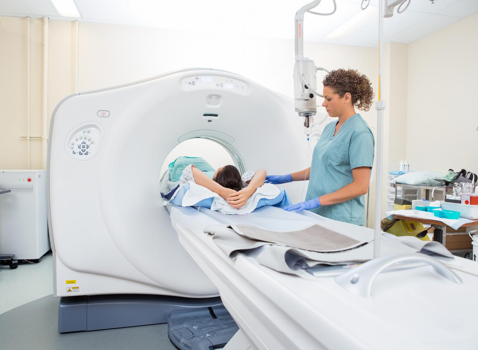
A CT scan (computed tomography) is a type of Xray imaging that uses a combination of X-rays and computer technology to create detailed crosssectional images of the body.
During the test, the patient lies on a table that moves through a doughnut-shaped machine that takes pictures of the body from different angles. The images can help detect the presence and extent of uterine cancer, as well as any spread to nearby organs or lymph nodes.
A biopsy is a procedure in which a small sample of tissue is removed from the uterus or another part of the body and examined under a microscope for signs of cancer. A biopsy is the only definitive way to diagnose uterine cancer.
In most cases, a biopsy for uterine cancer is performed during a procedure called a dilation and curettage (D&C), in which the cervix is dilated and a small instrument is inserted into the uterus to remove a sample of tissue. A biopsy may also be performed during a hysteroscopy, which is a procedure that uses a thin, lighted tube inserted through the vagina and cervix to examine the inside of the uterus and collect tissue samples
The tissue samples collected during a biopsy are examined by a pathologist, who will determine whether cancer cells are present and, if so, what type of cancer. The pathologist will also provide information on the stage of the cancer, which refers to how advanced it is and whether it has spread to other parts of the body.

Hysteroscopyisaprocedurethatallowsahealthcareproviderto examinetheinsideoftheuterususingathin,lightedtubecalleda hysteroscope. The hysteroscope is inserted through the vagina andcervixandintotheuterus,allowingtheprovidertoviewthe lining of the uterus and collect tissue samples for further examination.
During the procedure, the healthcare provider may identify abnormalitiesintheliningoftheuterus,suchaspolyps,fibroids,or othergrowths,whichmaybecancerous.Theymayalsocollect tissuesamplesforbiopsytoconfirmorruleoutthepresenceof uterinecancer.