The Moredun Foundation News Sheet Vol. 8 | No. 5 | February 2025
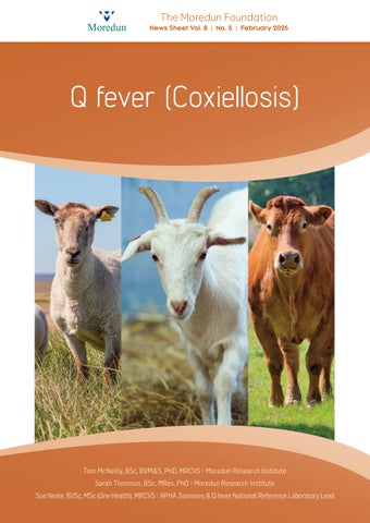
Q fever (Coxiellosis)
Tom McNeilly, BSc, BVM&S, PhD, MRCVS Moredun Research Institute Sarah Thomson, BSc, MRes, PhD Moredun Research Institute Sue Neale, BVSc, MSc (One Health), MRCVS APHA Zoonoses & Q fever National Reference Laboratory Lead