Discover IMPLANTAT,the educational habitat of S.I.N. An online teaching platform created to make more professionals accelerate their career and increase their success.
Access IMPLANTAT.GLOBAL or scan the QR Code and begin your journey of knowledge now!
Scientific Evidence
�
Research and development of products in partnership with renowned universities and institutes around the world as: Aarhus University - Denmark, Chalmers University - Sweden, KU Lueven - Belgium, Malmö University - Sweden, UNESP - Brazil, USP - Brazil, UFU - Brazil, SLmandic - Brazil.
Production Excellence
�
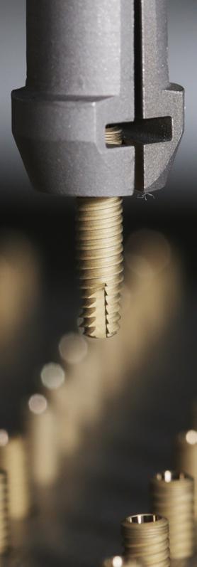
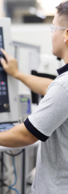
Large investments in technological updating of our manufacturing facilities over the past three years in state-of-the art equipment.
�
Annual production of over 5 million items.
Get to know our Smile Factory. Scan the QR code with your cell phone camera and take a 360º tour of S.I.N.
One of the most important implant companies worldwide. Wide international presence.
Guaranteed Quality and Certifications
Rigorous quality control of process, from the arrival of the raw material to the delivery of the final product, proven through national and international certifications.
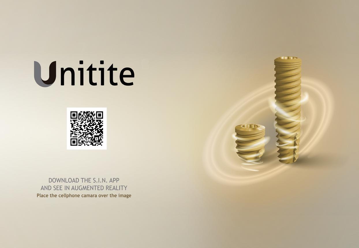
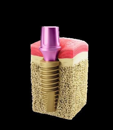
REDEFINING CONCEPTS IN IMPLANTOLOGY.
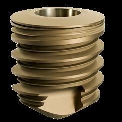
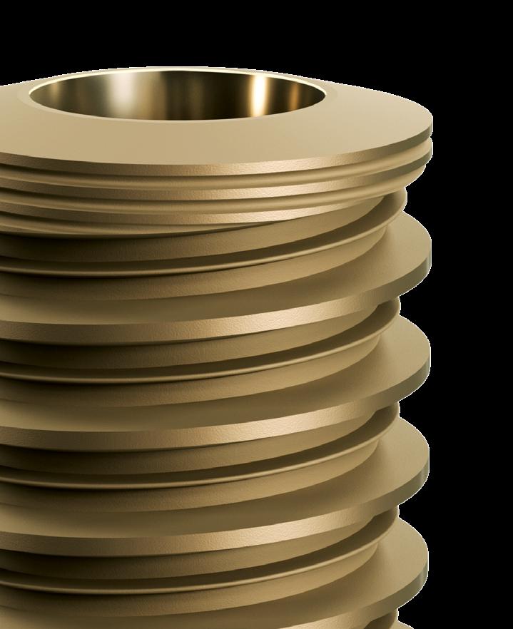
From the synergy between the exclusive macrogeometry and the most advanced surface nanoactivation emearges the UNITITE®, an implant line that has revolutionized the world market due to its originality, innovation, and high performance.
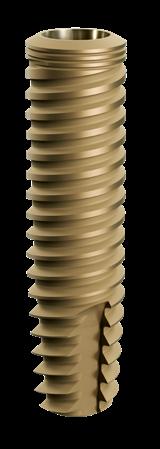
EXPLORE THE BEST IMPLANT OF THE PRESENT.
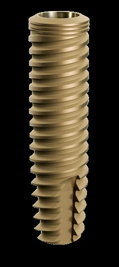
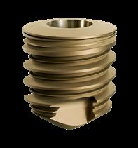
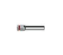
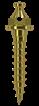
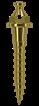
UNITITE ® compact
Exclusive HAnano® surface: developed at Chalmers University, in Sweden, HAnano® was evaluated by more than 50 preclincial and clinical studies, which verify a faster osseointegration, besides promoting a superior bone quality.
● Healing Chambers: only the external threads touches the bone tissue, while the internal threads are kept apart, promoting a very high quality hybrid healing.
● Faster with more bone: the high hydrophilicity, which is generated by an ultrafine and homogeneous layer of hydroxyapatite, increases the activity of the proteins involved in the process of osseointegration.
● Distinctive hybrid macrogeometry: accuracy of the drilling system and the design of the external threads give high stability, and minimize the compression of the healing bone tissue.
●
● Scientific evidence: more than 10 years of research and development with the renowned scientists in at leading universities worldwide.
COMPLETE SOLUTIONS
Unitite® brings you what is the most modern in the world of implantology. Using Unitite® Slim, and Unitite® Compact your surgical planning has more possibilities for innovative and high-performance solutions.
One concept, several possibilities.
Hydroxyapatite (HA), which is the main mineral present in the natural bone structure, when applied on the surface of nanostructured titanium implants, forms a homogeneous and stable coating functioning as a scar catalyst that speeds up osseointegration when compared to conventional surfaces. From 2005 on, HAnano® surfaces have been developed by researchers from leading universities in Gothenburg (Sweden). Scientists from several countries have tested and approved its effectiveness, the results of which have been published in dozens of articles in world-renowned scientific journals.
The HAnano® coating is formed by hydroxyapatite nanocrystals, with size and shape similar to those of human bone, sintered on a microrough titanium measuring 20 nm thick that promotes a change on surface energy, increasing the hydrophilicity
and providing substrate that stimulates a greater osteoblasts multiplication. The HAnano® present on the surface of the Unitite® and Strong SW Plus implants has shown an improvement in scar response in molecular tests of signal transduction, where the proteins involved in the scar process recorded a substantial increase in concentration, presenting the coating positive effect on the interaction with the pre-osteoblastic cells. Likewise, there was an increase in the concentration of important osteogenic markers, such as alkaline phosphatase and ostecalcin, in a clear signaling of the mineralization process acceleration. Among the most relevant aspects, with the greatest clinical significance, is the bone mechanical quality which is formed around this highly hydrophilic Unitite® and Strong SW Plus surface, which derives from the resulting ionic potential of the HAnano®.
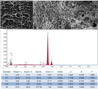
The chart and table above corresponds to an EDS analysis on the Unitite® surface, bringing the purity and stability of the implant
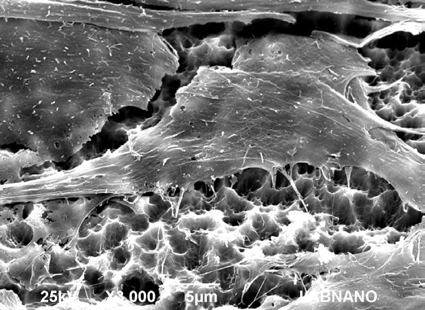
The image below shows the Unitite® surface at an increase of 5.000x / 10,000x / 100,000x respectively. The moderately rough Ti surface with the PLUS of a nano-layer of Hydroxyapatite.
surface closer.
SCIENTIFIC PUBLICATIONS
The positive and superior results of HAnano® have been evaluated and proven by numerous scientific studies in several recognized universities and research institutions worldwide. You can check some of them on the QrCodes below:
NANO HYDROXYAPATITE STRUCTURES INFLUENCE EARLY BONE FORMATION.
Meirelles L, Arvidsson A, Andersson M, Kjellin P, Albrektsson T, Wennerberg A.
Journal of Biomedical Materials Research Part A Volume 87A, Issue 2,2008, pp. 299-307
THE EFFECT OF CHEMICAL AND NANOTOPOGRAPHICAL MODIFICATIONS ON THE EARLY STAGES OF OSSEOINTEGRATION.
Meirelles L, Currie F, Jacobsson M, Albrektsson T, Wennerberg A.
The International Journal of Oral and Maxillofacial Implants Volume 23, Issue 4, 2008, pp. 641-647
NANO HYDROXYAPATITE-COATED IMPLANTS IMPROVE BONE NANOMECHANICAL PROPERTIES.
Jimbo R, Coelho PG, Bryington M, Baldassarri M, Tovar N, Currie F, Hayashi M, Janal MN, Andersson M, Ono D, Vandeweghe S, Wennerberg
A.J Dent Res. 2012;91(12):1172-7
Scanning Electron Microscopy demonstrating osteoblastic cell on HAnano® surface. Courtesy: Cavalcanti JH, Tanaka M, Bezerra FJ, CBPF RJ.
UNITITE® HIGH LEVEL OF EXCELLENCE
Unitite® was developed based on more than 10 years of studies in important universities of the world. That is how we have been able to verify its efficacy through clinical and scientific results.
In the following chart we observed the results of Unitite® with respect to marginal bone loss performed in an animal study. In this study, Unitite® was compared to implants SLActive (Straumann), TiUnite (Nobel Biocare) and Nanotite (Biomet 3i), with lower bone loss two to four weeks after implant placement.
Source: modified from Bonfante et al.
The Unitite® demonstrated excellent results for bone maintenance in finite element analysis.
Source: modified from Shunmugasamy et al.
By analyzing the results demonstrated below, it was found that the dissipation of forces in the bone tissue of the Unitite® is comparable to the main brands of dental implants.
Source: modified from Shunmugasamy et al.
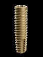
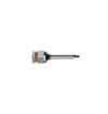
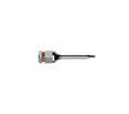
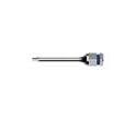
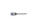
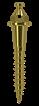
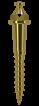
UNITITE® SLIM
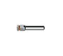
UNITITE ® slim
INDICATIONS FOR CLINICAL USE:
� 2.9 – Central and lateral incisors (mandible) lateral incisors (maxilla)
Offers three different lengths for your surgical planning.
Only 2.9 mm diameter: Unitite® Slim provides rehabilitation in narrow areas and limited interdental spaces, such as the upper lateral incisors, and lower incisors areas.
More safety: the reduced dimension protects vital oral structures, and their vascularization.
Morse Taper: biomechanical superiority of prosthetic connections with internal angle of 3 degrees.
Produced with Cold-Worked grade 4 Titanium: This production technique offers long-term stability and mechanical strength for thin-walled implants.
> Indicated for all type of bones
> Recommended 1.5 mm infra-bone installation.
> Speed of the initial drills: 1200 rpm.
> Speed of the drill 2.7mm: 800 rpm.
> Speed of the bone tap 2.9mm: 20 rpm*.
> Insertion speed: 20 to 40 rpm.
> Maximum Torque: 45 N.cm.
> Includes cover screw of 2.0 mm.
> Suitable for late loading: As from 60 days.
* For bone types I and II, the bone tap is required to ensure the correct healing process.
DRILLING
SEQUENCE GUIDE
● For bone types I and II, the bone tap is required to ensure the correct healing process.
TECHNICAL INFORMATION
Scan to see step by step
UNITITE® SLIM PROSTHETIC SEQUENCE
UNIVERSAL ABUTMENT SEQUENCE (ANALOG AND DIGITAL)
Cemented Single
TITANIUM HEALING CAP
For installation and removal of PEEK healling caps compatible with Unitite® Slim, it is necessary to purchase the CICS and CRCS keys separately.
Digital sequence
UNITITE® SLIM PROSTHETIC SEQUENCE
MICRO MULTI-UNIT ABUTMENT (ANALOG AND DIGITAL)
Single and Multiple screw retained prosthesis
For installation and removal of PEEK healling caps compatible with Unitite® Slim, it is necessary to purchase the CICS and CRCS keys separately.
Mucosa meters: Available for the complete line, helps in the measurement and choice of prosthetic components.
Ease of clinical use through color-coding.
Compact format facilitates sterilization even in smaller autoclaves.
Reduced number of drills required for osteotomy.
Accurate fits of all parts regardless of the position or movement.
included.
Inclined tray for easy viewing during the surgical procedure.
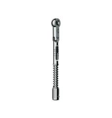
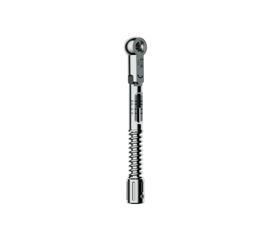
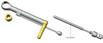
Torque wrench. Bidigital adapter
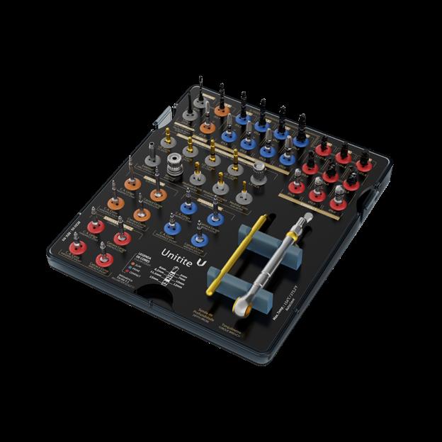
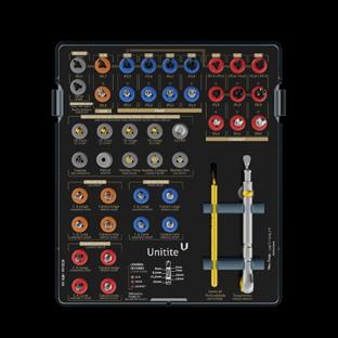
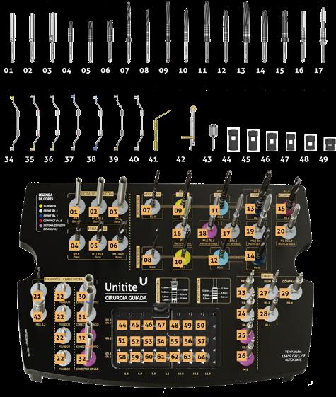
ORGANIZING BOX
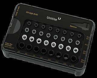
UNITITE® SAFE DRILL KIT
MAKING YOUR SURGERIES SIMPLER AND MORE PRACTICAL
Removable tray for easy cleaning. Helps in reducing the time of surgery.
Plastic rings with perfect fits guarantees speed and safety in the surgical procedures.
Prevents injuries of noble structures such as nerves, sinus, upper jaw and nasal cavity.
Perforations for irrigation. Drilling depth
Stoppers available for each diameter of the drills. All Unitite® drills are prepared to be used with the depth stoppers found in the Safe Drill Kit.
Scan to see how to use the kit
Greater safety of clinical use for cases with limited bone availability.
The Unitite® Safe Drill Kit is only compatible with the Unitite® Surgical Kit. For the morse taper infrabone installation, it is required to use the 1.5mm ring higher than the desired implant height (except for Unitite® Compact).
SHORT DRILL KIT
UNITITE® MILLING SYSTEM IS COMPLETE.
Drill length: 27 and 28.5mm.
Stainless steel and DLC coating (Diamond Like Carbon): increased cutting power, ensuring less bone heating.
Possibility of autoclaving in its case.
Milimetric markings: until 10 mm.
Compatible with UNITITE® implant system.
INDICATION
In cases requiring drills with shorter length for patients with limited mouth opening.
SHORT DRILL KIT: KSDU
* The kit is shipped with the cartridge and the component blister.
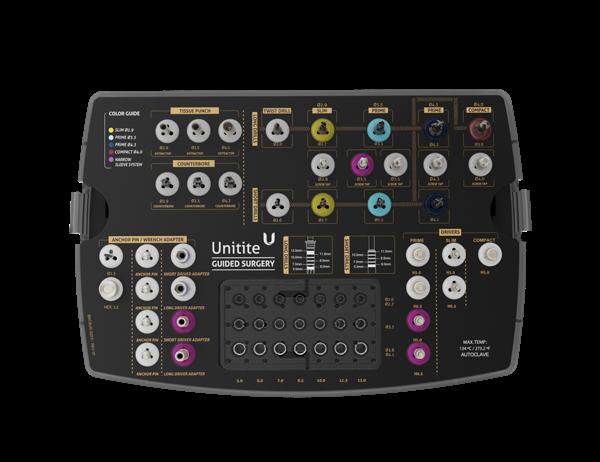
UNITITE® GUIDED SURGERY KIT
COMPLETE AND COMPACT KITS
Developed with high-tech innovation and superior industrial quality, S.I.N. Guided Surgery provides several benefits throughout the dental implant installation procedure. Now you can offer your patients a more comfortable surgery, accurate precision, reduced surgical time and
Color coding modern and easy to browse through.
Integrated Safe Drill system limiters that allow precise control of the alveolus depth.
Options of installation in several diameters*, lengths and prosthetic platforms of the implant lines.
With the S.I.N. Guided Surgery technique, you will have:
Shorter surgery time, as there is greater precision in implant installation.
More predictability and accuracy in planning.
High implant survival rate.
Reduced bleeding.
Faster recovery for patient.
Long
and short drill system
> Greater range of options according to the clinical case.
Standard drills 42.5 mm
> Millimetric depth markings;
> Safe Drill fitting;
> Recommended for all types of procedure.
Flexible sleeve positioning system
> It allows the PLACEMENT OF THE SURGICAL GUIDES IN TWO DIFFERENT POSITIONS in relation to the bone level.
Better postoperative recovery.
Preservation of bone tissue volume around the implant.
Better maintenance of soft tissue.
Possibility of immediate installation of the prosthesis through a digital. workflow.
Short Drills: 37.5 mm
> Indicated for patients with poor mouth opening/posterior regions;
> Allows the installation of implants of 7mm / 8.5mm / 10mm / 11.5mm**;
> It does not have a fitting for the Safe Drill limiter.
*In condition H6.5 with short drill, the maximum implant length to be installed should be 10mm.
Narrow sleeve system
> It AVOIDS COLLISION BETWEEN GUIDE SLEEVES and orientation errors at short mesio-distal distances.
PARA FIXADOR DE GUIA Ø 1.4 mm
PARA FIXADOR DE GUIA Ø 5.0 mm
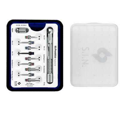
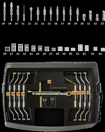
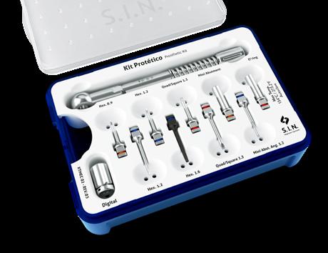
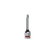
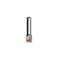
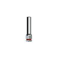
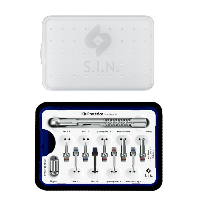
PROSTHETIC KIT
FUNCTIONAL, PRACTICAL AND COMPACT.
35% lighter than other kits on the market.
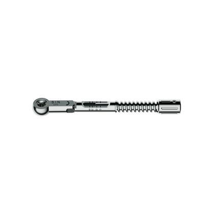
Detachable wrench with torque meter: safety when installing components.
Used for insertion, removal and torque of prosthetic components.
Full lock: keys do not moveregardless of position and movement.
CODE: KTMEC 02
Ease to assemble: all descriptions already engraved on the tray.
Transparent lid for identification without the need of opening and loss of asepsis after autoclaving.
Silicone rings color-coded according to the each key.
Functionality: Instrumental with better retention in the use of the torque ratchet.
Thinner driver, specific for angled abutment.
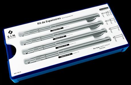
BONE EXPANDER KIT
Ideal for performing lateral bone expansion, the Bone Expander Kit is the essential tool for its clinical ease, in addition to avoiding the need to use bone grafts.
Expanders made from surgical steel.
Compact kit that can be sterilized in smaller autoclaves.
CODE: KEXP
ORGANIZING BOX: COEXP
CODE
SXPS 01
SXPS 02
SXPS 03
SXPS 04
Stops included.
DESCRIPTION
Expansor with stop 1 - ø 1.65 mm Tip
Expansor with stop 2 - ø 1.90 mm Tip
Expansor with stop 3 - ø 2.85 mm Tip
Expansor with stop 4 - ø 3.15 mm Tip
COEXP Expander Organing Box
Dimension specifications of the tips printed on the tray.
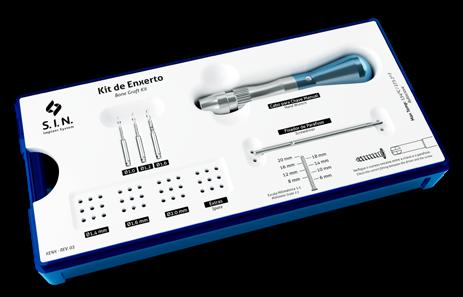
BONE GRAFT SURGICAL KIT
Used for stabilization of bone grafts in block and for guided bone regeneration surgery, the Bone Graft Kit has a driver with a cross-fit, in order to give more precision when making use of the screws.
Manual driver used for fixing the screws.
Compact kit that can be sterilized in smaller autoclaves.
CODE: KENX
ORGANIZING BOX: COENX
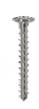
BONE GRAFT SCREWS
NOTE: Screws are sold separately
CODE DESCRIPTION
CDM 02 Hand Wrench
CPEX Screwdriver
FH 1015
drill ø 1.0 mm x 15.0 mm
FH 1215 Helical drill ø 1.2 mm x 15.0mm
FH 1615 Helical drill ø 1.6 mm x 15.0mm
COENX Bone graft organizing box
Head with cross-fit.
Dimensions of the screws printed on the tray.
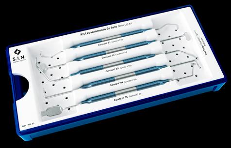
SINUS LIFT KIT
Indicated for sinus lift surgery, the Sinus Lift Kit enables the sinus membrane to be displaced, as well as curettage and compaction of the bone graft.
Instrumentals made from surgical steel.
Compact and lightweight box design that allows sterilization in smaller autoclaves.
CODE: KLEV 02
ORGANIZING BOX: COLEV
Cables with handles and channels that facilitate their grip.
Sinus Lift Organizing Box
Lighter curettes for easier handling.
CRT 01
Curette 01
Curette 02
Curette
Curette 04
Curette
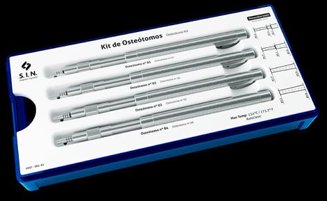
OSTEOTOME KIT
It enables the performance of atraumatic maxillary sinus elevation, which results in vertical bone gain, the Osteotome Kit is the ideal tool for its cases and avoids the need for bone grafting.
Specifications of tip dimensions printed on the tray.
Stops included.
Compact kit that can be sterilized in smaller autoclaves.
CODE: KOST
ORGANIZING BOX: COOST
SOST 01
SOST 02
SOST 03
SOST 04
COOST
Osteotome Summer W/ Stop 1 - ø 1.60 mm Tip
Summer W/ Stop 2 - ø 1.90 mm Tip
Osteotome Summer W/ Stop 3 - ø 2.90 mm Tip
Osteotome Summer W/ Stop 4 - ø 3.20 mm Tip
Osteotome Organizing Box
Osteotome
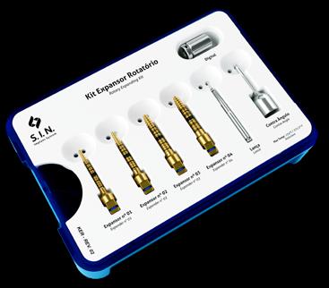
ROTARY EXPANDING KIT
Indicated for situations of little bone thickness, besides having 3 options, being ratchet, contra-angle and digital key. Recommended for bone expansion and compaction and avoids the need for bone grafting.
Expanders made of titanium.
CODE: KER
ORGANIZING BOX: COER
CODE
DESCRIPTION CPQ 02 Digital Adapter
CQCA 27 Contra-angle square drive
COER Rotary Expanding Box
Drills and drivers made from surgical steel.
Compact kit that can be sterilized in smaller autoclaves.
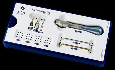
ORTHODONTIC KIT
Kit with surgical simplicity for installation and removal of orthodontic implants, aiding in orthodontic treatment.
Digital driver used for fixing the screws.
CODE: KOR
Screws dimensions specifications printed on the tray.
ORGANIZING BOX: COOR
CODE DESCRIPTION
CMPO 70 Manual Driver - High Utility
CCPO 24 Handpiece - High Utility
FML 70 Manual lance-type drill
FH 1015 Helical Drill 1,0 x 15 mm
CDM 02 Manual Driver
CDPO 24 Digital Key for Orthodontic Screw (for final screw installation only)
COOR Orthodontic Kit Box
NOTE: Screws are sold separately.
Cross head manual driver.
Compact format that facilitates sterilization in smaller autoclaves.
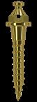
ORTHODONTIC IMPLANTS
> Easy installation and removal.
> Immediate loading can be done after surgical application.
> Easy connection with orthodontic accessories.
> Hole diameter : 0.6 mm.
AUTO DRILLING APEX:
INSTALLATION TECHNICAL INFORMATION
Lengths:
Gingival depth = 0, 1, 2 and 3 mm.
Length = 6, 8 and 10 mm. (6 and 8: lower jaw / 10: bone type IV).
Diameter:
mm
mm
mm
SELF-DRILLING WITHOUT TRANSMUCOSAL PROFILE
SELF-DRILLING WITH SHORT
SELF-DRILLING WITH TRANSMUCOSAL PROFILE (2MM)
SELF-DRILLING WITH
COMPLEMENTARY KITS INSTRUMENTAL
DIGITAL DRIVERS
ITEM CODE
DESCRIPTION LENGTH
INDICATION
CDA 20 ABUTMENT DRIVER 20,0MM SHORT Used to set the mini-abutment and conical abutment screw
CDA 24 ABUTMENT DRIVER 24.0MM LONG Used to set the mini-abutment and conical abutment screw
CDH 0920 HEXAGONAL DIGITAL DRIVER 20,0MM SHORT Used for installation of Externa Hex. Tryon implant cover, two-pieces straight universal abut and angled universal abut
CDH 0924 HEXAGONAL DIGITAL DRIVER 24.0MM LONG Used for installation of Externa Hex. Tryon implant cover, two-pieces straight universal abut and angled universal abut
CDH 1220 HEXAGONAL DIGITAL DRIVER 20,0MM SHORT
CDH 1224
HEXAGONAL DIGITAL DRIVER 24.0MM LONG
CDHA 1220 HEX. DIGITAL DRIVER 20.0MM ANG. MINI-ABUTMENT SHORT
Used to set the mounting piece, healing, transfer, retaining screw (PTL 16, PT 2006, PT 2008, PRH 20 and PRH 30) and lab screws. 1.2mm hexagonal tip
Used to set the mounting piece, healing, transfer, retaining screw (PTL 16, PT 2006, PT 2008, PRH 20 and PRH 30) and lab screws. 1.2mm hexagonal tip
Used to set the angular mini-abutment screw 1.2mm hexagonal tip (except for the Unitite angular mini-abutment).
CDHA 1224 HEX. DIGITAL DRIVER 24.0MM ANG. MINI-ABUTMENT LONG Used to set the angular mini-abutment screw 1.2mm hexagonal tip (except for the Unitite angular mini-abutment).
CDHA 1237 HEX. DIGITAL DRIVER 37.0MM ANG. MINI-ABUTMENT EXTRA LONG
CDQ 1220 SQUARE DIGITAL DRIVER 20.0MM SHORT
SURGICAL HAMMER
ITEM CODE
Used to set the angular mini-abutment screw 1.2mm hexagonal tip (except for the Unitite angular mini-abutment).
Used to set the square-fit retaining screws (PTQ 2008, PTQH 18 and PTQ 2006). 1.3mm tip
DESCRIPTION
MART 1 > Surgical-grade stainless steel used with Osteotome and Expander kits. > Contact end made of synthetic material that provides improved sensitivity, less impact and reduced trauma during use
DIGITAL DRIVERS
CDQ 1224
CDQ 1237
SQUARE DIGITAL DRIVER 24.0MM LONG
SQUARE DIGITAL DRIVER 37.0MM EXTRA LONG
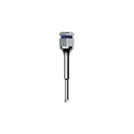
CLH 1277 HEX. DRIVER 77,0MM EXTRA LONG
CLQ 1277 HEX. DRIVER 77,0MM EXTRA LONG
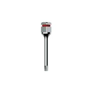
CRC 16
CRC 18
PROVISIONAL CYLINDER REMOVAL DRIVER SHORT
PROVISIONAL CYLINDER REMOVAL DRIVER SHORT
Used to set the square-fit locking screws (PTQ 2008, PTQH 18 and PTQ 2006). 1.3mm tip
Used to set the square-fit locking screws (PTQ 2008, PTQH 18 and PTQ 2006). 1.3mm tip
Lab screwdriver. Used to set retaining screws (PTL 16, PT 2006, PT 2008, PRH 20 and PRH 30) and lab screws. 1.2mm hexagonal tip
Lab screwdriver. Used to set the square-fit retaining screws (PTQ 2008, PTQH 18 and PTQ 2006). 1.3mm tip
Used to remove 1.6mm Cone Morse Strong SW provisional cylinder
Used to remove the 1.8 mm Cone Morse Strong SW provisional cylinder
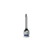
CDH 1620 HEX DIGITAL DRIVER 1.6MM SHORT Used to set Multifunctional Abutment. 1.6mm Hex lid
CDH 1624 HEX DIGITAL DRIVER 1.6MM
CCH 1620 RATCHET HEX DRIVER 1.6MM SHORT
CCH 1624 RATCHET HEX DRIVER 1.6MM MEDIUM
BONE PROFILING MILLING CUTTERS
Used to set Multifunctional Abutment. 1.6mm Hex lid
Used to set and torque of the Multifunction Abutment. 1.6mm Hex lid
Used to set and torque of the Multifunction Abutment. 1.6mm Hex lid
PO 4150 Platform 4.1 mm – External Hex. Opens bone profile to 5.0 mm
PO 5055 Platform 5.0 mm – External Hex. Opens bone profile to 5.5 mm
COUNTER-ANGLE DRIVER
ITEM
CTA 1224
CTH 0924
CTH 1220
CTH 1224
CTH 1230
CTHA 1220
CTHA 1224
CTQ 20
CTQ 24
CTQ 30
CTH 1620
CTH 1624
ABUTMENT TORQUE DRIVER 24.0MM
LONG
COUNTER-ANGLE HEXAGONAL TORQUE DRIVER 24.0MM LONG
COUNTER-ANGLE
HEXAGONAL TORQUE
DRIVER 20,0MM SHORT
COUNTER-ANGLE
HEXAGONAL TORQUE DRIVER 24.0MM
COUNTER-ANGLE
LONG
HEXAGONAL TORQUE DRIVER 30,0MM EXTRA LONG
ANGULAR MINI-ABUTMENT
COUNTER-ANGLE
HEXAGONAL TORQUE
DRIVER 20,0MM
ANGULAR MINI-ABUTMENT
COUNTER-ANGLE
HEXAGONAL TORQUE
DRIVER 24.0MM
SQUARE TORQUE
DRIVER 20,0MM
SQUARE TORQUE
DRIVER 24.0MM
SQUARE TORQUE
DRIVER 30,0MM
COUNTER-ANGLE HEX
DRIVER 1.6MM
COUNTER-ANGLE HEX
DRIVER 1.6MM
SHORT
LONG
SHORT
LONG
EXTRA LONG
Used to set the mini-abutment and conical abutment screw.
Used for installation of Externa Hex. Tryon implant cover, two-pieces straight universal abut and angled universal abut.
Used to set the mounting piece, healing, transfer, retaining screws (PTL 16, PT 2006, PT 2008, PRH 20 and PRH 30) and lab screws. 1.2mm hexagonal tip.
Used to set the mounting piece, healing, transfer, retaining screws (PTL 16, PT 2006, PT 2008, PRH 20 and PRH 30) and lab screws. 1.2mm hexagonal tip.
Used to set the mounting piece, healing, transfer, retaining screws (PTL 16, PT 2006, PT 2008, PRH 20 and PRH 30) and lab screws. 1.2mm hexagonal tip.
Used to set the angular mini-abutment screw 1.2mm hexagonal tip (except for the Unitite angular miniabutment).
Used to set the angular mini-abutment screw 1.2mm hexagonal tip (except for the Unitite angular miniabutment).
Used counter-angle to set square-fit retaining screws (PTQ 2008, PTQH 18 and PTQ 2006). 1.3mm tip.
Used counter-angle to set square-fit retaining screws (PTQ 2008, PTQH 18 and PTQ 2006). 1.3mm tip
Used counter-angle to set square-fit retaining screws (PTQ 2008, PTQH 18 and PTQ 2006). 1.3mm tip.
SHORT
Used in contra-angle to set Multifunction Abutment.
MEDIUM
Used in contra-angle to set Multifunction Abutment.
HELICAL MILLING CUTTERS
ITEM CODE MEASUREMENTS
FH 2010 ø 2.0x 10,0 mm
FH2020 ø 2.0x 18,0 mm
FH3010 ø 3.0x 10,0 mm
FH3020 ø 3.0x 18,0 mm
DESCRIPTION
> Surgical-grade stainless steel
> Thermal treatment
> Laser markings
> Used as a sequence to make the alveolus
TREPHINE MILLING
ITEM CODE
MEASUREMENTS
FTR 02 ø 2.0 mm
FTR04 ø 4,2 mm
FTR 05 ø 5,1 mm
FTR 06 ø 6,1 mm
FTR 08 ø 8,0 mm
DESCRIPTION
> Surgical-grade stainless steel
> Thermal treatment
> Laser markings
> May be used to remove implants, remove bone, and bone biopsy
> Measures refer to the inner diameter of the part
SUPERIOR QUALITY AND TECHNOLOGY
WE WARRANT, BECAUSE WE ARE PROUD OF OUR PRODUCTS.
S.I.N.’s main priority is assuring the quality and safety to our clients. Offering the best for implants, components, surgical kits and tooling is the base of all our action.
INSPECTION IN A 100% OF THE BATCHES MANUFACTURED.
The quality control is made in all S.I.N. products, to assure the success in the surgeries of all our clients, to meet the best quality standards, as well as to add value to all the ones who chose to give a smile back to people.
IMPLANTS WITH WARRANTY FOR LIFE*
5 YEARS OF WARRANTY PROSTHESIS COMPONENTS*
*SCAN THE LATERAL QR CODE TO ACCESS S.I.N WARRANTY TERMS OR ACCESS THE LINK HTTPS://BIT.LY/39DW3CF
S.I.N. ORIGINAL
COMPONENTS
S.I.N. ensures the quality of your implants and original components. Our manufacturing process has strict quality control and safety, approved by various national and international certifications.
Learn about the advantages of using implants and original components S.I.N.:
� The compatibility of the components tested in mechanical studies.
� Production of the components corresponds exactly to the internal designs of the implant.
� Accurate fit prevents bone loss and loosening or screw fracture.
� Guarantee the use of high quality raw material.
� Mechanical resistance to occlusion forces.
� Greater safety by providing quality products to yours patients.
� The pink color of Unitite® components makes the appearance of the prosthesis in the transmucosus much more natural even when there is retraction, salcerization or periimplant changes.
S. I .N .
MORE EASE AND SAFETY FOR YOUR CLINICAL PROCEDURES
S.I.N. packaging are practical, maintaining the products in their integrity, facilitating the handling and the identification.
The package is easy to open and handle even with gloves on.
Transparency of package for optimal visibility of the implant.
Separate compartments in same package for implant and cover.
With a proper connector, capture the implant with the counter angle key and move it until it reaches the perfect fit.
Snap-on top opening system ensures sterilization of the implant.
The only implant system that offers the cover screw in the same packaging. To capture it, remove the cover screw of the tube with in the 1.2 mm hexagonal digital key.
The implant should
be captured with the ratchet wrench.
GENERAL INSTRUCTIONS
Special care and clarification on surgical instruments.
CLEANING THE KIT CASE
1. Manually remove all surgical instruments from the kit. Wash the kit trays separately.
2. Prepare the enzymatic detergent according to the detergent manufacturer’s recommendation.
3. Immerse all parts of the product into the prepared detergent solution and leave for 5 minutes. Then, using a soft bristle brush, scrub the parts for at least 2 minutes until complete remove organic matter from the products.
4. Remove the parts from the detergent solution and rinse with tap water for 1 minute until the residue is completely removed. Repeat the rinse two more times.
5. Visually inspect each part to check for process residues or organic residues from the used of the product.
6. If residue in detect in the product, repeat the cleaning process until the residue is completely removed.
7. Dry with a soft, clean, dry cloth or disposable paper.
STERILIZATION
Product reusable and provided non-sterile. It must be clean and sterilized in autoclave before use.
Dry all instruments before the steam sterilization cycle.
The product is to be enclosed in a steam sterilizable wrap.
Steam sterilize in cycles from 121°C to 1 ATM pressure for 30 minutes or from 134°C to 2 ATM pressure for 20 minutes. Drying time 30 minutes
Always accommodate the case in autoclave over a plane surface and away of device walls.
Never stack objects or other cases.
CLEANING THE SURGICAL INSTRUMENTS
1. Prepare the enzymatic detergent according to the detergent manufacturer’s recommendation.
2. Immerse all parts of the product into the prepared detergent solution and leave for 5 minutes. Then, using a soft bristle brush, scrub the parts for at least 2 minutes until complete remove organic matter from the products.
3. Remove the parts from the detergent solution and rinse with tap water for 1 minute until the residue is completely removed. Repeat the rinse two more times.
4. Visually inspect each part to check for process residues or organic residues from the used of the product.
5. If residue in detect in the product, repeat the cleaning process until the residue is completely removed.
6. Dry with a soft, clean, dry cloth or disposable paper.
7. Proceed to the sterilization process.
CLEANING OF TORQUE WRENCHES
1. Pull the steering reversing rod back.
2. Remove the ratchet from the socket with your head.
3. Rotate the fixing door counterclockwise.
4. Remove the central shaft of the Torque Ratchet.
5. Remove the torque grading rod.
6. Prepare the enzymatic detergent according to the manufacturer’s instructions.
7. Immerse all parts of the product in the prepared detergent solution and leave for at least 5 minutes, then using a soft bristle brush, scrub the parts to remove organic matter from the products.
8. Remove the pieces from the detergent solution and rinse with running water for 1 minute, repeat the rinse two more times, totaling 3 rinses of 1 minute each.
9. Visually inspect each piece to check whether there is residue from the cleaning process or organic residues from the use of the product.
10. If the presence of residue on the product is confirmed, repeat the cleaning process until the residue is completely removed.
11. Dry with a soft, clean, dry cloth or disposable paper.
CLEANING RECOMMENDATION
a. Wear appropriate clothing (gloves, masks, glasses, hats, etc.).
b. Begin cleaning immediately after surgical use.
c. Never let the instrument dry containing organic residues after surgical use.
d. Never let the instrument dry naturally after cleaning.
e. Never use saline solutions, especially sodium hypochlorite and saline, disinfectants, hydrogen peroxide or alcohol to clean or rinse surgical instruments and Kit trays.
f. Never use steel wool or sponges or abrasive products, so that the instruments are not damaged.
g. Do not accumulate instruments in large quantities on top of each other to avoid deformation of smaller and delicate pieces.
STERILIZATION RECOMMENDATIONS
• Sterilize the products in the same day or one day earlier the procedure.
• The chemical sterilization is not recommend, once some products may cause the discoloration and damages to the case.
• Do not use temperature higher than 60°C to drying process.
• Do not use dry heat stoves for sterilization of the instruments and S.I.N kits.
TORQUE WRENCH – CLEANING
PROCEDURES
The ratchet must be disassembled and cleaned immediately after every use. For proper cleaning, disassemble multi-piece instruments into their single parts. No tools are necessary for this process.
Pull the steering reversing rod back.
Remove the ratchet from the socket with your head.
Remove the central shaft of the Torque Ratchet.
Rotate the fixing door counterclockwise.
Begin the washing procedure.
Remove the torque grading rod.
WHAT THE SPECIALISTS SAY
UNITITE IMPLANTS HAVE MADE THE RESULTS OF CURRENT MAJOR CLINICAL DEMANDS MORE PREDICTABLE, SUCH AS SHORTENING THE TIME BETWEEN IMPLANT INSTALLATION AND FINAL PATIENT REHABILITATION, MAINTAINING PERI-IMPLANT BONE HEIGHT, WHICH HAS A LARGE IMPACT ON LONG-TERM AESTHETIC PREDICTABILITY AND THE REHABILITATION OF AREAS WITH POOR BONE AVAILABILITY IN AN EFFICIENT AND MINIMALLY INVASIVE WAY, IN MANY CASES AVOIDING THE NEED FOR BONE GRAFTS. I AM VERY FLATTERED TO HAVE PARTICIPATED ACTIVELY IN THIS PROJECT.
“
Researcher in the Bme - KULeuven, Belgium. Post-PhD in Biomechanics by the FEMEC/UFU and Researcher in the Bme KULeuven, Belgium. PhD in Periodontics/Dental Implant - FOAr/UNESP - Araraquara, Brazil. Master in Oral Rehabilitation - FOUFU - Uberlândia, Brazil.
SURFACE COATING HANANO®, USED IN THE UNITITE IMPLANT, AND 20 NANOMETERS THICK, HOMOGENEOUSLY COATING THE ENTIRE SURFACE, SIGNIFICANTLY INCREASES SURFACE ENERGY, HYDROPHILICITY AND SCAR RESPONSE IN THE EARLY STAGES OF THE OSSEOINTEGRATION PROCESS. THE POSITIVE IMPACT OF ITS BIOAVAILABILITY HAS BEEN DEMONSTRATED BY DIFFERENT ADVANCED METHODS OF RESEARCH, SUCH AS SIGNAL TRANSDUCTION AND ATOMIC FORCE MICROSCOPY. HIGHER PROTEIN ADSORPTION, ASSOCIATED TO A STATISTICALLY SIGNIFICANT PRESENCE OF PROTEINS RELATED TO THE BONE HEALING PROCESS IN THE PRESENCE OF A BIOLOGICAL CATALYST FOR MINERALIZATION, MAKE THIS SURFACE ONE OF THE MOST ADVANCED IN THE IMPLANTS GLOBAL MARKET.
“
A Graduate of Bauru School of Dentistry - USP Specialist in Periodontics, Bauru School of Dentistry - USP Specialist in Implantology by INEPO - SP Master in Implantology by UNIP - São Paulo Doctor in Biotechnology by IBB - UNESP
Roberto Pessoa
Fabio Bezerra
“ “
OUR RESEARCH GROUP HAS WORKED WITH THE HANANO® SURFACE FOR OVER 10 YEARS. UNTIL NOW THIS RESEARCH HAS RESULTED IN TWO DOCTORAL THESES AND ANOTHER ONE IS IN PROGRESS. OUR EXPERIMENTAL RESULTS IN 17 IN VIVO STUDIES, MOSTLY ON RABBITS, USUALLY SHOWS AN IMPROVED BONE RESPONSE FOR THE TITANIUM WITH THE HANANO® SURFACE AND PEEK IMPLANTS WHEN COMPARED WITH IMPLANTS WITHOUT THIS SURFACE.
DDS/PhD and Director of the Department of Dental Prosthesis at the Malmö University, Sweden. Specialized in Implant Surface and author of more than 220 scientific articles published in renowned magazines on this subject.
WITH THE NEW SURFACE OF UNITITE, WE HAVE NOTICED THROUGH STUDIES THAT PRIMARY STABILITY IS ACTUALLY OBTAINED. THE MACROGEOMETRY OF THE IMPLANT ITSELF ALLOWS THE BLOOD TO FLOW THROUGH THE ENTIRE IMPLANT, AND THERE IS A COMPLETE OSSEOINTEGRATION FROM THE APEX OF THE IMPLANT TO THE CENTRAL WALLS, AND EVEN TO THE CERVICAL AREA OF THE IMPLANT ITSELF. THE UNITITE IS, WITHOUT A DOUBT, A MAJOR STEP FORWARD IN THE WORLD OF IMPLANTOLOGY, NOT ONLY ACCORDING TO THE MULTICENTER STUDIES, BUT ALSO THE RESULTS AND THE RADIOGRAPHIC AND CLINICAL CONTROLS THAT WE HAVE, WHICH ARE VERY ENCOURAGING.
“
PhD and Masters in Oral and Maxillofacial Surgery at the Eastman Dental Institute –University of London – and Professor at the Instituto Superior de Saúde do Alto Ave (ISAVE) in Portugal.
Per Kjellin
THE HANANO® SURFACE IS AN ULTRATHIN LAYER OF SYNTHETIC BONE ON THE SURFACE OF THE IMPLANT. EACH CRYSTAL OF SYNTHETIC BONE IS EXTREMELY SMALL, 10 TO 14 NM IN LENGTH AND ABOUT 5NM IN THICKNESS. WHAT MAKES THESE CRYSTALS SO SPECIAL IS THAT THEY HAVE THE SAME SIZE AND SHAPE AS THOSE FOUND IN HUMAN BONE AND ARE RECOGNIZED BY THE BONE CELLS, AS WELL AS BY THE BONE TISSUE, WHICH ACTIVATES THE CATALYZER AND STARTS A HUGE PROCESS OF BUILDING BONE AROUND THE IMPLANT. THIS EFFECT HAS BEEN PROVEN IN MORE THAN 20 PRE-CLINICAL STUDIES WITH THE BEST RESEARCHERS IN THE WORLD IN THE AREA OF IMPLANTS.
“
CTO of Promimic, Co-inventor of the HAnano® surface, PhD in Materials and Chemical Surfaces by the Chalmers University in Gothenburg, Sweden, and author of several studies in the area of nanomaterials.
Ann Wennerberg
Fernando Duarte
SCIENTIFIC PUBLICATIONS
Arvidsson A, Currie F, Kjellin P, Sul YT, Stenport V. Nucleation and growth of calcium phosphates in the presence of fibrinogen on titanium implants with four potentially bioactive surface preparations. An in vitro study. J Mater Sci: Mater Med 2009; 20:1869–1879
Arvidsson A, Franke-Stenport V, Andersson M, Kjellin P, Sul YT, Wennerberg A. Formation of calcium phosphates on titanium implants with four different bioactive surface preparations. An in vitro study. J Mater Sci: Mater Med 2007; 18:1945-1954
Barkarmo S, Wennerberg A, Hoffman M, Kjellin P, Breding K, Handa P, Stenport V. 2013. Nanohydroxyapatite-coated PEEK implants: A pilot study in rabbit bone. J Biomed Mater Res A 2013; 101A:465–471
Bezerra F, Pessoa RS, Zambuzzi WF. Carregamento funcional imediato ou precoce de implants com câmara de cicatrização e nano-superfície: estudo clínico prospectivo longitudinal. Innov Implant J, Biomater Esthet. 2015;9(2/3):13-7
Bezerra F, Lenharo A, Pessoa RS, Duarte LRS, Granjeiro JM. Avaliação do impacto do edentulismo total mandibular e da reabilitação fixa sobre implants com carga imediata na qualidade de vida de pacientes idosos. Rev Dental Press Periodontia Implantol. 2011 jul-set;5(3):101-10
Bezerra F, Ribeiro EDP, Bittencourt S, Lenharo A. Influência da experiência do operador na estabilidade primária de implants com diferentes macro-geometrias – estudo in vitro. Int J Dent 2010; 9(2):63-67
Bezerra F, Ribeiro EP, Bittencourt S, Lenharo A. Influência da macrogeometria na estabilidade primária dos implants em diferentes densidades ósseas. Implant News 2010;7(5):671-6.
Bezerra F, Ribeiro EP, Bittencourt S, Lenharo A. Influência da macrogeometria na estabilidade dos implants. Innov Implant J 2010; 5:29-34
Bonfante EA, Janal MN, Granato R, Marin C, Suzuki M, Tovar N, Coelho PG. Buccal and lingual bone level alterations after immediate implantation of four implant surfaces: a study in dogs. Clin. Oral Impl. Res. 2013; 24:1375–1380
Bonfante EA, Granato R, Marin C, Suzuki M, Oliveira SR, Giro G, Coelho PG: Early bone healing and biomechanical fixation of dual acid-etched and as-machined implants with healing chambers: an experimental study in dogs. The International Journal of Oral & Maxillofacial Implants 2011; 26: 75-82
Campos FEB, Jimbo R, Bonfante EA, Barbosa EA, Oliveira MTF, Janal MN, Coelho PG. Are insertion torque and early osseointegration proportional? A histologic evaluation. Clinical Oral Implants Research 2014 Jul 4. doi: 10.1111/clr.12448. [Epub ahead of print]
Campos FEB, Jimbo R, Bonfante EA, Oliveira MTF, Moura C, Barbosa DZ, Coelho PG. Drilling dimension effects in early stages
of osseointegration and implant stability in a canine model. Med Oral Patol Oral Cir Bucal. 2015 Apr 10. [Epub ahead of print]
Arvidsson A, Currie F, Kjellin P, Sul YT, Stenport V. Nucleation and growth of calcium phosphates in the presence of fibrinogen on titanium implants with four potentially bioactive surface preparations. An in vitro study. J Mater Sci: Mater Med 2009; 20:1869–1879
Arvidsson A, Franke-Stenport V, Andersson M, Kjellin P, Sul YT, Wennerberg A. Formation of calcium phosphates on titanium implants with four different bioactive surface preparations. An in vitro study. J Mater Sci: Mater Med 2007; 18:1945-1954
Barkarmo S, Wennerberg A, Hoffman M, Kjellin P, Breding K, Handa P, Stenport V. 2013. Nanohydroxyapatite-coated PEEK implants: A pilot study in rabbit bone. J Biomed Mater Res A 2013; 101A:465–471
Bezerra F, Pessoa RS, Zambuzzi WF. Carregamento funcional imediato ou precoce de implants com câmara de cicatrização e nano-superfície: estudo clínico prospectivo longitudinal. Innov Implant J, Biomater Esthet. 2015;9(2/3):13-7
Bezerra F, Lenharo A, Pessoa RS, Duarte LRS, Granjeiro JM. Avaliação do impacto do edentulismo total mandibular e da reabilitação fixa sobre implants com carga imediata na qualidade de vida de pacientes idosos. Rev Dental Press Periodontia Implantol. 2011 jul-set;5(3):101-10
Bezerra F, Ribeiro EDP, Bittencourt S, Lenharo A. Influência da experiência do operador na estabilidade primária de implants com diferentes macro-geometrias – estudo in vitro. Int J Dent 2010; 9(2):63-67
Bezerra F, Ribeiro EP, Bittencourt S, Lenharo A. Influência da macrogeometria na estabilidade primária dos implants em diferentes densidades ósseas. Implant News 2010;7(5):671-6.
Bezerra F, Ribeiro EP, Bittencourt S, Lenharo A. Influência da macrogeometria na estabilidade dos implants. Innov Implant J 2010; 5:29-34
Bonfante EA, Janal MN, Granato R, Marin C, Suzuki M, Tovar N, Coelho PG. Buccal and lingual bone level alterations after immediate implantation of four implant surfaces: a study in dogs. Clin. Oral Impl. Res. 2013; 24:1375–1380
Bonfante EA, Granato R, Marin C, Suzuki M, Oliveira SR, Giro G, Coelho PG: Early bone healing and biomechanical fixation of dual acid-etched and as-machined implants with healing chambers: an experimental study in dogs. The International Journal of Oral & Maxillofacial Implants 2011; 26: 75-82
Campos FEB, Jimbo R, Bonfante EA, Barbosa EA, Oliveira MTF, Janal MN, Coelho PG. Are insertion torque and early osseointegration proportional? A histologic evaluation. Clinical Oral Implants Research 2014 Jul 4. doi: 10.1111/clr.12448. [Epub ahead of print]
Campos FEB, Jimbo R, Bonfante EA, Oliveira MTF, Moura C, Barbosa DZ, Coelho PG. Drilling dimension effects in early stages of osseointegration and implant stability in a canine model. Med Oral Patol Oral Cir Bucal. 2015 Apr 10. [Epub ahead of print]
Coelho PG, Marin C, Granato R, Bonfante EA, Lima CP, Oliveira S, Ehrenfest DMD, Suzuki M. Alveolar Buccal Bone Maintenance After Immediate Implantation with a Surgical Flap Approach: A Study in Dogs. The International Journal of Periodontics & Restorative Dentistry 2011;31:e80–e86
Coelho PG, Granjeiro JM, Romanos GE, Suzuki M, Silva NR, Cardaropoli G, et al. Basic research methods and current trends of dental implant surfaces. J Biomed Mater Res B Appl Biomater. 2009;88(2):579-96.
Coelho PG, Jimbo R. Osseointegration of metallic devices: current trends based on implant hardware design. Archives of biochemistry and biophysics. 2014;561:99-108
Coelho PG, Jimbo R, Tovar N, Bonfante EA. Osseointegration: hierarchical designing encompassing the macrometer, micrometer, and nanometer length scales. Dent Mater. 2015;31(1):37-52
Ehrenfest DMD, Coelho PG, Kang BS, Sul YT, Albrektsson T. Classification of osseointegrated implant surfaces: materials, chemistry and topography. Trends in Biotechnology 2009; 198-206
Jimbo R, Coelho PG, Bryington M, Baldassarri M, Tovar N, Currie F, et al. Nano hydroxyapatite-coated implants improve bone nanomechanical properties. J Dent Res. 2012;91(12):1172-7.
Jimbo R, Coelho PG, Bryington M, Baldassarri M, Tovar N, Currie F, et al. Nano hydroxyapatite-coated implants improve bone nanomechanical properties. J Dent Res. 2012;91(12):1172-7
Jimbo R, Sotres J, Johansson C, Breding K, Currie F, Wennerberg A. The biological response to three different nanostructures applied on smooth implant surfaces. Clin Oral Implants Res. 2012;23(6):706-12.
Martins LM, Bonfante EA, Zavanelli RA, Freitas Jr AC, Silva NRFA, Marotta L, et al. Fatigue reliability of three single-unit implantabutment designs. Implant Dent. 2011; 21: 67-71
Meirelles L, Albrektsson T, Kjellin P, Arvidsson A, Franke-Stenport V, Andersson M, Currie F, Wennerberg A. Bone reaction to nano hydroxyapatite modified titanium implants placed in a gap-healing model. Journal of Biomedical Materials Research A 2008; 625-631
Meirelles L, Albrektsson T, Kjellin P, Arvidsson A, Franke-Stenport V, Andersson M, Currie F, Wennerberg A. Bone reaction to nano hydroxyapatite modified titanium implants placed in a gap-healing model. Journal of Biomedical Materials Research A 2008; 625-631
Meirelles L, Arvidsson A, Andersson M, Jellin P, Albrektsson T, Wennerberg A: Nano hydroxyapatite structures influence early bone formation. J Biomed Mater Res A. 2008 Nov;87(2):299-307
Meirelles L, Currie F, Jacobsson M, Albrektsson T, Wennerberg A. The effect of chemical and nanotopographical modifications on the early stages of osseointegration. Int J Oral Maxillofac Implants 2008;23: 641-647
F, Andersson M, Albrektsson T, Wennerberg A. Effect of Hydroxyapatite and Titania Nanostructures on Early In Vivo Bone Response. Clinical Implant Dentistry and Related Research 2008; 10(4): 245-254
Meirelles L, Melin L, Peltola T, Kjellin P, Kangasniemi I, Currie F, Andersson M, Albrektsson T, Wennerberg A. Effect of Hydroxyapatite and Titania Nanostructures on Early In Vivo Bone Response. Clinical Implant Dentistry and Related Research 2008; 10(4): 245-254
Pessoa RS, Coelho PG, Muraru L, Marcantonio Jr E, Vaz LG, Sloten JV, Jaecques SVN: Influence of implant design on the biomechanical environment of immediately placed implants: computed tomography-based nonlinear three-dimensional finite element analysis. Int J Oral Maxillofac Implants 2011;26:1279–1287
Pessoa RS, Souza RM, Pereira LM, Neves FD, Jaecques SVN, Sloten JV, Quirynen M, Teughels W, Spin-Neto R. Remodelação óssea de implants com conexão hexágono externo e elementos de retenção no módulo da crista sob carregamento imediato –estudo clínico prospectivo longitudinal de um ano. ImplantNews 2015;12(4):E2-E7
Pessoa RS, Sousa RM, Pereira LM, Silva TD, Bezerra FJB, SpinNeto R. Avaliação da estabilidade dos tecidos duros e moles em implants imediatos com carga imediata em área estética: estudo clínico. Dental Press Implantol. 2015 Apr-Jun;9(2):100-9
Lenharo A, Granjeiro JM, Leão L, Bezerra F, Oliva MA. Estudo prospectivo longitudinal multicêntrico avaliando o sucesso clínico de uma nova macrogeometria de implants osseointegráveis: acompanhamento de 06 a 12 meses. Revista Fluminense de Odontologia 2010; 34: 43-48
Göransson A, Arvidsson A, Currie F, Franke-Stenport V, Kjellin P, Mustafa K, Sul YT, Wennerberg A. An in vitro comparison of possibly bioactive titanium implant surfaces. Journal of Biomedical Materials Research A 2008; 1037-1047
Shunmugasamy VC, Gupta N, Pessoa RS, Janal MN, Coelho PG. Influence of clinically relevant factors on the immediate biomechanical surrounding for a series of dental implant designs. J Biomech Eng. 2011;133(3):031005.
Svanborg LM, Meirelles L, Franke-Stenport V, Kjellin P, Currie F, Andersson M, Wennerberg A. Evaluation of Bone Healing on Sandblasted and Acid Etched Implants Coated with nanocrystalline Hydroxyapatite: An In Vivo Study in Rabbit Femur. International Journal of Dentistry 2014; 1-7
Westas E, Gillstedt M, Lönn-Stensrud J, Bruzell E, Andersson M: Biofilm formation on nanostructured hydroxyapatite-coated titanium. J Biomed Mater Res A. 2014 Apr;102(4):1063-70
OUR GLOBAL PRESENCE
MONTENEGRO
CROATIA
NETHERLANDS
LUXEMBOURG
BELGIUM
BULGARIA
ROMANIA
UKRAINE
EL SALVADOR
COLOMBIA
DOMINICAN REPUBLIC ECUADOR
ITALY
EGYPT
TURKEY
BOLIVIA
CHILE PERU
LEBANON
SPAIN
ARGENTINA
BRAZIL PARAGUAY
AZERBAIJAN
PORTUGAL
MOROCCO
HEADQUARTERS
2140 Vereador Abel Ferreira Av
Jardim Anália Franco
São Paulo – SP - Brazil
FACTORY
421 Soldado Ocimar Guimarães da Silva St - Jardim Anália Franco
São Paulo – SP - Brazil
PORTUGAL BRANCH
General Ferreira Martins St, 10 8D1495-137 Algés - Portugal