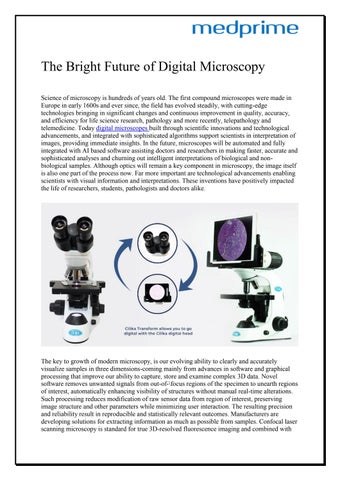
The Bright Future of Digital Microscopy Science of microscopy is hundreds of years old. The first compound microscopes were made in Europe in early 1600s and ever since, the field has evolved steadily, with cutting-edge technologies bringing in significant changes and continuous improvement in quality, accuracy, and efficiency for life science research, pathology and more recently, telepathology and telemedicine. Today digital microscopes built through scientific innovations and technological advancements, and integrated with sophisticated algorithms support scientists in interpretation of images, providing immediate insights. In the future, microscopes will be automated and fully integrated with AI based software assisting doctors and researchers in making faster, accurate and sophisticated analyses and churning out intelligent interpretations of biological and nonbiological samples. Although optics will remain a key component in microscopy, the image itself is also one part of the process now. Far more important are technological advancements enabling scientists with visual information and interpretations. These inventions have positively impacted the life of researchers, students, pathologists and doctors alike.
The key to growth of modern microscopy, is our evolving ability to clearly and accurately visualize samples in three dimensions-coming mainly from advances in software and graphical processing that improve our ability to capture, store and examine complex 3D data. Novel software removes unwanted signals from out-of-\focus regions of the specimen to unearth regions of interest, automatically enhancing visibility of structures without manual real-time alterations. Such processing reduces modification of raw sensor data from region of interest, preserving image structure and other parameters while minimizing user interaction. The resulting precision and reliability result in reproducible and statistically relevant outcomes. Manufacturers are developing solutions for extracting information as much as possible from samples. Confocal laser scanning microscopy is standard for true 3D-resolved fluorescence imaging and combined with