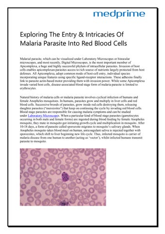
Exploring The Entry & Intricacies Of Malaria Parasite Into Red Blood Cells Malarial parasite, which can be visualized under Laboratory Microscopes or binocular microscopes, and most recently, Digital Microscopes, is the most important member of Apicomplexa, a huge and highly successful phylum of intracellular parasites. Invasion of host cells enables apicomplexan parasites access to rich source of nutrients largely protected from host defenses. All Apicomplexa, adopt common mode of host-cell entry, individual species incorporating unique features using specific ligand-receptor interactions. These adhesins finally link to parasite actin-based motor providing them with invasion power. While some Apicomplexa invade varied host cells, disease-associated blood stage form of malaria parasite is limited to erythrocytes. Natural history of malaria cells or malaria parasite involves cyclical infection of humans and female Anopheles mosquitoes. In humans, parasites grow and multiply in liver cells and red blood cells. Successive broods of parasites, grow inside red cells destroying them, releasing daughter parasites (“merozoites”) that keep on continuing the cycle by invading red blood cells. Blood stage parasites are responsible for causing malaria symptoms and can be studied under Laboratory Microscopes. When a particular kind of blood stage parasites (gametocytes occurring in both male and female forms) are ingested during blood feeding by female Anopheles mosquito, they mate in mosquito gut initiating growth cycle and multiplication in mosquito. After 10-18 days, a form of parasite called sporozoite migrates to mosquito’s salivary glands. When Anopheles mosquito takes blood meal on human, anticoagulant saliva is injected together with sporozoites, which shift to liver beginning new life cycle. Thus, infected mosquito is carrier of malaria disease from one human to another (acting as ‘vector’), whilst infected humans transmit parasite to mosquito.
Exploring The Entry & Intricacies Of Malaria Parasite Into Red Blood Cells
Issuu converts static files into: and more. Sign up and create your flipbook.