The applicability of human iPSC-derived sensory neurons for the modelling of peripheral neuropathy in vitro
Dr Eve Corrie1, Dr Emily Johnson2, and Dr Emma V. Jones1
1Medicines Discovery Catapult, Alderley Park, Cheshire, SK10 4ZF, UK
2ApconiX, Alderley Park, Cheshire, SK10 4TG, UK


Human modelsof peripheralsensoryneurons
• The human pain sensing system is known to differ significantly from that of the rodent, for instance in ion channel expression, neuropeptide signalling and receptor pharmacology. Regulatory bodies are increasingly encouraging the use of New Approach Methodologies (NAMs) to improve human relevance in drug development, leading to a shift away from animal models and towards human models.
• iPSC-derived sensory neurons offer a human-relevant platform that recapitulate key features of peripheral sensory neurons and may offer a way to study human pain mechanisms in vitro and allow the testing of compounds developed to treat pain, particularly peripheral pain such as chemotherapy-induced peripheral neuropathy (CIPN), diabetic neuropathy or inflammatory pain.

• iPSC-derived peripheral sensory neurons (RealDRG ) were purchased from Anatomic Incorporated and functionally assessed for their suitability for neuropathic pain modelling in the context of analgesics R&D.
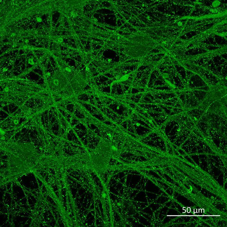
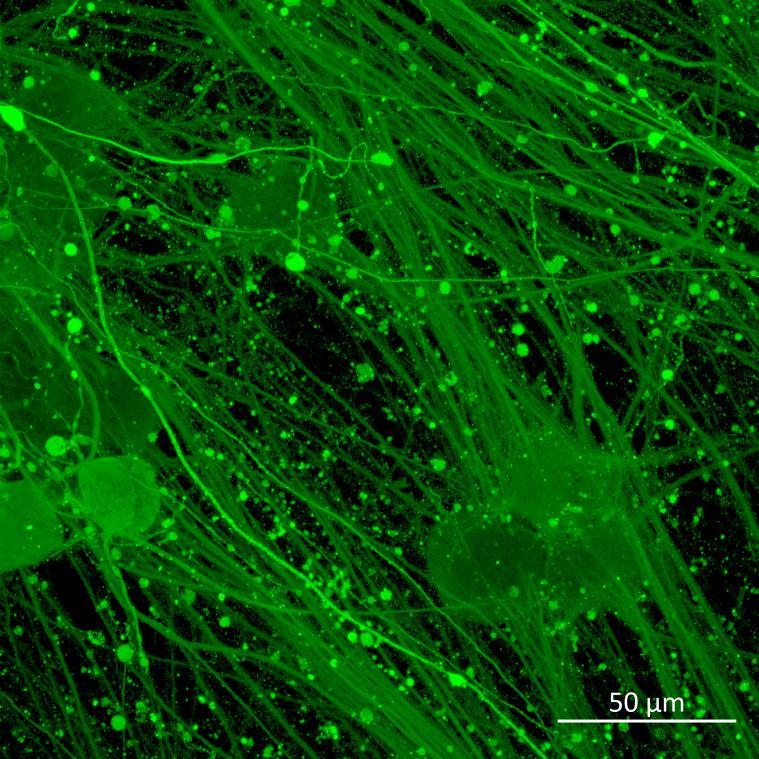
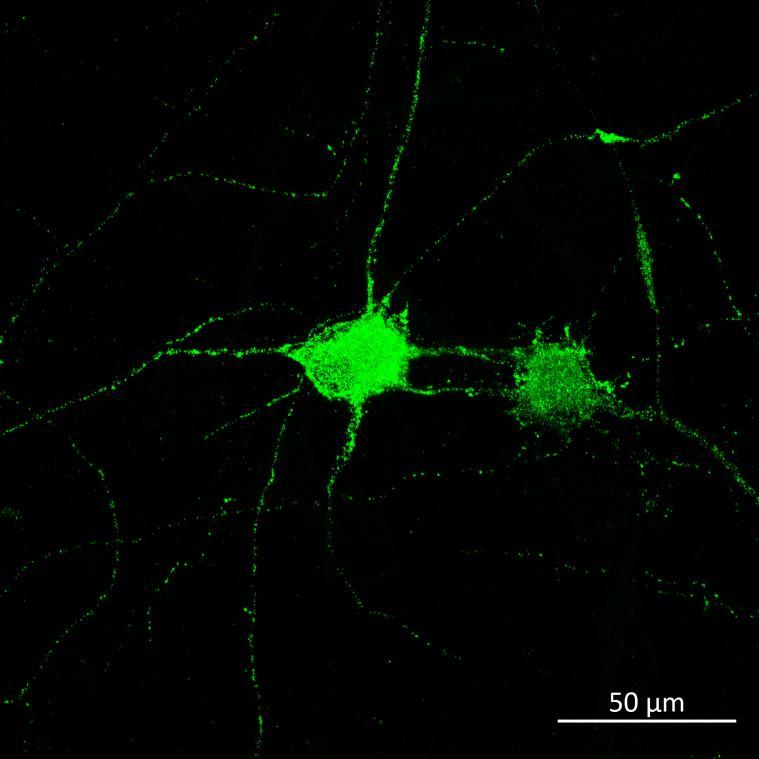
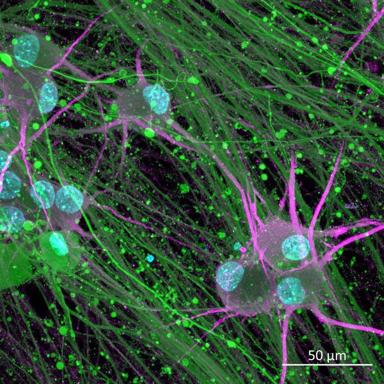
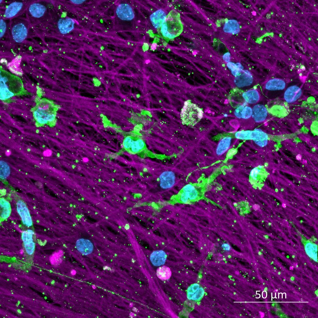
• iPSC-derived sensory neurons were labelled with well-characterised markers of peripheral sensory neurons by immunocytochemistry.
• Strong expression of the voltage gated sodium channel NaV1.8 and the nociceptor-specific marker peripherin was shown.
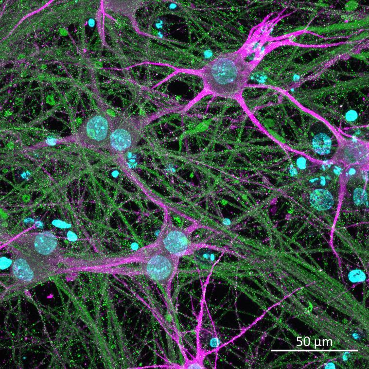
A subpopulation of neurons strongly labelled with the neuropeptide CGRP, which is also expressed by a subpopulation of human DRG.
Assessingfunctionalresponseswithhigh-throughput,singlecell calciumimaging
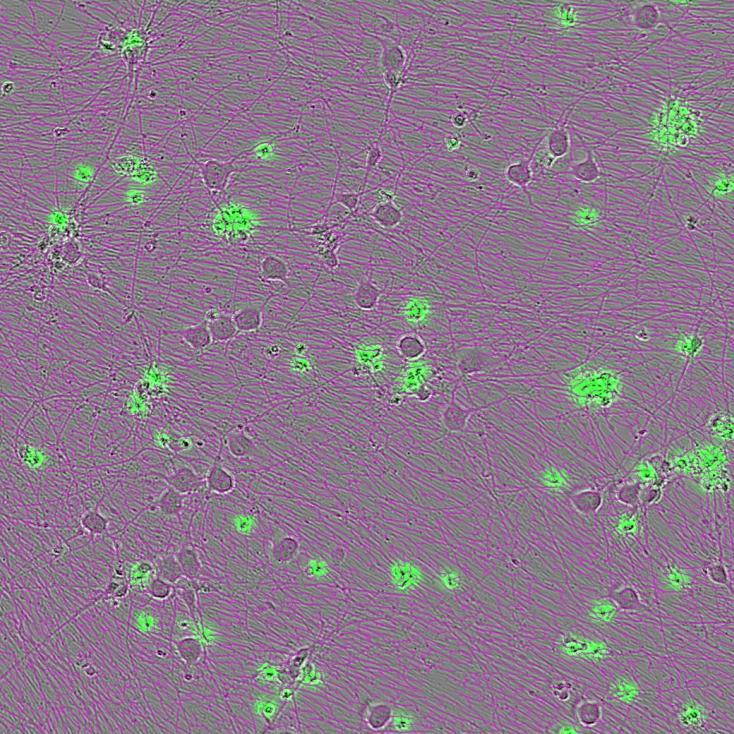
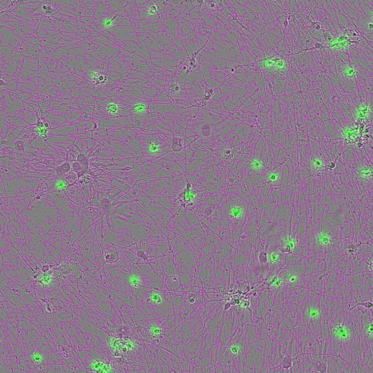
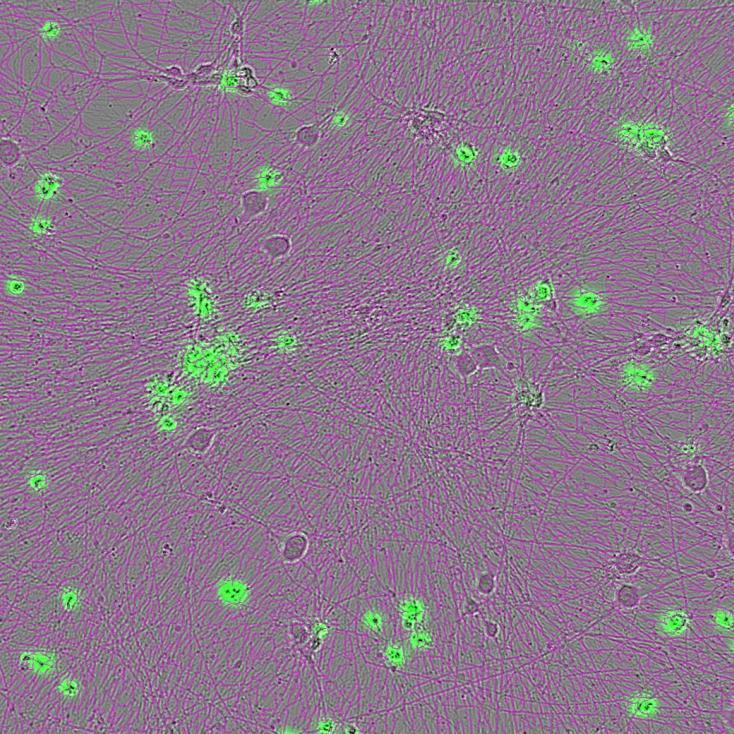
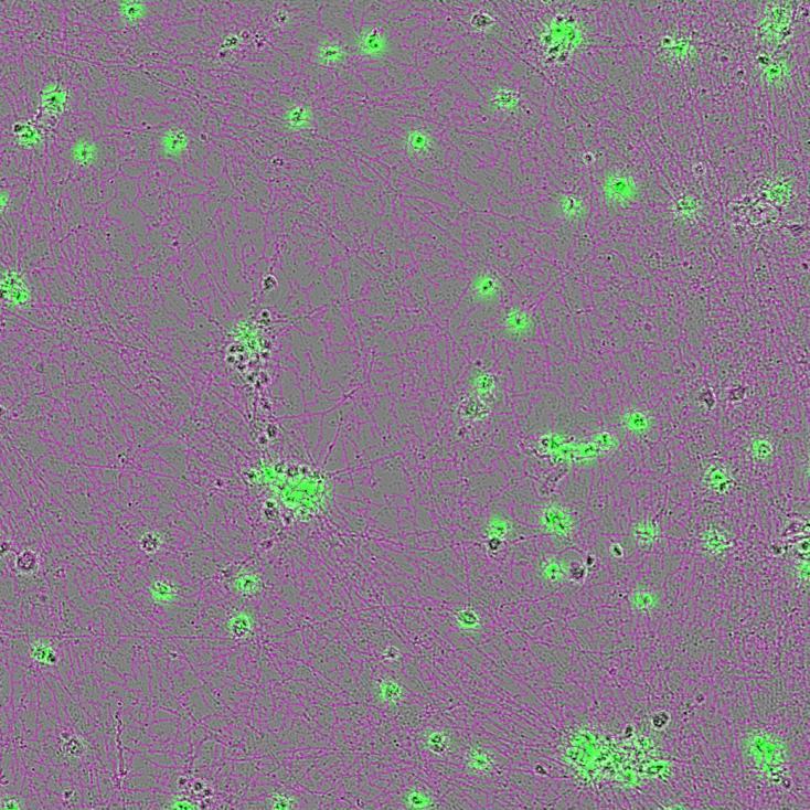
• We observed the effect of the CIPN-causing drugs paclitaxel and oxaliplatin on sensory neurons in vitro using an Incucyte SX5, analysing neurite length and cytotoxicity over time.
• No cytotoxicity was observed with paclitaxel, but neurite length decreased in a dose-dependent manner.
• In contrast, the highest concentration of oxaliplatin induced cytotoxicity and neurite degeneration, but there was no effect on neurite length or cytotoxicity at lower concentrations.
• Neurite degeneration was associated with Neurofilament Light (NF-L) release into the media as assessed by AlphaLISA, recapitulating in vivo findings of NF-L as a biomarker of CIPN severity and progression.
• These findings model the neurite degeneration and NF-L release seen in CIPN, but also demonstrate some key differences in mechanism of action between different chemotherapeutic agents.
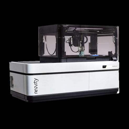
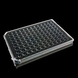
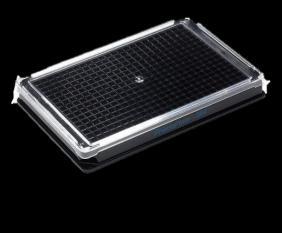
• To assess responses to nociceptive agonists we developed a 96- and 384-well format calcium imaging assay on an Opera Phenix microscope with pipettor (Perkin Elmer).
• A machine learning-based ROI selection and automated analysis pipeline allowed analysis of calcium flux over time during agonist addition in single cell format.
• This assay allows us to measure the amplitude of calcium responses and the proportion of neurons responding (classed as a post-stimulus amplitude >Mean+10SDs of the baseline and a ΔF/F0>0.2).
• This CGRP presence was further confirmed by ELISA of the cell lysate. ROI selection Size exclusion Background subtraction Normalisation to baseline
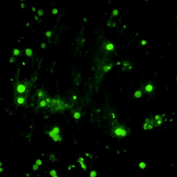

• Sensory neurons were responsive to a range of nociceptive agonists, with calcium influx in response to agonism of TRPV1 (capsaicin), P2X3 (α,β-methylene ATP), TRPM8 (WS-12), TRPM3 (CIM0216) and histamine receptors.
• No responses were seen with DMSO addition or TRPA1 agonism (cinnamaldehyde).
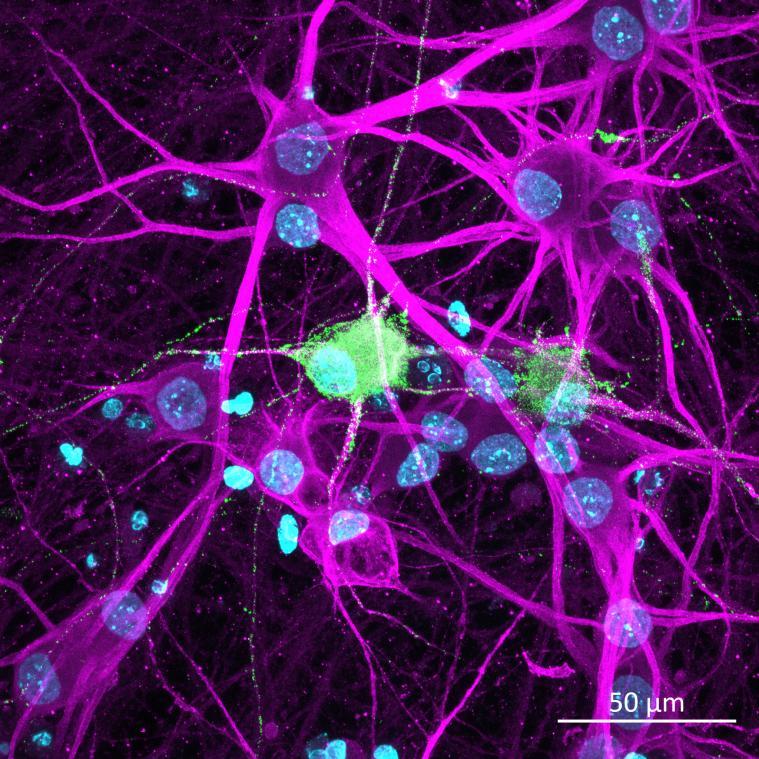
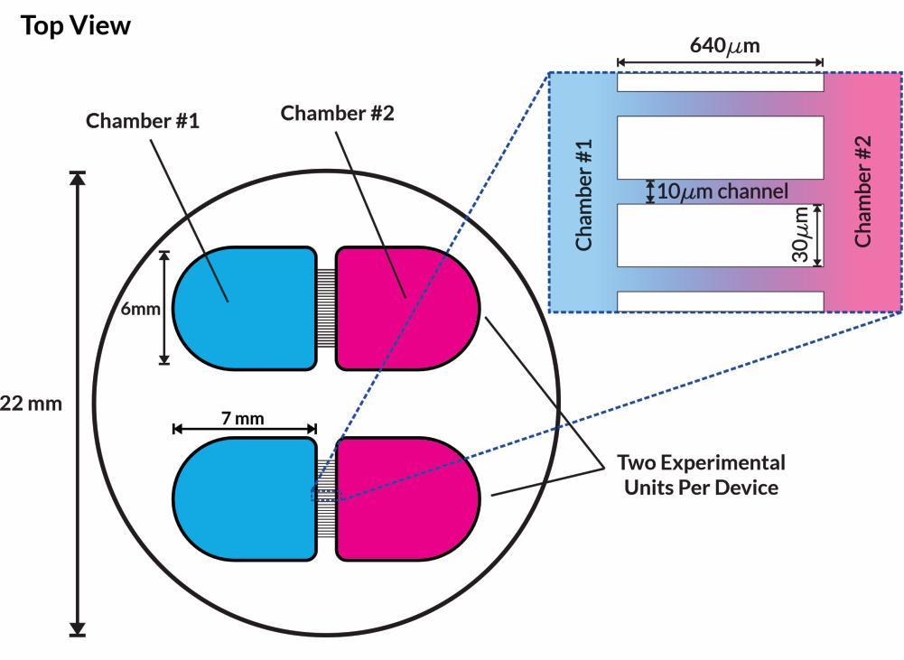
• To improve the translational relevance of our cell-based systems we investigated the amenability of sensory neurons to culture in microfluidics devices and coculture with iPSC-derived microglia.
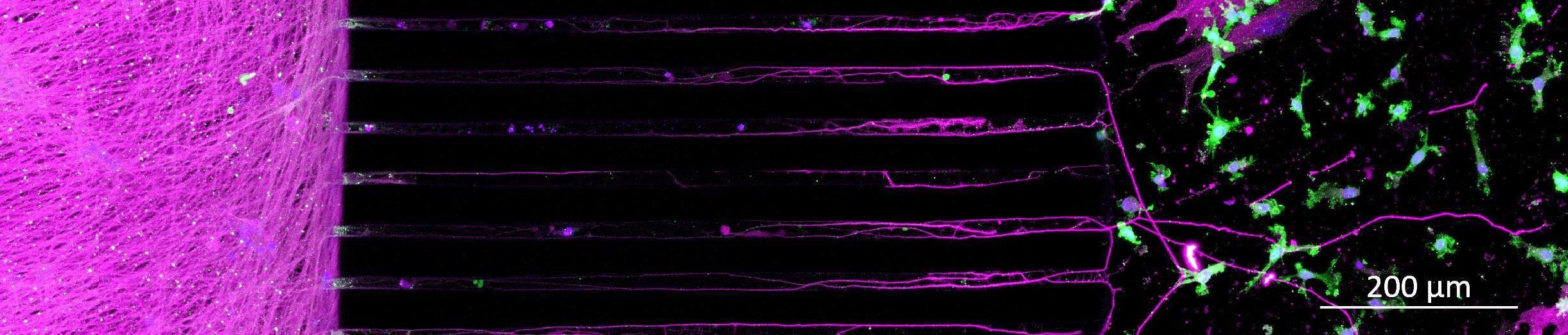
• Immunocytochemistry showed successful coculture of sensory neurons with microglia.
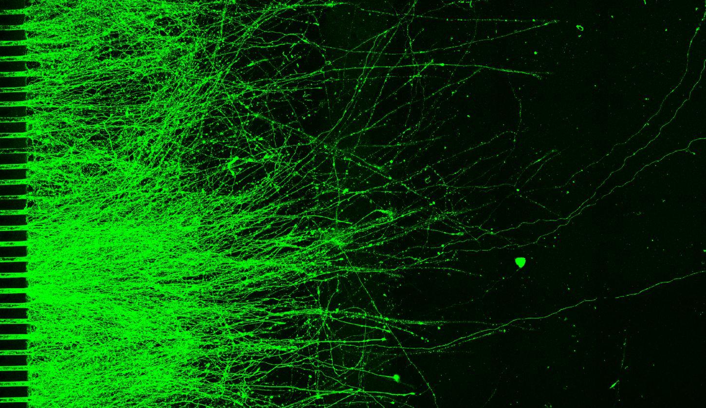
• In microfluidics, neurons projected through microchannels and into the connecting chamber, and microglia were able to be cultured in this chamber as a compartmentalised coculture system.
• This increased complexity allows improved modelling of the sensory system such as the contribution of spinal cord microglia to chronic pain and neuropathy.
• This calcium imaging approach was used to generate concentration response curves to α,β-methylene ATP, a P2X3 agonist.
• The mean response amplitude of all neurons as well as the proportion of responders both generated response curves with an EC50 between 100 and 250nM.
• The clinically developed small molecule P2X3 antagonists eliapixant and gefapixant were then added prior to α,β-methylene ATP application.
• Both compounds reduced the calcium response amplitude, validating calcium imaging in sensory neurons as a useful tool for analgesic development.
Microfluidics device
• Human-relevant in vitro models of the sensory system are essential for the development of novel analgesics, offering improved translational potential compared to traditional animal models.
• iPSC-derived sensory neurons provide a biologically relevant platform for studying peripheral pain mechanisms and should be increasingly used to identify and evaluate new therapeutic strategies in preclinical drug discovery.