A human iPSC platform for studying pathogenic protein aggregation in neurodegeneration
Eve Corrie1, Anna-Lena Zepernick1, Matthieu Trigano1, and Emma V. Jones1
1Medicines Discovery Catapult, Block 35, Alderley Park, Cheshire, SK10 4ZF, UK
Introduction
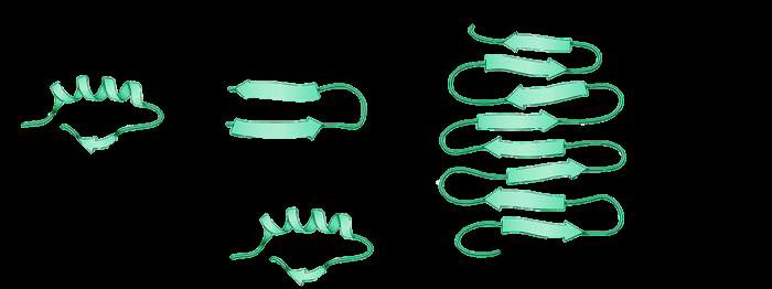
• Protein misfolding, phosphorylation, and aggregation
• These pathogenic proteins can seed further misfolding and propagate pathology across anatomically
• These processes contribute to cellular dysfunction and death, and are increasingly
• MDC has developed human in vitro systems to study the pathology of
using high content imaging assays and analysis pipelines
Live imaging of tau aggregation using lentiviral systems
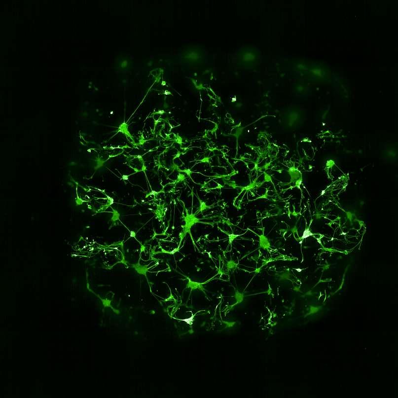
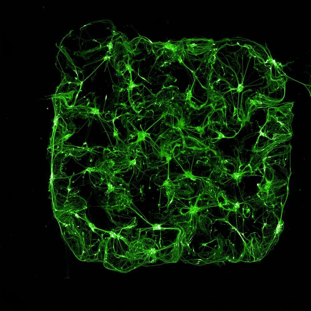
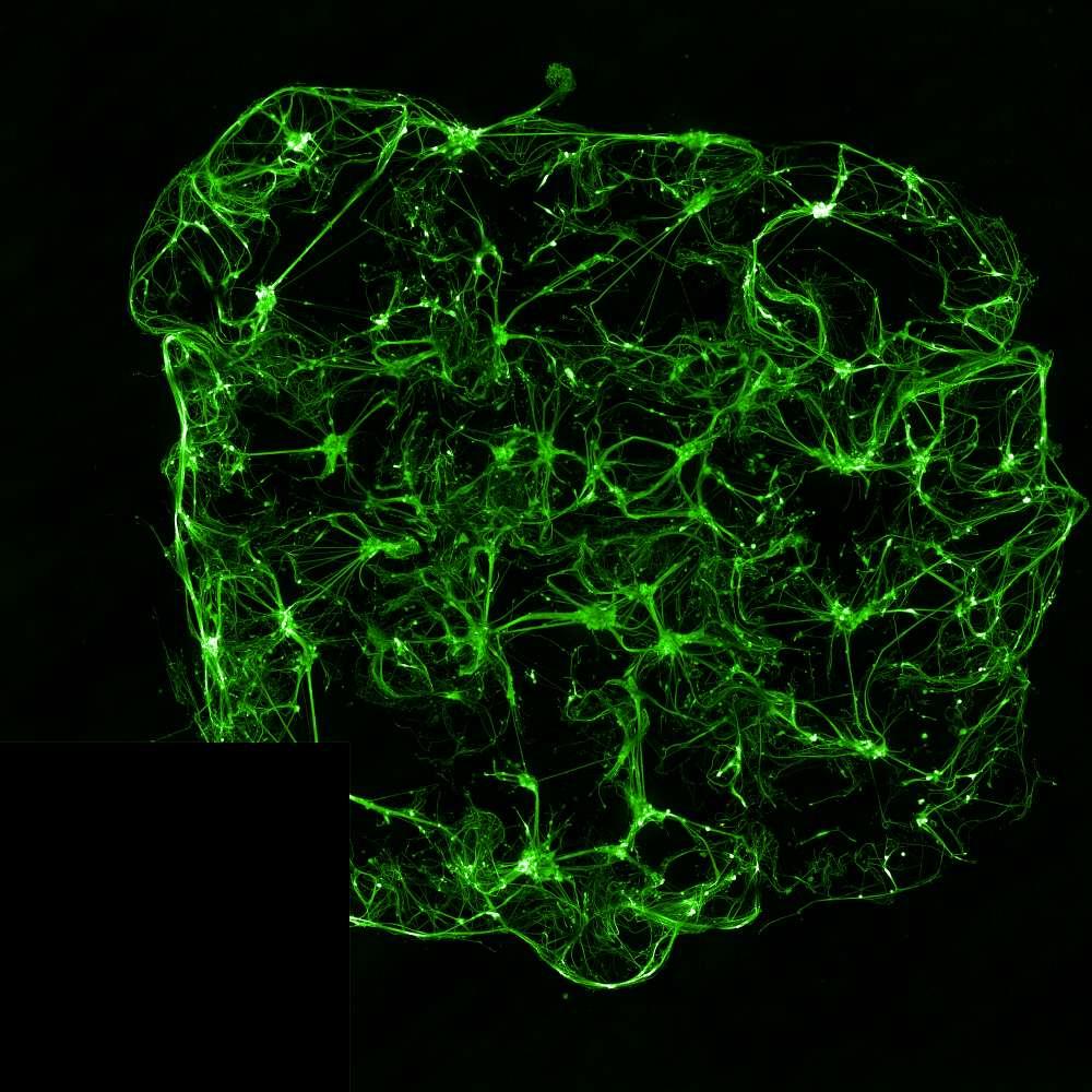
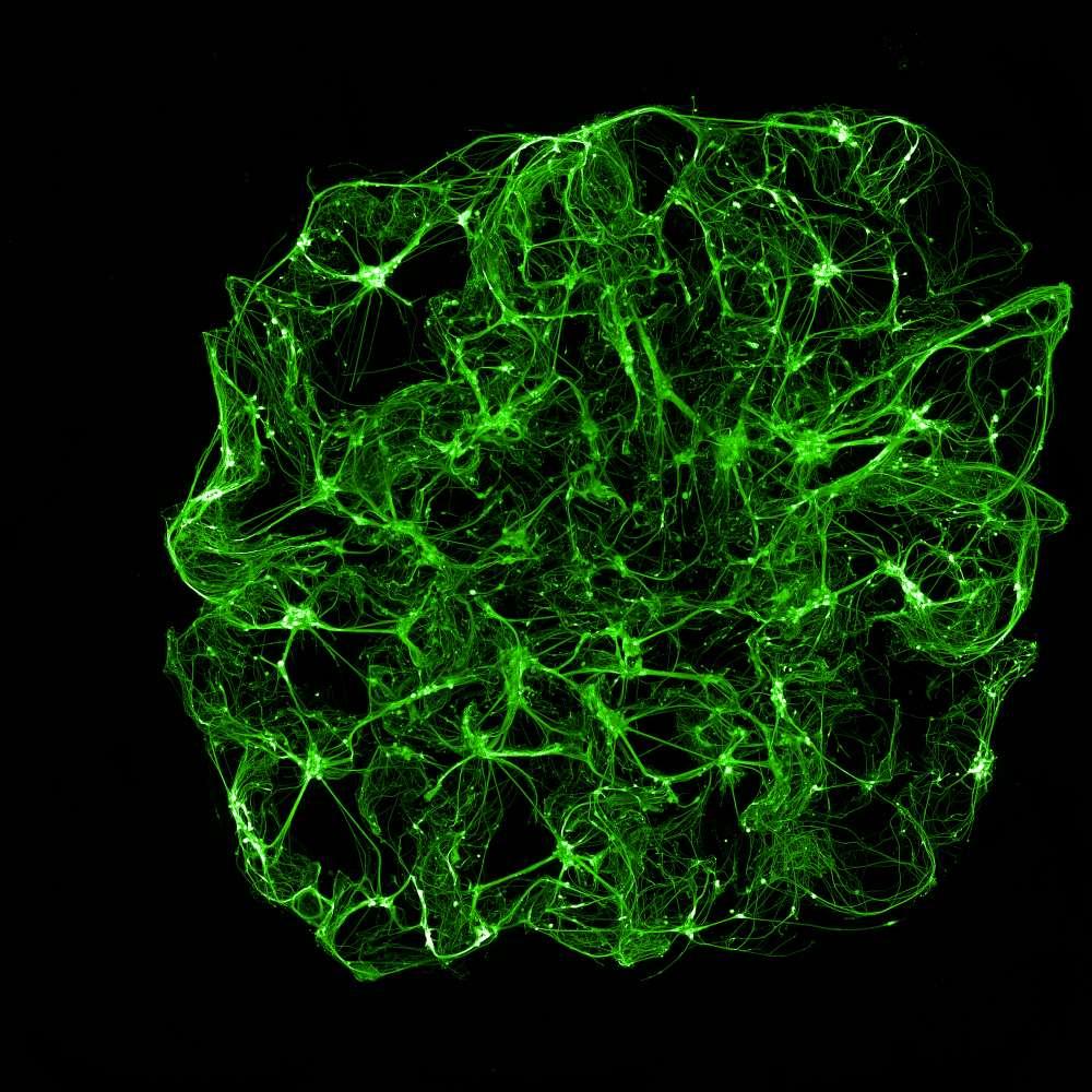
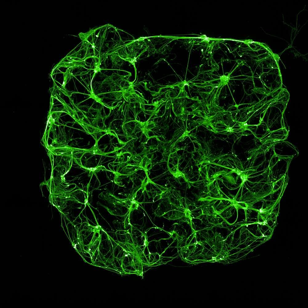
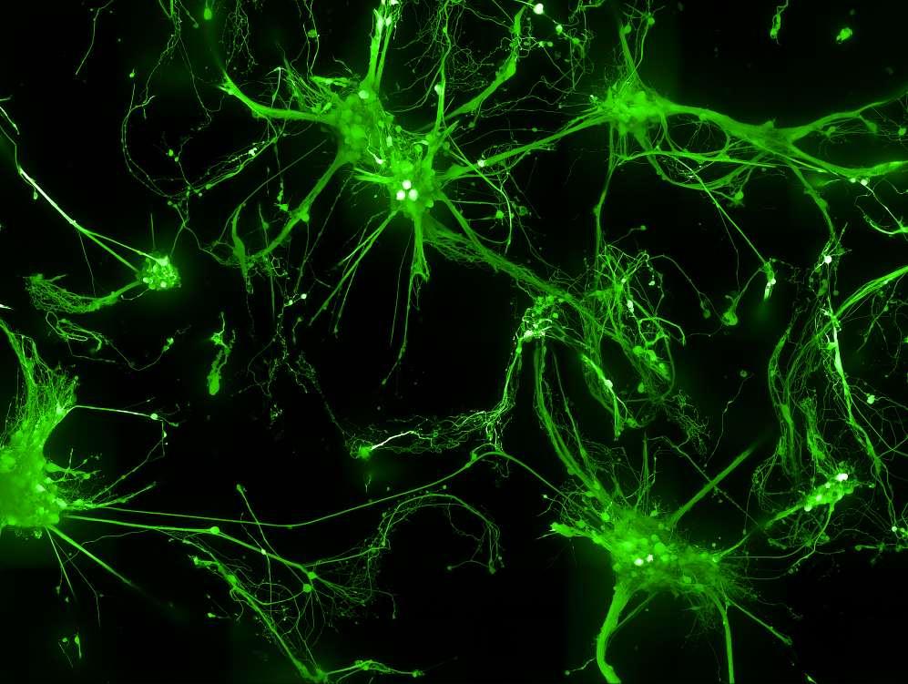
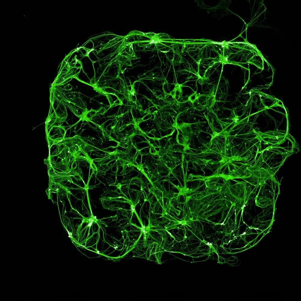
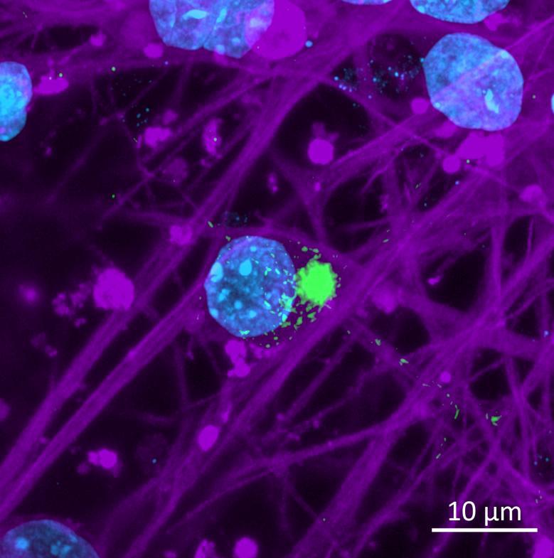
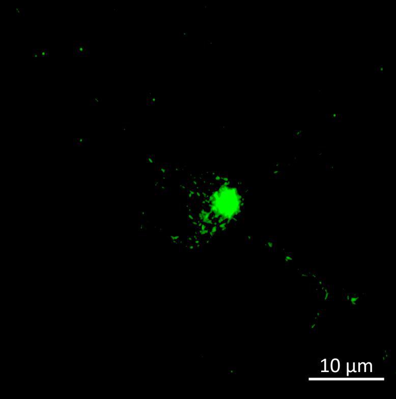
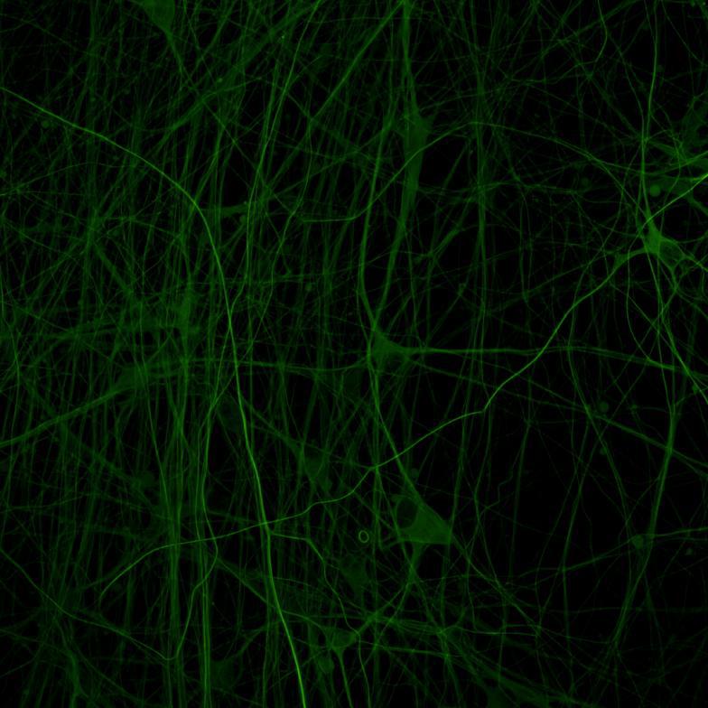
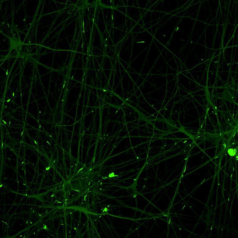
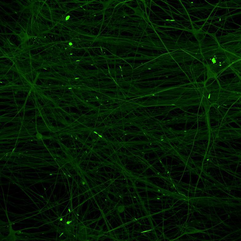
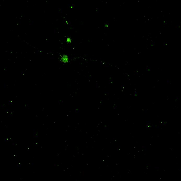
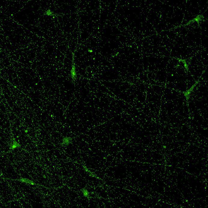
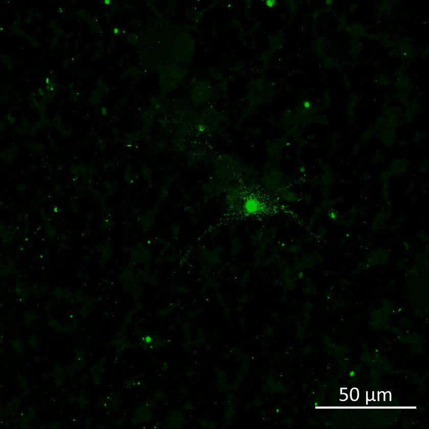
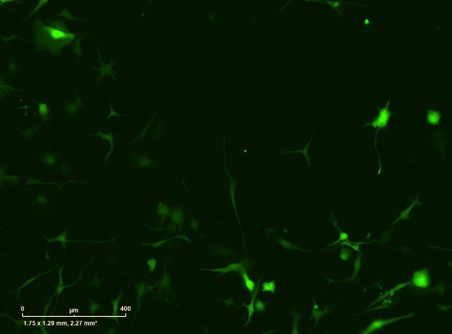
• Wildtype neurons were transduced with a lentivirus to express a full-length 4R tau isoform with a familial frontotemporal dementia P301L mutation labelled with EGFP
• Neurons were then exposed to recombinant tau preformed fibrils (PFFs) and imaged
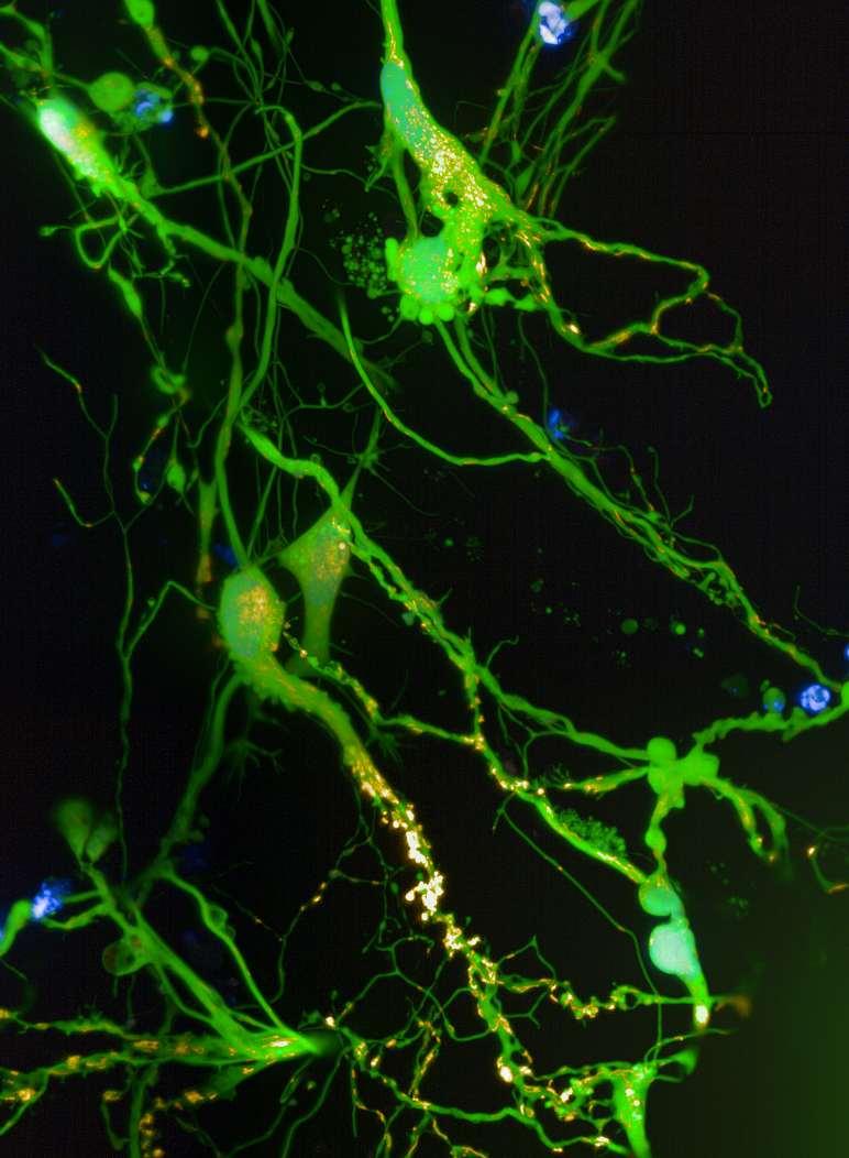
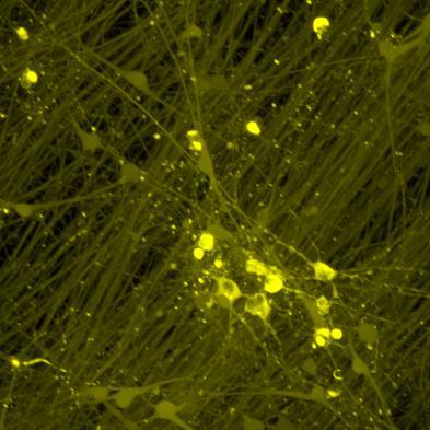
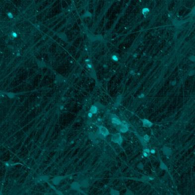
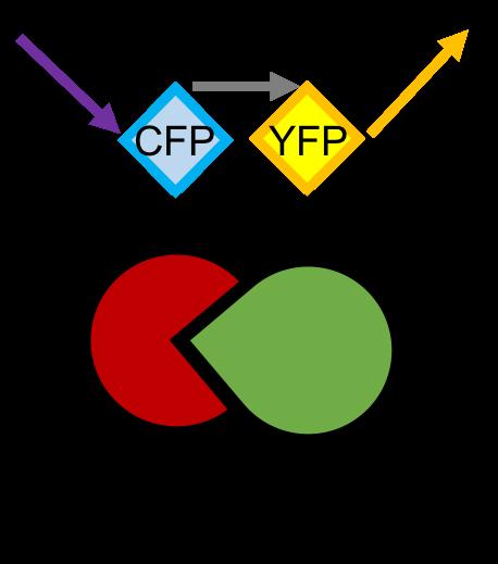
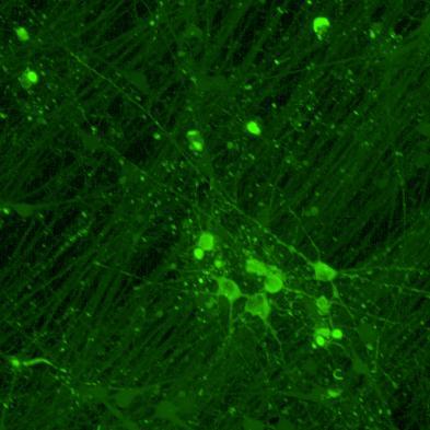
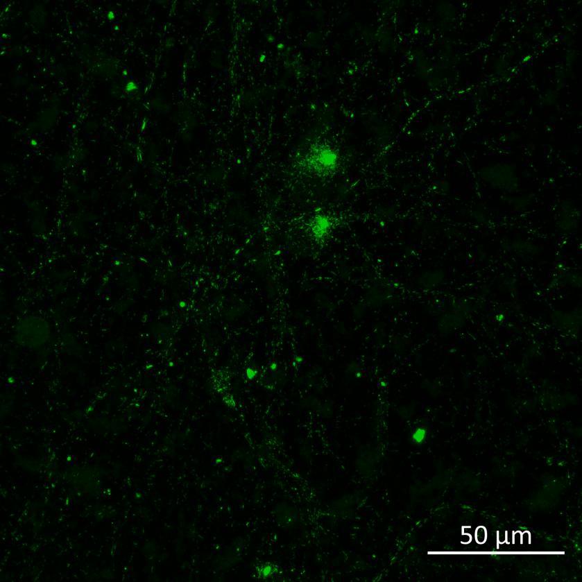
• Neurons were transduced with lentiviruses to express 4R microtubule binding domain (K18) fragments with P301L mutation labelled with EYFP or Cerulean
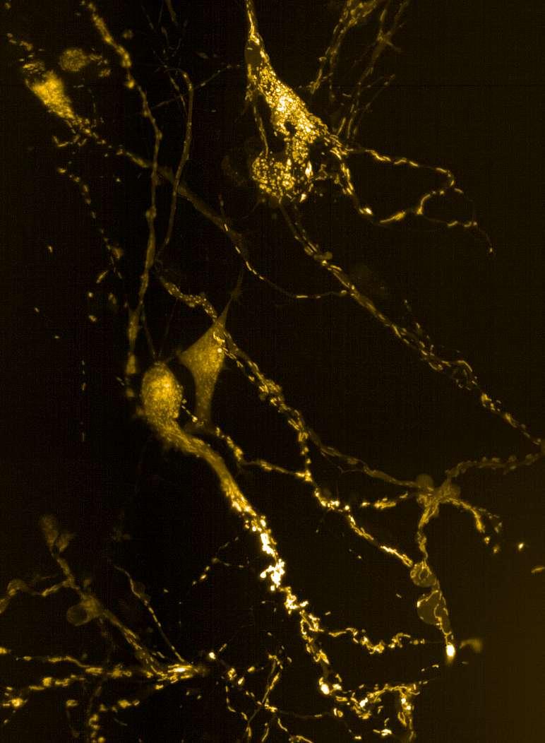
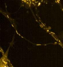
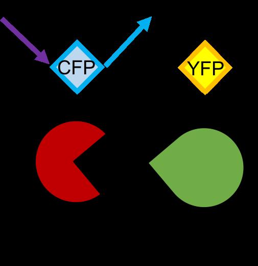
• When aggregation occurs, the fluorophores become close enough for FRET to occur
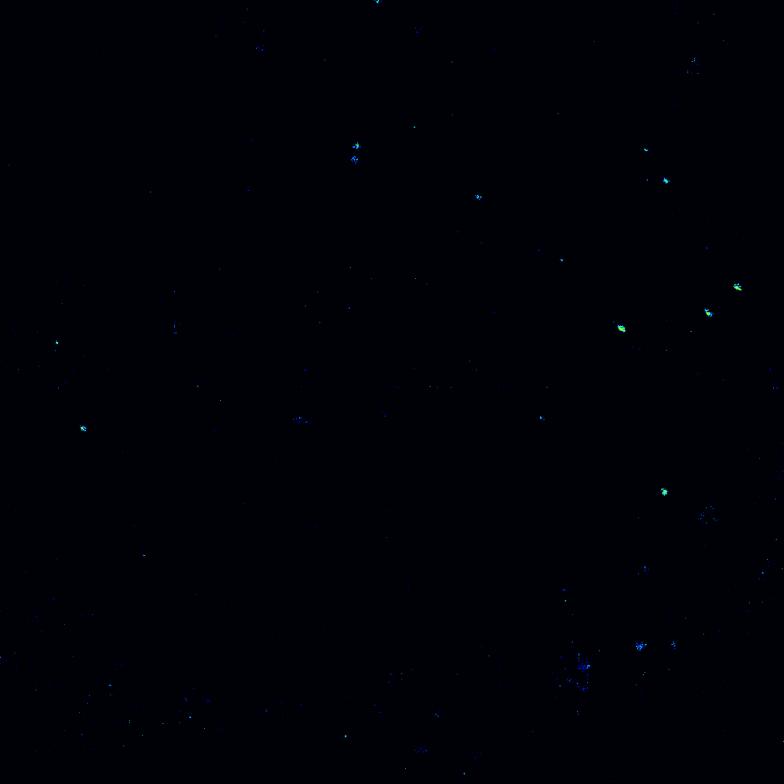
• The transduced neurons were then exposed to tau PFFs and FRET imaging performed
Phosphorylation and aggregation of
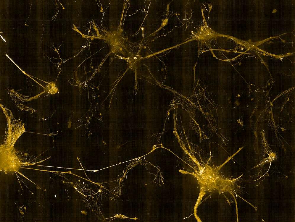
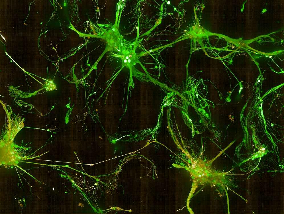
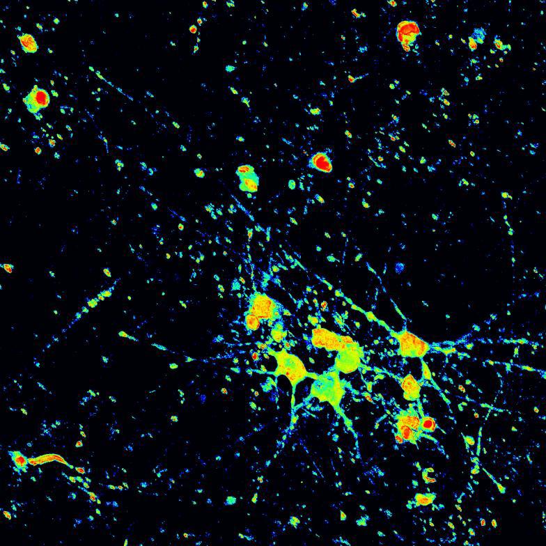
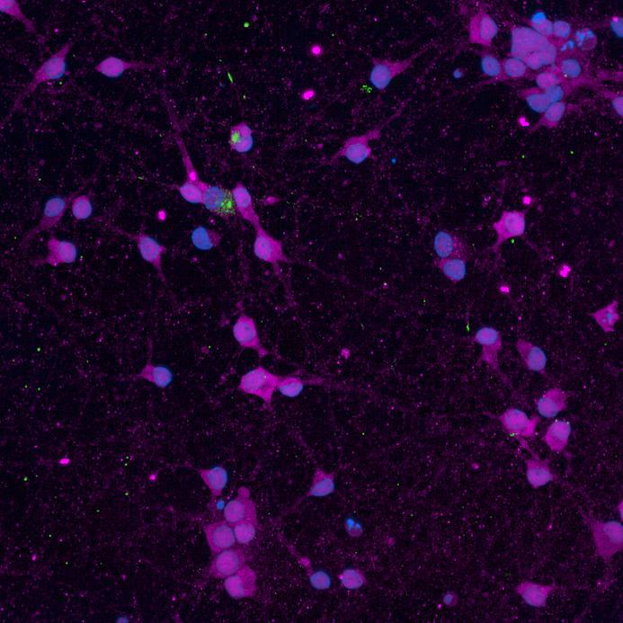
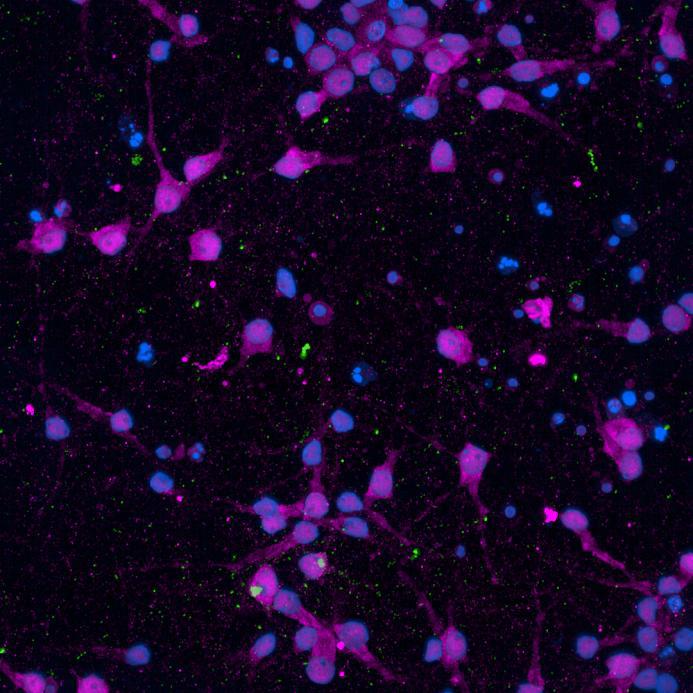
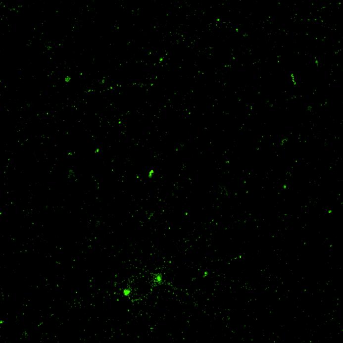
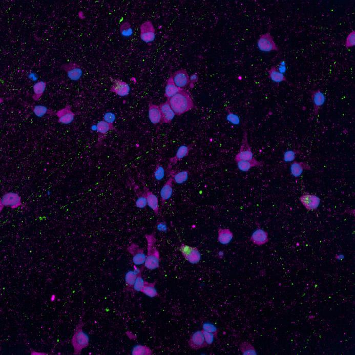
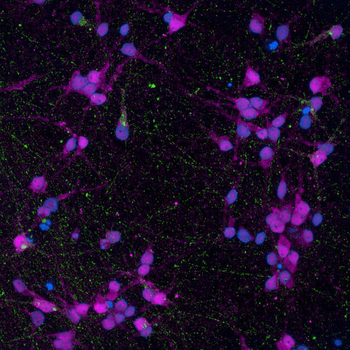
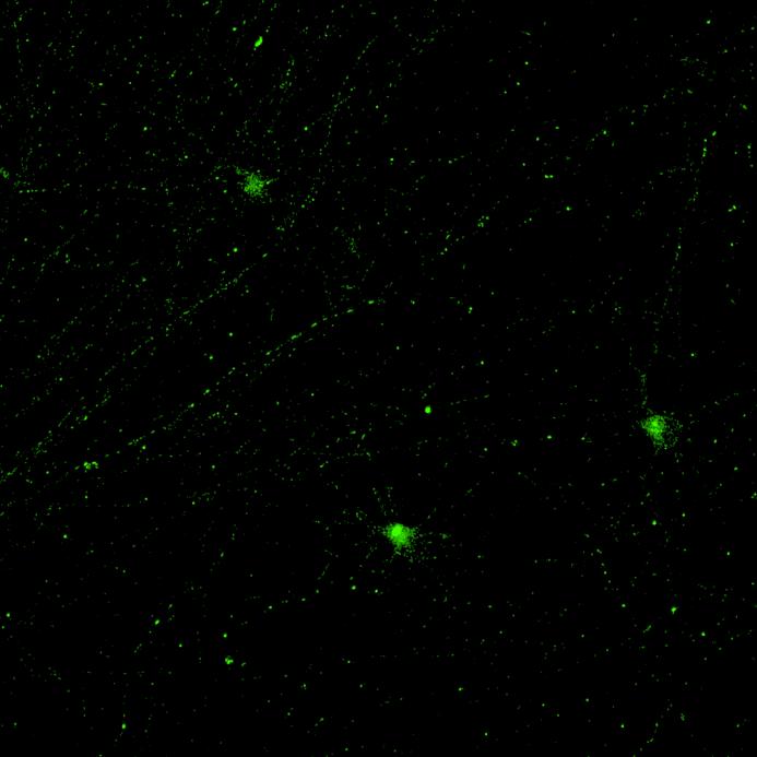
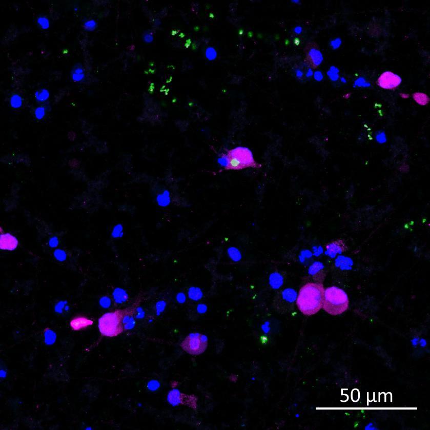
• Isogenic control and early onset Parkinson’s disease SNCA A53T mutation neurons were exposed to α-synuclein PFFs with A53T mutation
• Neurons were fixed and labelled for α-synuclein phosphorylated at serine 129 (pS129)
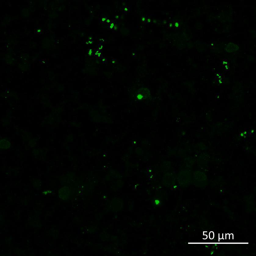
• PFF treated neurons show Lewy body-like perinuclear aggregates of pS129 α-synuclein
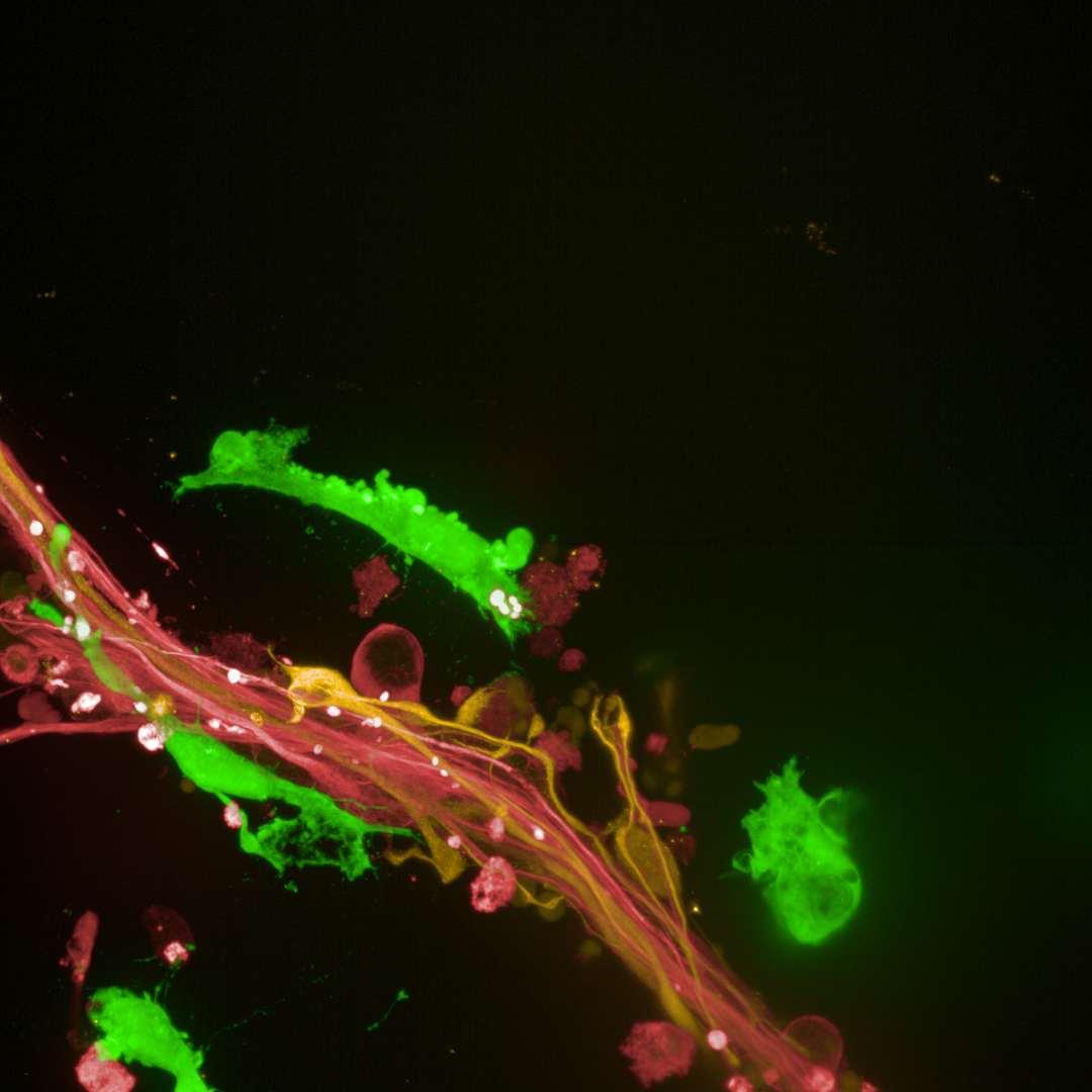
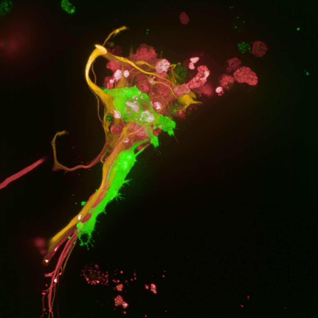
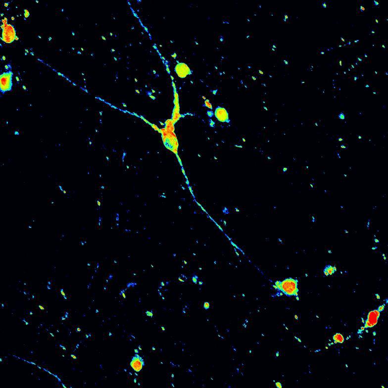
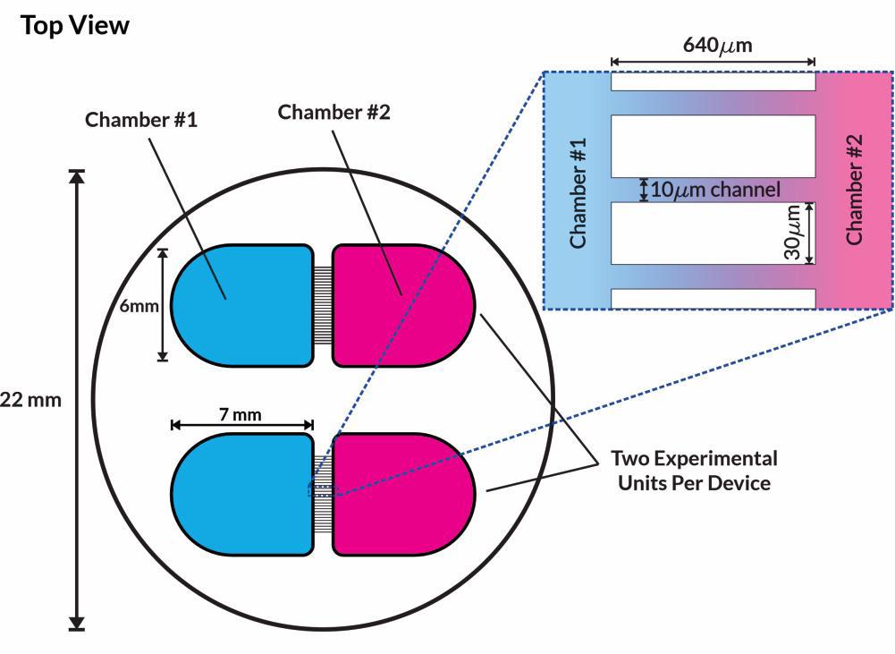
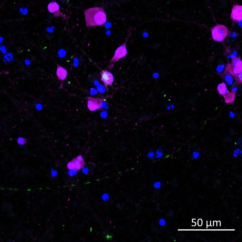
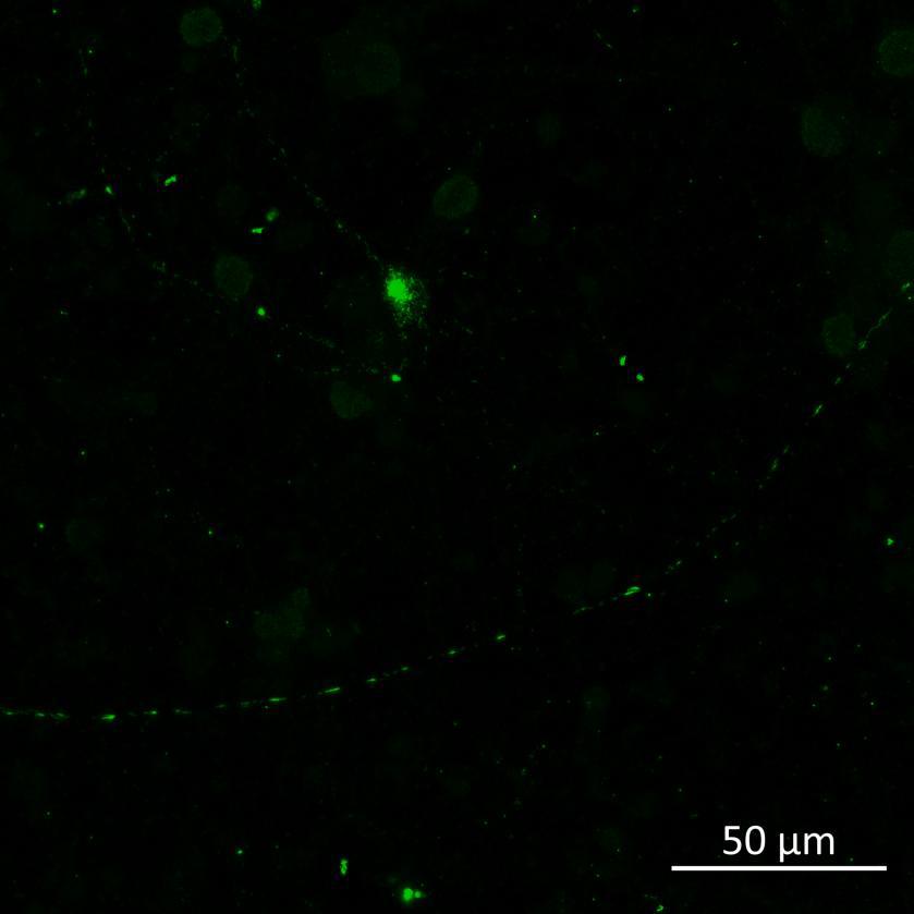
• Isogenic control or SNCA A53T mutation neurons were plated in both chambers of a microfluidics device then cultured to allow the neurons to connect via the microchannels
• PFFs were added to the donor chamber only before fixing and labelling for pS129 α-synuclein
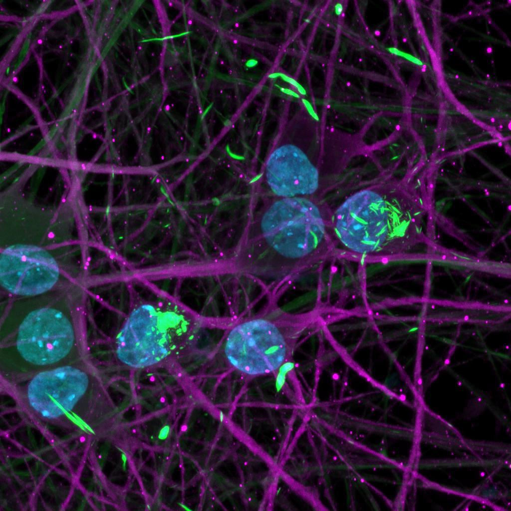
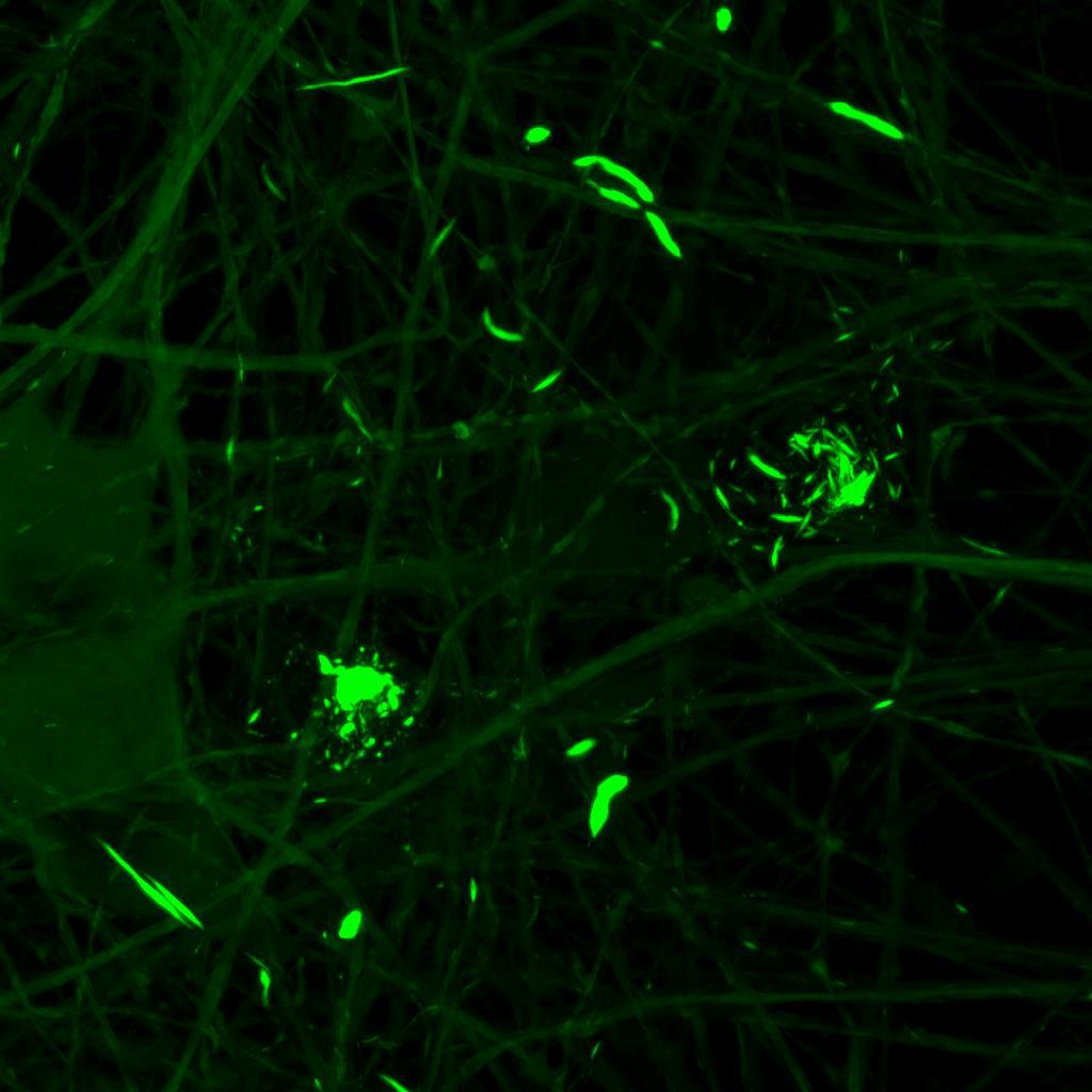
• Increasing over time, PFF exposure induced areas of bright EGFP-positive aggregates of full-length tau, and aggregation of the K18 fragment FRET pair as seen by positive normalised FRET (NFRET) signal
• K18 PFFs with P301L mutation induced aggregation more readily than
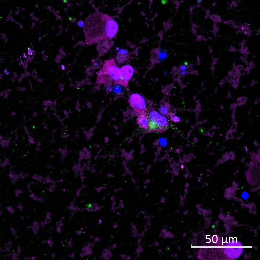
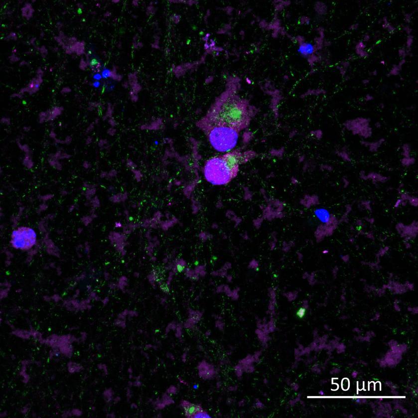
• pS129 positive neurons were detected in donor chambers due to directly applied PFFs and acceptor chambers due to seeded PFFs, though automated analysis failed to detect the small proportion of positive wildtype neurons in acceptor chambers
Projection of neurites through microchannels at 2 weeks
• A53T mutation increases pS129 pathology in both donor and acceptor chambers
0N4R-EGFP tau 2 weeks post-PFF
K18 FRET biosensor system 2 weeks post-PFF
Development of a human reactive astrocyte platform for drug discovery
Introduction
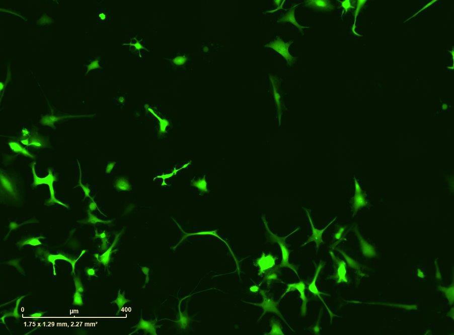
Use of high content imaging and functional analysis to study astrocyte activation and observe responses to anti-inflammatory drugs
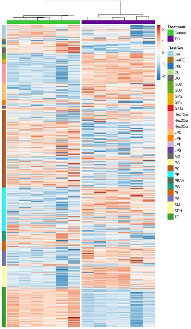
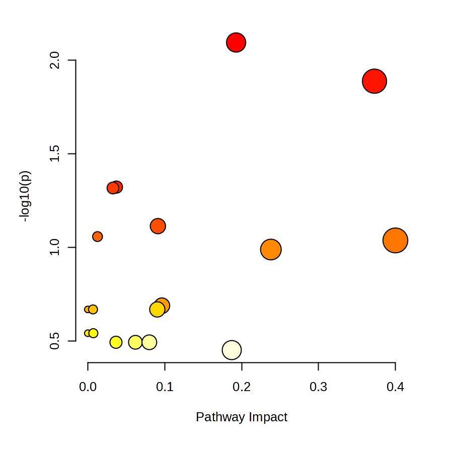
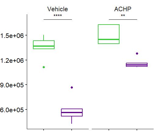
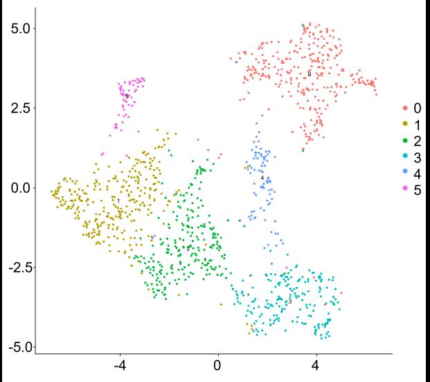
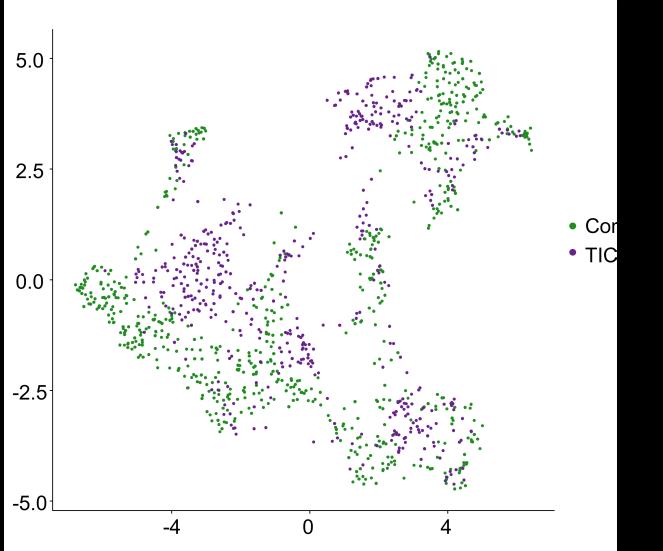
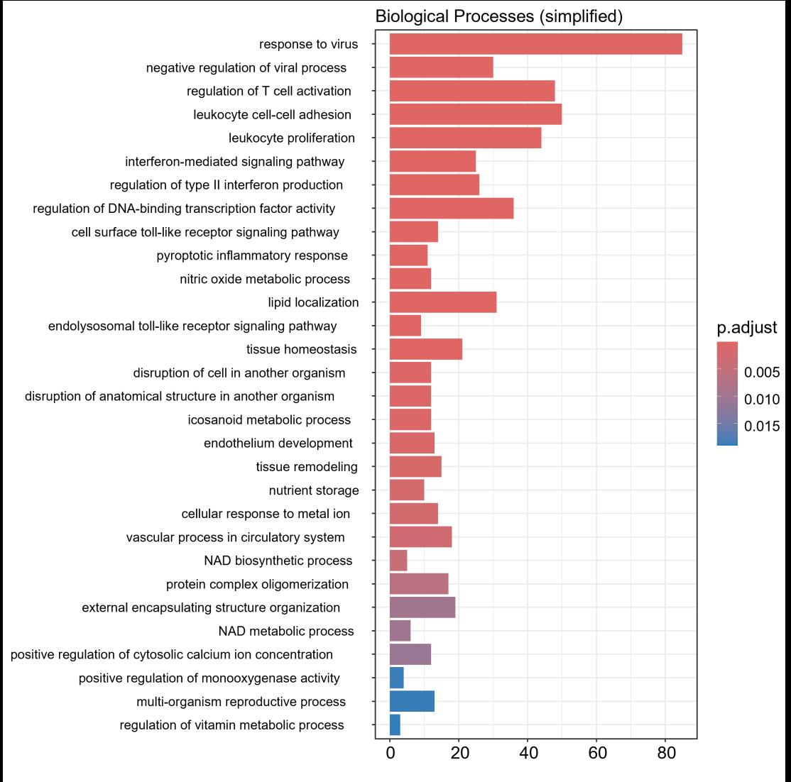
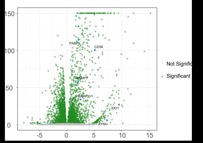
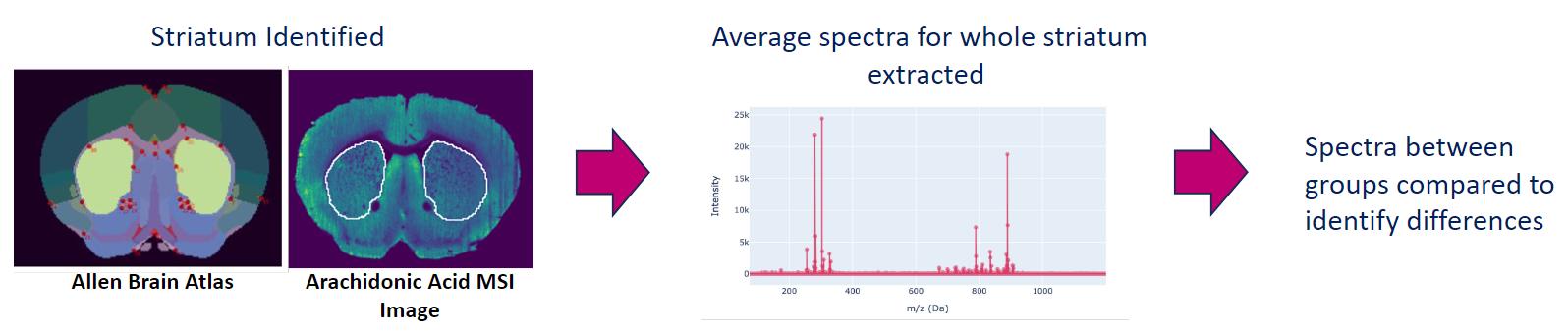
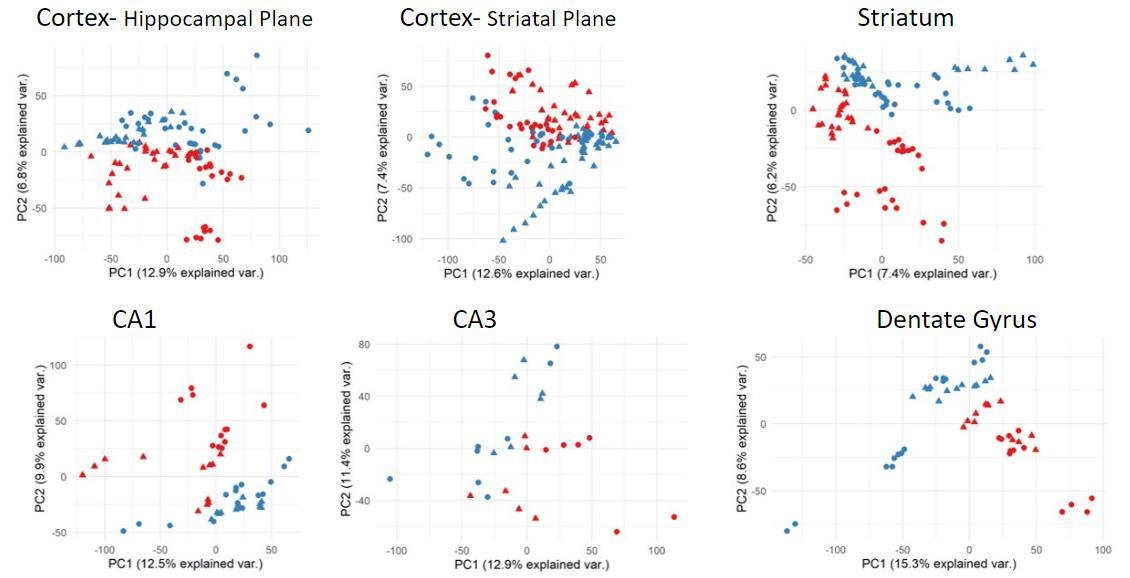
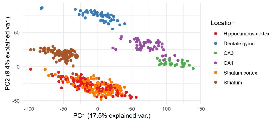
LC-MS and scRNA-seq identify changes in the lipidome, metabolome and transcriptome of reactive astrocytes
Results and conclusion ≤
Anna-Lena Zepernick, Tara J Bowen, Robert Pedley, Eve Corrie, Matthieu Trigano, Emma V. Jones¹
¹ Medicines D scovery Catapu t, Block 35, Alderley Park, Cheshire, SK10 4ZF, UK
Use of Innovative Multi-Omics
Platforms to Study Neurodegenerative Disease
Kerry Shea1,3, Lucy, Frost1, TJ Allen1, Eleanor Platt1, Irma O’Meara1, Ana-Maria Nastase1, Tara Bowen1, Maike Langini1, Régine Anderson1, Caitlin Shaw1, Paul Sharp1, Jim McCarthy2, Donna Finch2, Gayle Marshall1