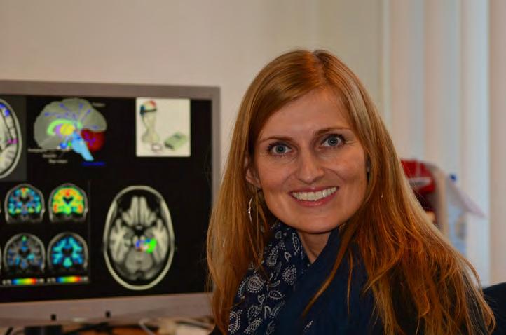
Diagnosing and Treating Epilepsy, Other Disorders Originally used only for research purposes, magnetoencephalography has, in more recent decades, been introduced into clinical care. With applications in epilepsy already benefiting from its use, and still others on the horizon, the technique is helping advance diagnosis and treatment for a range of diseases, disorders and injuries. The Martinos Center for Biomedical Imaging offers a clinical MEG service to aid in many of these applications. Led by Steven Stufflebeam, medical director for the Martinos Center, it is the only such service in the Northeast. Its most common application is presurgical evaluation and surgical planning in epilepsy patients: localizing epileptic discharges, determining the language-dominant hemisphere and mapping the eloquent cortex. Here, MEG is often combined with MR imaging, which provides structural guidance for the analysis of the MEG data. By localizing epileptic discharges, MEG helps identify the sources of seizures through analyses of spontaneous brain activity. Measurements are typically performed during rest using whole-head MEG combined with electroencephalography (EEG), a closely related method that provides complementary information about electrical activity in the brain.
134
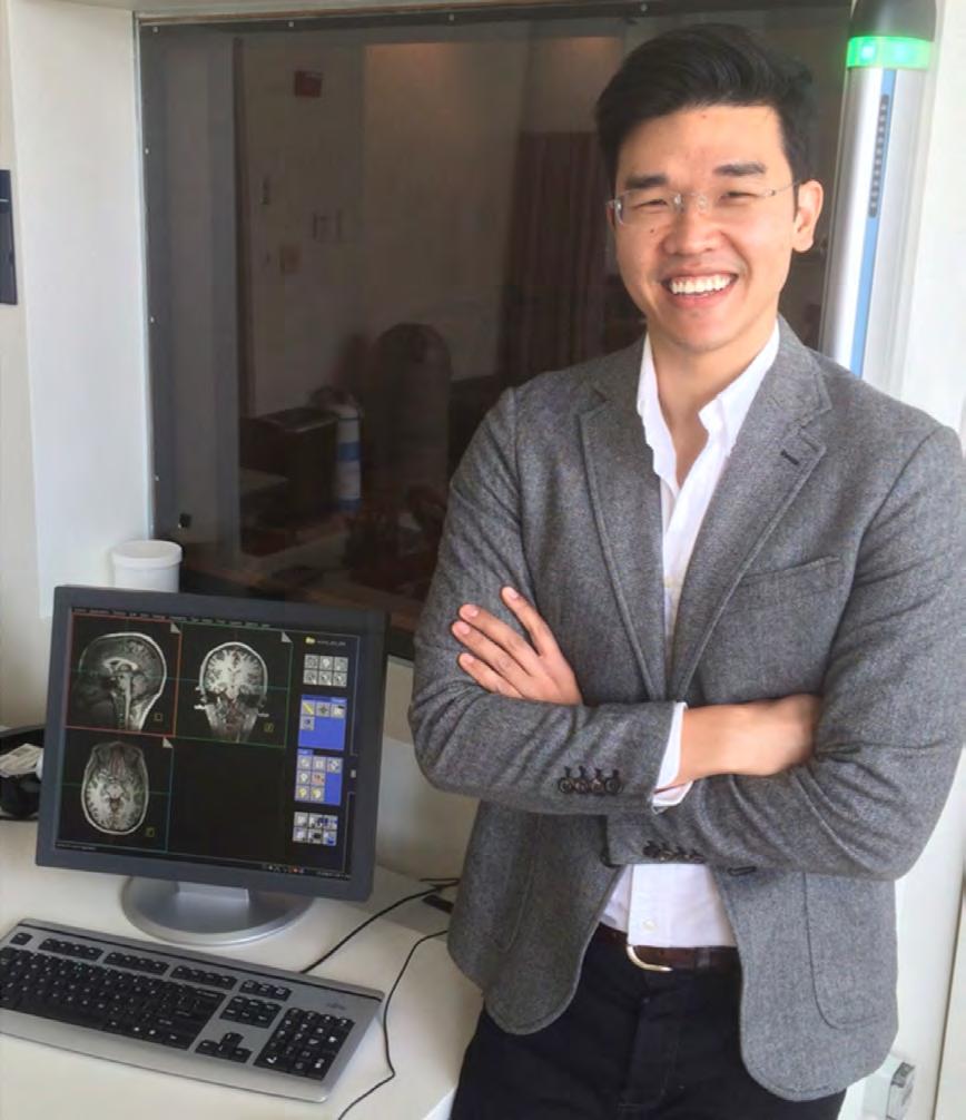
After a decision is made to remove the tumor or other lesion responsible for the seizures, MEG can map the eloquent cortex to help avoid functional deficits as a result of surgery. A critical use is determining the language-dominant hemisphere of the brain. This measurement is particularly important for patients scheduled for a left anterior temporal lobectomy (ATL), a widely used procedure for medial temporal lobe epilepsy. Such lateralization is vital for preserving quality of life as verbal memory and language can be impacted by ATL. Beyond determining the language-dominant hemisphere, clinicians use MEG to delineate the language cortex and other areas of the eloquent cortex (for example, motor and visual) to outline the regions they want to avoid during resection. The technique can localize both receptive and productive areas of language with high temporal resolution; the measurements are often combined with fMRI data to improve spatial resolution. Studies have shown that presurgical evaluation of epilepsy patients with MEG leads to improved outcomes in these patients, with greater surgical success and fewer postsurgical deficits.