
8 minute read
The ‘Unassuming’ Ken Kwong and a Pivotal fMRI Breakthrough

In early 1992 the neuroscience community was flush with excitement. Jack Belliveau had recently published his pioneering work with functional MRI and the possibilities of the approach seemed truly limitless.
Researchers were particularly inspired by the potential for brain mapping that was evident in Belliveau’s work. They could now see, more or less in real time, changes in the brain occurring in response to particular stimuli or tasks. There was just one problem: the need to use an injected contrast agent limited the potential of fMRI in human subjects, as any medically unnecessary injection poses some degree of risk.
Advertisement
As it happened, another Center investigator, a postdoctoral fellow named Kenneth Kwong, had found a way around this problem. In a paper published in June 1992 in Proceedings of the National Academy of Sciences, Kwong reported a means to measure intrinsic contrast—that is, contrast occurring naturally in the brain—with fMRI, thus removing the need to use an external agent. In doing so, he positioned the technique for much broader application than would have been possible otherwise, opening the door to the many extraordinary advances we have seen in the nearly 30 years since.
Kwong isn’t one to tout his accomplishments. The book Quiet, something of a treatise on the power of introverts, of those who have little need for attention and little if any use for the (over)stimulation of the outside world, describes him as “a brilliant but unassuming scientist” who never asks for recognition for his extraordinary achievements. And indeed, in conversations about the heady early days of functional MRI, he often either downplays the creativity of his insights or deflects attention by pointing to the moments of chance or serendipity that led him to these insights.
Try as he might, though, to understate the significance of his contributions, the results of his work speak loudly enough.
Still, for all the impact his research has had, Kwong didn’t actually set out to find the key to performing noninvasive functional MRI. He had come to the Center several years before, to work with MIT graduate student Daisy Chien. In 1990, he was seeking new ways to measure cerebral perfusion—essentially, blood flow in the brain. It was this search that started him down the fMRI path.
In a 1982 report, Thulborn had shown in scans of blood samples that MRI could measure changes in the amount of oxygen in the blood. In doing so, he anticipated a phenomenon described by researcher Seiji Ogawa of Bell Laboratories in a 1990 paper, which would come to be known as blood oxygen level dependency, or BOLD.
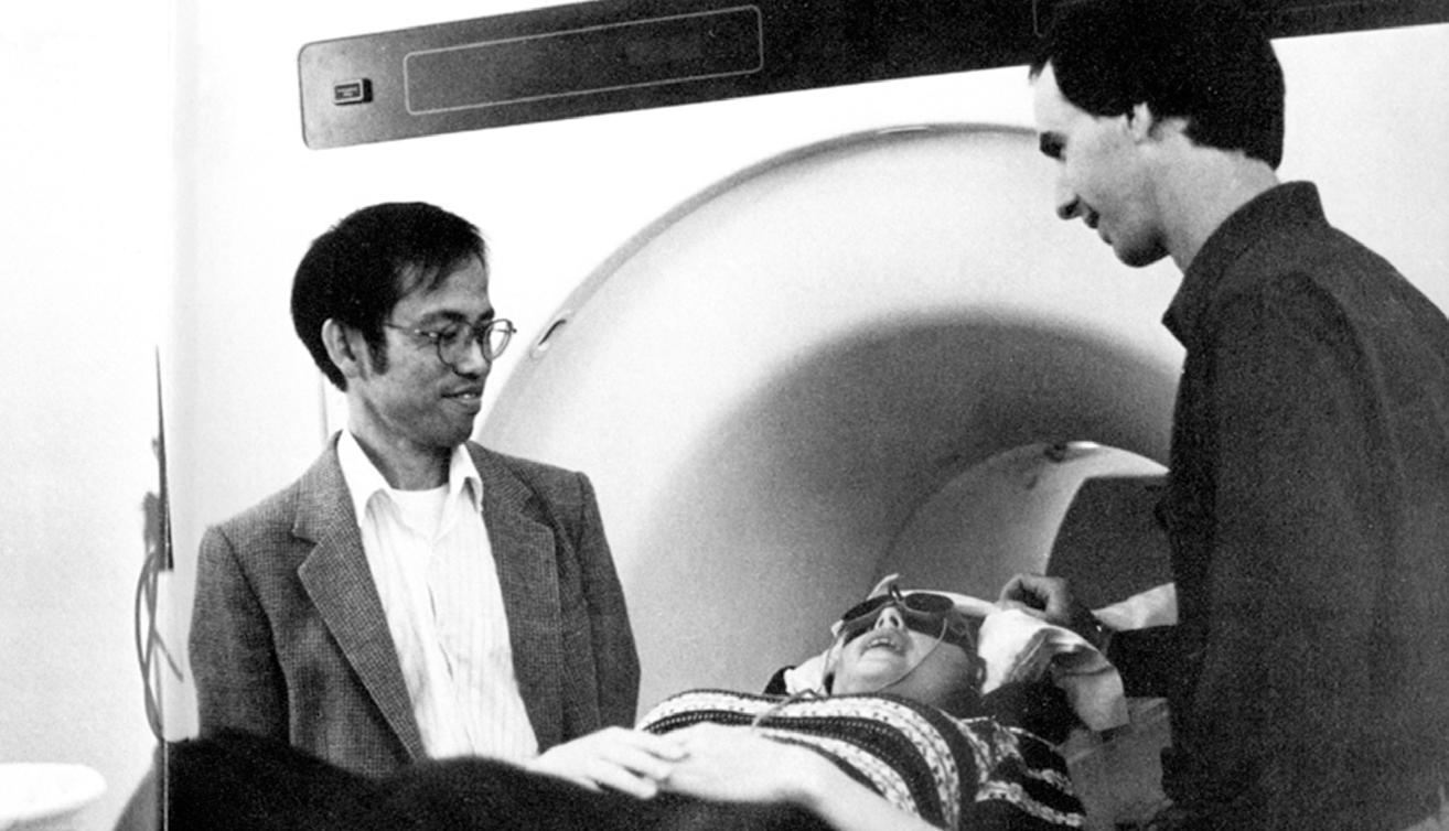
Ken Kwong (left) and John Baker (right) prepare a volunteer for an fMRI experiment, circa 1991. The subject is wearing the now-famous stimulation goggles, which activate the visual cortex by flashing patterns of light.
The first moment of serendipity he mentions came early in his search, when a brief snippet of conversation with Keith Thulborn, an MGH radiology resident at the time, alerted Kwong to a possible means of measuring perfusion. A very brief snippet, as it happened. “I heard one sentence,” Kwong says. “I wasn’t even sure he was talking to me.”
“Had I not caught Keith’s sentence I would not have made any link between deoxyhemoglobin and the MR signal,” Kwong says today. (Hemoglobin is the oxygen-transport protein found in red blood cells. When it is not bound with oxygen, it is called deoxyhemoglobin.) “It was not my area of expertise, and at the time I wasn’t aware of Ogawa’s work.” But he had caught it. And now he wanted to know whether measuring deoxyhemoglobin was an option he could put to use.
The first step: designing an experiment. For help with this, he once again turned to something he had overheard in the hallway.
In 1990, even as Kwong was exploring options for measuring perfusion, Jack Belliveau was developing a means to image brain activity with MRI and performing his experiments using visual stimulation to induce changes in activity—the same experiments that would be reported in Science in November 1991. Kwong wasn’t involved in the functional MRI project but he had heard through the proverbial grapevine what Belliveau was up to. And he saw in it a natural approach to eliciting changes in perfusion.
He talked to Belliveau about his idea to use the visual stimulation paradigm for his own experiments. Belliveau was supportive, offering suggestions about how to go about it and even loaning Kwong a pair of visual stimulation goggles—the now-famous red goggles that researcher Peter Fox had used in the early 1980s and that Belliveau had borrowed and used for his own experiments. (With apologies to Fox, the never-returned goggles are now on display in Mass General’s Paul S. Russell Museum of Medical History and Innovation.)
Kwong performed his first experiment with the new approach on the evening of May 9, 1991, in what is now Bay 3 at the MGH Martinos Center for Biomedical Imaging. It’s tempting to imagine here a sense of import in the air, a knowing understanding of the significance of what was about to happen. But the fact is, for Kwong, it was a scanning session like any other, especially as he had no expectation of the approach working right out of the gate.
And yet it did. Kwong processed the data acquired during the scan and, “lo and behold, I saw a bright blob coming out of the visual cortex.” More to the point, he noted a clear change in MRI signal due to changes in blood deoxyhemoglobin, which suggested that hemodynamic change during neuronal activation could be observed with MRI. Even with just a single run in a single subject, he knew the experiment had been a success.
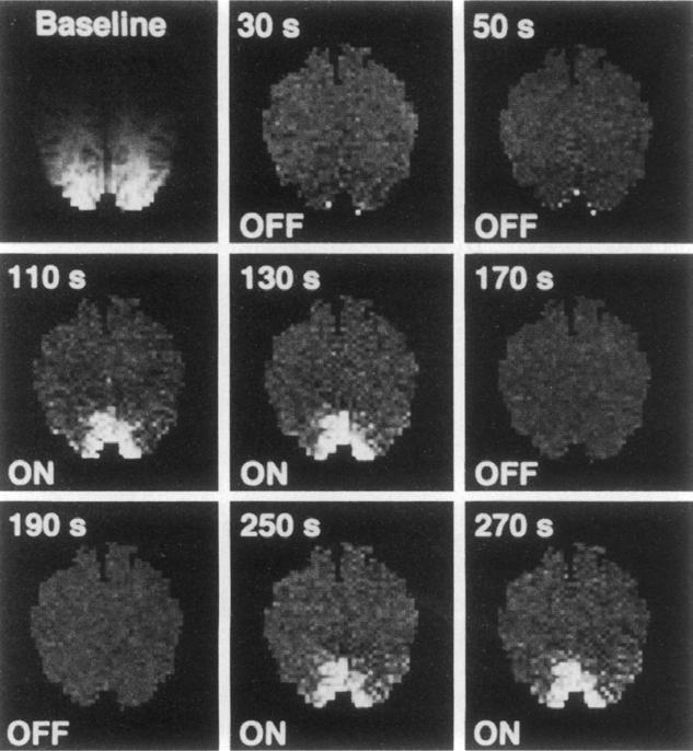
Images from Kwong’s 1992 Proceedings of the National Academy of Sciences paper showing activation in the visual cortex in response to stimulation.
So, what next? What do you do when you’re reasonably sure you’ve just made a breakthrough scientific discovery, one that could have far-reaching implications, even beyond what you can imagine, for basic science research and even clinical practice? How do you react? Kwong remembers joking with colleagues in the following days, telling them he had demonstrated cold fusion: a “too good to be true” kind of a result, and a potentially misleading one. Then he rolled up his sleeves and got to work.
Keeping his enthusiasm in check, he focused on whether the signal differences he had seen were artifacts (that is, anomalies in the image). Indeed, this question occupied him over the next several months as he ran further experiments and analyzed the results, trying to confirm that the changes he observed—both in the original experiments and subsequently—were in fact due to visual activation. Finally, he was confident that they were. Now all that remained was to tell the world what he had found. out isn’t always as easy as it might seem. Even if you’ve just shown that you can measure brain activity entirely noninvasively.
Reporting the Findings; Or, the Third Time’s the Charm
Disseminating scientific findings is of course an integral part of the research endeavor. It enables other investigators to absorb the findings, to incorporate them, validate them, challenge them. And it conveys to the world at large what is now possible thanks to the efforts of the study’s authors, as well as of those who came before them. But as researchers everywhere know all too well, getting the word out isn’t always as easy as it might seem. Even if you’ve just shown that you can measure brain activity entirely noninvasively.
In the wake of his experiments, Kwong planned to submit an abstract describing “work in progress” movies of brain activation to the 10th annual meeting of the Society for Magnetic Resonance in Medicine (SMRM), to be held in San Francisco in August 1991. As was the custom in those pre-online submission days, he hand-delivered the package to FedEx just minutes before the midnight deadline. Somehow, though, tragically, the package never made it; it was “lost in the mail,” ending up wherever it is that missing letters and packages go. This left the announcement of Kwong’s groundbreaking findings to a mention by Bernice Hoppel during a paper presentation and a short video in a plenary lecture by Tom Brady.
Kwong naturally would have preferred to present his findings in full, but even this brief, tantalizing glimpse of what he had achieved created quite the stir at the meeting. Many in the audience immediately appreciated its potential. Some went back to their labs and initiated similar experiments. Despite the FedEx setback, dissemination of the findings was already underway.
In the meantime, Kwong and colleagues wrote a more comprehensive paper detailing the work. They submitted it to Nature in October 1991. A few months later the journal rejected it. Why? Said one of the reviewers: “If the point of this paper is that MRI can be used to map the brain, this point has been made in the Science paper [by Belliveau et al.]. If the point of this paper is that MRI can shed new light on the regulation of cerebral hemodynamics and metabolism by neural activity, I am not yet convinced.”
The authors were disappointed, even a bit frustrated. The reviewer seemed to have missed the major advance that the study offered: dynamic mapping of the brain using only intrinsic contrast. “I was surprised when Nature rejected the original paper,” Kwong says, in his unassuming way. “I thought that, while one of the reviewers raised some good questions, another didn’t understand the significance of the findings.”
Still, the researchers soldiered on. They expanded the scope of the report and submitted it to Proceedings of the National Academy of Sciences in early 1992. As the old saw goes, the third time’s the charm. This paper, “Dynamic magnetic resonance imaging of human brain activity during primary sensory stimulation,” was accepted. The journal published it in June.
With these experiments, Kwong and colleagues demonstrated that imaging of cerebral activation was possible using only naturally occurring contrast, that they could observe changes in the brain following sensory stimulation without having to inject the subject with any kind of external agent. It remained now to explore more fully the potential of the technique, to discover what about the complexities of the brain and the human body as a whole could be learned in applying it. As it turned out, investigators around the world were eager to do just that.
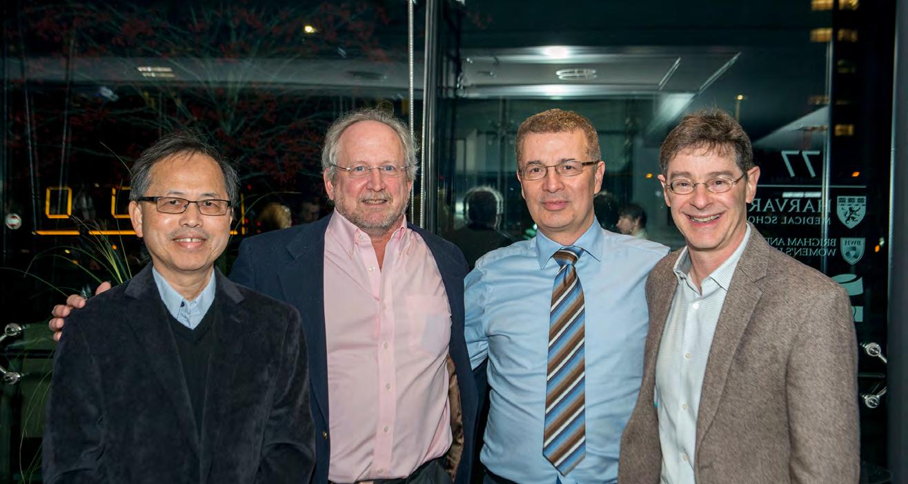
fMRI pioneers and Martinos researchers past and present: Ken Kwong, David Kennedy, Arno Villringer and Bruce Rosen at the fMRI25 symposium in 2016. The daylong symposium celebrated the first quarter-century of the technique.
