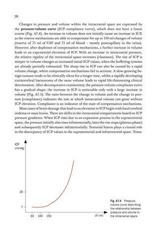
98 Changes in pressure and volume within the intracranial space are expressed by the pressure-volume curve (ICP compliance curve), which does not have a linear course (Fig. A7.4). An increase in volume does not initially cause an increase in ICP, as the reserve mechanisms are able to compensate for up to 150 ml changes of volume (reserve of 75 ml of CSF and 75 ml of blood – mostly postcapillary in the veins). However, after depletion of compensation mechanisms, a further increase in volume leads to an exponential elevation of ICP. With an increase in intracranial pressure, the relative rigidity of the intracranial space increases (elastance). The rise of ICP is steeper in volume changes at increased initial ICP values, when the buffering systems are already partially exhausted. The sharp rise in ICP can also be caused by a rapid volume change, where compensation mechanisms fail to activate. A slow-growing benign tumour tends to be clinically silent for a longer time, whilst a rapidly developing extracerebral haematoma of the same volume leads to rapid life-threatening clinical deterioration. After decompressive craniectomy, the pressure-volume compliance curve has a gradual shape; the increase in ICP is noticeable only with a large increase in volume (Fig. A7.5). The ratio between the change in volume and the change in pressure (compliance) indicates the rate at which intracranial volume can grow without ICP elevation. Compliance is an indicator of the state of compensation mechanisms. Most cases of brain damage that lead to an elevation in ICP begin with focal cerebral oedema or mass lesion. There are shifts in the intracranial compartments based on ICP pressure gradients. When ICP rises due to an expansion process in the supratentorial space, the pressure initially also rises infratentorially, later the rise stops (plateau phase) and subsequently ICP decreases infratentorially. Tentorial hiatus plays a crucial role in the discrepancy of ICP values in the supratentorial and infratentorial space. Trans-
ICP
(mmHg)
20
7 50
150
150
ΔV (ml)
Fig. A7.4 Pressurevolume curve describing the relationship between pressure and volume in the intracranial space
A7 Intracranial Pressure
ICP
99
B
(mmHg)
C
A
D
Fig. A7.5 Various pressure-volume curves: A – typical shape with a gradual increase in volume; B – steep course at higher initial ICP values; C – steep course with a rapid increase in volume; D – gradual course after decompressive craniectomy
20
7 50
150
150
ΔV (ml)
tentorial herniation causes obliteration of the perimesencephalic cisterns, resulting in anatomical separation of the two compartments. The supratentorial space with the mass lesion is isolated; compression of the aqueduct creates obstructive hydrocephalus with a consequent fatal increase in ICP. Until occipital conus develops, the infratentorial space communicates with the spinal canal through the foramen magnum. Rapid recognition of these clinical syndromes along with targeted surgical treatment forms the basis for brain resuscitation. It is important to realise that brain herniations can occur without a significant rise in global ICP. This occurs predominantly in the cases of focal oedema.
A7.4 CEREBRAL BLOOD FLOW AND CEREBRAL PERFUSION PRESSURE
Cerebral blood flow (CBF) can be generally defined as the amount of blood flowing through the brain. Although brain tissue represents only 2% of body weight, it consumes 15% of cardiac output. According to the measurement method used, the average CBF is around 60 ml/100 g of brain tissue/minute (in the grey matter the CBF is higher – 75 ml, in the white matter it is lower – 45 ml). A decrease in cerebral perfusion to values between 12–18 ml/min/100 g leads to reversible brain dysfunction (penumbra zone), CBF values below 12 ml/min/100 g cause irreversible structural brain changes. The brain metabolism is aerobic; therefore the brain needs a constant supply of blood and oxygen to ensure homeostasis. An uninterrupted constant blood flow, which is ensured by autoregulation of cerebral vessels, is essential. Cerebral autoregulation is a homeostatic process that regulates and maintains constant CBF.
100 Cerebral blood flow is closely related to cerebral perfusion pressure (CPP). CPP represents the vascular pressure gradient across the cerebral vascular bed. The CPP value is obtained by calculating the difference between mean arterial pressure (MAP) and venous pressure in the area of the cortical and bridging vein outlets into the upper sagittal sinus. Because venous pressure is difficult to determine and exceeds intracranial pressure only slightly, the intracranial pressure (ICP) value is used to calculate CPP. CPP is defined as: CPP = MAP – ICP
CBF (ml/100 g/min)
The relationship between CPP and CBF is expressed by the autoregulation curve (Fig. A7.6). Cerebral blood flow does not passively follow changes in the blood pressure but is constant in the perfusion pressure range of approximately 50–150 mmHg (plateau phase). At CPP values of below 50 mm Hg, CBF decreases dramatically and increases exponentially at CPP above 150 mmHg. Constant flow in the plateau phase is enabled by cerebral autoregulation. Autoregulation of CBF is considered to be a multifactorial complex process that has not yet been reliably elucidated. It is maintained by myogenic, metabolic, and neurogenic mechanisms. Cerebrovascular resistance (vascular flow resistance), which is inversely proportional to the square of the vessel radius and to the drop pressure (pressure difference at the beginning and end of the tube), and is directly proportional to the dynamic viscosity of the fluid (Hagen-Poiseuille law), is the essence of autoregulation. As CPP decreases, the arterioles dilate, the vascular resistance decreases and the blood flow through the brain remains intact. By contrast, with increasing CPP, constant CBF is ensured by increased cerebrovascular resistance due to vasoconstriction. Vasodilation compensates for the decrease in perfusion pressure caused by an increase in ICP in the initial phase only. The lumen of the blood vessels is also affected by the level of carbon dioxide in the blood. Hypocapnia causes vasoconstriction (decrease in ICP) and hypercapnia causes vasodilation (increase in intracranial blood volume leads to ICP elevation).
50 PaCO2
diameter of brain arterioles 50
CPP (mmHg)
150
Ukázka elektronické knihy
Fig. A7.6 Autoregulation curve. Constant CBF in the CPP of 50–150 mmHg range is maintained by changes of the arteriole lumen, which is related to PaCO2