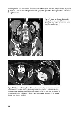
hydronephrosis and subsequent inflammation, or to rule out possible complications, especially abscess. CT also serves to guide renal biopsy or to guide the drainage of fluid collections or nephrostomy. Fig. 2.57 Renal carcinoma of the right kidney. Renal carcinoma (black arrows) in the right kidney on CT scan (coronal multiplanar reconstruction).
Fig. 2.58 Urinary bladder rupture. CT scan of urinary bladder rupture on transversal (on the left) and sagittal (on the right) multiplanar reconstructions. Contrast medium in urinary bladder (MM) leaks through the defect (white arrow, left) out from the bladder to the peritoneal cavity (white arrows, right). The urinary bladder was filled by the urinary catheter with contrast medium.
98
Ukázka elektronické knihy, UID: KOS506547