4
ULTRASOUND EXAMINATION OF THE LOWER LIMBS 1. oligosymptomatic DVT 2. the risk of future DVT through spread of SVT 3. therapeutic work‑up related to 1 and 2 above The deciding technique is color duplex sonography. Compression ultrasound (B‑mode) determines the extent of the superficial vein thrombosis, highlights the risk of propagation to deep veins and excludes DVT. Color flow mapping and dopplerometry are of ancillary importance: to clarify the extent of thrombotic obliteration and determine reflux sites in insufficient venous segments (Chapter 4.4.2) (Musil, 2013). D‑dimer assessment is of little value. In most patients with SVT, D‑dimer levels are markedly increased but determining them does not enable a distinction to be made between a primary thrombotic from a primary inflammatory process. D‑dimer values cannot predict the spread of thrombosis or other thrombotic complications (see above). Investigation of other coagulation parameters (e.g. factor VIII concentrations) has not to date, been introduced into clinical practice (Blättler, 2008). Venography here has been replaced by ultrasound. Venography relying on contrast medium used during the examination can induce thrombophlebitis. Screening for thrombophilia is not routinely performed; it should be considered in patients with recurring primary thrombophlebitis. Screening for occult primary illness (e.g. malignancy or vasculitis) is carried out if there are suspected clinical symptoms and/or signs of given diseases. or disease with respect to migratory, recurrent and/or multifocal non‑varicose vein SVT, recurrent varicose phlebitis, or current extensive DVT not directly related to superficial thrombosis. Screening for risk factors. These are generally similar: older age and extensive, long term untreated varices or chronic venous insufficiency (varices and trophic changes of the skin and subcutaneous tissue), local physical damage, heat, wearing
→
↑
↑
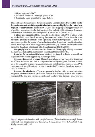
Fig. 4.3 Organized thrombus with calcified deposits ( ↑) in the GSV on the thigh, lumen width 7.2 mm (longitudinal and transverse, B-mode, linear probe 5.7 and 7.0 MHz, display depth 6 and 5 cm) 98
Ukázka elektronické knihy, UID: KOS260136