– 98 –
Ukázka elektronické knihy, UID: KOS257533
56
18
21 (7)
41 0–79
95
34
91 18–182
20
1
5 (3)
53
0.768
118
0.270
UCL95%
PI-II
0.798
40.1
18.9
29.5
27.5 6–74
37 (13)
26 (25)
60 (55)
25 (15)
32 (32)
7 (6)
74 (41)
18 (16)
29 (53)
6 (1)
29 (43)
21 (17)
31 (46)
18 (19)
Period with 1‰ < S-EtOH < 1.5‰, % of time (h)
0.519
41.7
20.8
31.3
31 7–70
42 (14)
12 (9)
7 (6)
70 (43)
18 (18)
14 (11)
26 (15)
14 (12)
40 (73)
37 (7)
55 (80)
43 (40)
24 (35)
36 (37)
Period with S-EtOH > 1.5‰, % of time (h)
0.317
22.6
4.7
13.6
11 0–59
21 (7)
Chi2
22 (21)
11 (10)
5 (3)
4 (4)
59 (48)
0
0
16 (29)
24 (4)
11 (16)
14 (12)
3 (4)
0.7 (0.7)
Period with S-EtOH < 1‰ and S-MetOH > 6.2 mmol/L, % of time (h)
0.528
20.4
5.3
12.8
10 0–38
32 (11)
17 (16)
11 (10)
ND
6 (6)
13 (11)
ND
0
0
38 (7)
4 (6)
18 (16)
6 (9)
9 (9)
Period with S-EtOH < 1‰ and S-Formate > 0.4 mmol/L, % of time (h)
Died
VS,CS
CS
VS,CS
VS
VS,CS
–
–
–
–
–
–
–
–
Outcome
42.1
45.4
37.3
54.5
53.0
45.6
39.1
53.0
62.1
66.4
53.6
37.8
53.2
69.7
Highest S-EtOH, mmol/L
0.184
56.9
44.9
50.9
53.0 37.3–69.7
test of difference between the Groups I, II. Notes: Group I – survivors without sequelae, Group II – survivors with sequelae and died. PI-II – results of VS – visual sequelae, CS – central nervous system sequelae. (reproduced, with permission, from Zakharov et al. 2015c, 666–676).
39
26
93
70
Mean
LCL95%
62 (59)
33 (31)
61
92
8
50 (51)
79 (64)
0
68 (61)
31 (56)
57 (10)
16 (24)
36 (32)
45 (67)
46 (48)
Period with S-EtOH < 1‰, % of time (h)
10
81
90
17
101
182
15
3
18
2
147
14
89
12
148
6
11
Median (range)
Poor Outcome
Survived without sequelae
104
5
Duration of observation (h)
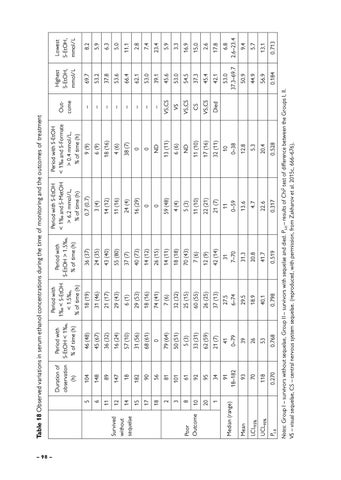
Table 18 Observed variations in serum ethanol concentrations during the time of monitoring and the outcomes of treatment
0.713
13.1
5.7
9.4
6.8 2.6–23.4
17.8
2.6
15.0
16.9
3.3
5.9
23.4
7.4
2.8
11.1
5.0
6.3
5.9
8.2
Lowest S-EtOH, mmol/L
80 70
Serum EtOH [mmol/L]
60 50 40 30 20 10 0
1
9 19* 2
3
7
8
10 13 20 4 5 6 11 12 14 15 16 17 18 21 Individual patients no.
Figure 23 Highest and lowest serum ethanol levels measured for each patient (reproduced, with permission, from Zakharov et al. 2015c, 666–676). Notes: Upper-end of boxes presents the highest serum ethanol concentrations measured during the observation period; lower-end of boxes presents the lowest serum ethanol concentrations measured during the observation period. Group I – survivors without sequelae (patients #4 – #21), Group II – patients with poor outcome (#1 – #19 – died; #2 – #20 – survived with sequelae). * – the patient died in hospital from infectious complications one month after the methanol poisoning.
sis sessions (4 cases) and the patients without follow-up examinations (3 cases), were excluded from the further analysis of possible association between serum ethanol fluctuations and the outcomes of treatment. The association between the outcomes of treatment and the duration of periods with sub-therapeutic or supra-therapeutic serum ethanol concentrations was not significant (r = 0.199, p = 0.387, and r = –0.157, p = 0.498, respectively). Further, the association between the outcomes and the duration of “unprotected” periods with sub-therapeutic serum ethanol concentrations and toxic serum levels of methanol or formate was not significant either (r = 0.433, p = 0.056, and r = 0.371, p = 0.129, respectively). The difference in mean duration of periods with sub-therapeutic, as well as with supra-therapeutic serum concentrations of ethanol was not significant between the two group of patients, the survivors without sequelae and the patients with poor outcome. 12. HOSPITAL TREATMENT OF ACUTE METHANOL POISONING WITH ANTIDOTES – 99 –
Ukázka elektronické knihy, UID: KOS257533
The complex pharmacokinetics of ethanol during the therapy makes it rather difficult to adapt the dosage and maintain the therapeutic range of its serum concentration consistently. However, in our study despite the wide fluctuations in serum ethanol concentration during the treatment, we found no association between serum ethanol fluctuations and treatment outcome. The results of our study support the findings of another study by Roy et al. (2003) in children with methanol poisoning treated with ethanol. Despite the wide variation of ethanol concentration during the time of observation, the total duration of “unprotected” periods with sub-therapeutic serum concentrations of ethanol and toxic serum methanol or formate levels was approximately 20% of the time of observation only. Therefore, for most of the time of observation, the patients were sufficiently protected by blocking the ADH enzyme and preventing further metabolism of methanol to formic acid. The episodes of significant decrease of serum ethanol were generally observed at the end and immediately after the termination of short sessions of IHD, indicating that most of ethanol administered as an antidote was effectively dialyzed. However, serum levels of methanol and formate decreased significantly too; therefore, the remaining sub-therapeutical concentrations of ethanol were apparently sufficient to block the ADH enzyme still effectively. The target level of serum ethanol of 22 mmol/L (1,000 mg/L) is not an evidence-based one, and the therapeutic level may actually be lower (Barceloux et al., 2002; Roy et al., 2003). Nevertheless, it has to be emphasized that in 3 of 7 cases, where the short sessions of IHD lasting 2–3 hours were applied, the patients had visual and CNS impairment 3–6 months after discharge. In another case, health sequelae could have been underdiagnosed, because the patient had pH 7.11 on admission, and the follow-up examination was not carried out. In the last 3 cases, the patients with short sessions of IHD survived without sequelae, but their serum formate levels on admission were low. Short sessions of IHD lasting 2–3 hours are generally insufficient to eliminate serum formate, as well as serum methanol, in high concentrations. The significant fluctuations in serum ethanol concentrations were associated with the change of route of administration from the intravenous to the oral one. In these cases, the serum methanol level was already low, serum formate was negative, and arterial blood pH was normal to the moment of the change of route of ethanol administration. Therefore, the fluctuations of serum ethanol in these cases had minimal negative impact on the outcome due to preceding intensive treatment and elimination of most of the toxic agents. In one case (patient #6), the ethanol was administered per os only during the treatment: This patient had low serum methanol and formate concentrations on admission, and short session of IHD was performed to rapidly eliminate most of the toxic agents. Generally, the intravenous route of ethanol – 100 –
Ukázka elektronické knihy, UID: KOS257533