
www.idosr.org Kyomukama et al
©IDOSR Publication International Digital Organization for Scientific Research
ISSN:2579-079X
IDOSR JOURNAL OF SCIENCE AND TECHNOLOGY 8(1):38-46, 2022. Prevalence of Pelvic Inflammatory Disease among Women Attending the Gynecology Clinic at Kampala International University Teaching Hospital, Uganda
1 and Ezera Agwu2
1Department of Obstetrics and Gynecology of Kampala International University, Uganda
2Department of Microbiology, Kampala International University, Uganda
ABSTRACT
Pelvic inflammatory disease (PID) is major health problem in developed and developing country involving more young women. Itis associated with high rate of female reproductive health morbidity; it can complicate with ectopic pregnancy, infertility and chronic pelvic pain. A poor response therapy increases the likelihood of these complications; this could be due to an increase in antimicrobial resistant pathogens. This was a cross-sectional study conducted among women who attended gynecology clinic at Kampala International University Teaching Hospital. Consecutive enrolment of 324 participants who consented to participate wasdonedailyuntilarequiredsamplesize wasrealizedfrom November2019to January 2020. Structured questionnaires were used to collect data on associated factors; endocervicalswabwastakenfrompatientclinicallydiagnosedwithPID.Culturingforcolony characteristics followed by Gram stain was used for provisional identity of pathogenic bacteria. Further identification was done by a set of biochemical tests. Antibacterial susceptibility pattern of isolated bacterial pathogens was determined by Kirby Bauer disc diffusion method, a rapid diagnostic test to detect Chlamydia antibody in the endocervical swab sample was also used to identify the Chlamydia trachomatis carriers among the patients. Data was analyzed using STATA VERSION 14.2. The prevalence of pelvic inflammatory disease was 19.1%. Not being educated, having two or more sexual partners, previous history of PID and induced abortion, also the previous use of contraceptives specificallytheuseofIUD,wereallsignificantlyassociatedwithPelvicinflammatorydisease (P value <0.05). The prevalence of PID is HIGH at KIU-TH. Keywords: prevalence, PID, women, pathogens
INTRODUCTION
Pelvicinflammatorydiseaseisaninfection which affects the upper female reproductive tract from the internal cervical opening, the uterus, fallopian tube, ovaries and pelvic peritoneum [1]. The oldest and clear description of pelvic inflammatory disease was done by Mauriceau in 1693 when he described puerperal infection with abscess in both sides of the uterus [2]. While the first description of the bacterial agent that causes pelvic inflammatory disease was after Neisser discovered Neisseria gonorrhea in 1879. This description was done by Westermark in 1886 when he demonstrated the presence of Neisseria gonorrhea in tubal pus and Wertheim in 1894 the first to demonstrate these
organisms invading the tube. Chronic pelvic inflammatory tumors unconnected withthepuerperalstate weredescribedby Simpson in 1843; which described the condition as pelvic cellulitis. Neisseria Gonorrhea was since then known as the leading cause of pelvic inflammatory disease (PID) and its history is strongly linked to the one of pelvic inflammatory disease.It was thought to becausedsolely by an inflammation occurring in the cellular tissue of the pelvis [3].
Virchow in 1862 referred to the term cellulitis, introduced parametritis or perimetritis. Parametritis was thought to result from a variety of causes such as a blow or fall, excessive excitement, the use of aphrodisiacs, and use of abortifacents.
www.idosr.org Kyomukama et al
The treatment of gonorrhea towards 1892 was then done by quack doctors by the introduction of potassium permanganate solutions for urethral irrigations, followed bytheuseofsilversalt[4].In1930agroup of specialists in Massachusetts decided that it was time to make the management of gonorrhea a proper function of respectable medicine and disclosed that sulphonamide drugs would cure gonorrhea. This was then used for a short span of five years as gonococcus proceeded to develop overwhelming resistance to every new member of this group of drugs and later penicillin was used as single or multiple dose in the treatment ofgonorrhea which led toa new assessment of this disease as a cause of serious morbidity in both males and females [4]. According to [5] in South
Study design
Africa, who did a study on antimicrobial resistance among women with pelvic infection, he reported that PID continues to manifest resistance to many classes of antibioticssuchastetracycline,macrolide, and quinolone. In order to overcome this, different treatment regimens have been developed and different studies have shown the different clinical and microbiological cure rate concerning Neisseria gonorrhea and other associated microorganism as the cause of PID is known to be in 70% of cases of the polymicrobial condition [6]
Purpose of the study
To determine the prevalence of pelvic inflammatory disease among women attending gynecology clinic at Kampala International University Teaching Hospital.
RESEARCH METHODOLOGY
This was a cross sectional study. Laboratory investigations were done to achieve the prevalence, the common bacteria isolates and antibacterial susceptibility pattern in women with pelvic inflammatory disease attending gynecologyclinicat Kampalainternational university teaching hospital. Association between PID and different factors was established.
Study area
The study was conducted at Kampala International University Teaching Hospital found in Ishaka Bushenyi Municipality at approximately 60km from Mbarara town along Mbarara Kasese highway.
Study population
The study populations were all women of reproductive age in the catchment area
Target population
All women of reproductive age attending gynecologyclinicat Kampalainternational university teaching hospital shall be considered for inclusion in this study.
Inclusion criteria
All the women at the reproductive age attending gynecology clinic of Kampala international university teaching hospital as well as emancipated minors.
Exclusion criteria
Women on antibiotics, pregnant women, unconscious patients who cannot consent and minors were excluded from the study.
Sample size determination
Objective number 1, the sample size was determined using [7] formula with the estimated prevalence of 50%, because the of lack of current prevalence of PID in Uganda
n = Desired sample size z =Standardnormal deviation at 95%level of confidence; z= 1.96

p = Prevalence of pelvic inflammatory diseaseinUganda,assumedat50%,andd= Level of precision=0.05 �� = (1.96)2 ×0.5(1 0.5) (0.05)2
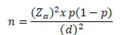
N1= 384 this is considered as an assumption of sample size.
Objective number 2: Musa et.al [8]inthestudydoneinMbarara RegionalReferralHospitalonscreeningfor
Chlamydia Trachomatis among women of reproductive, found that 26% of women were carriers of Chlamydia Trachomatis. [8]
Where; n = Desired sample size z=Standardnormaldeviateat95%levelof confidence; z= 1.96 p = prevalence of Chlamydia trachomatis inreproductiveagedwomen,usingastudy done in Mbarara by Musa and colleagues [8] d=Level of precision= 0.05 n= (1.96)2-0.2(1-0.2) (0.05)2 N2= 295
Specificobjective3:Mark etal[9] reported that women with a previous history of PID have a greatest risk of having subsequent PID,OR=5.9, theproportion(p)PIDamong woman with previous history of PID 13.1% and among women without previous history of PID was 3.6% r=ratio of previous history of PID to those without previous history of PID, r=13.1:3.6, r=3.6:1=3.6 Where; =z-statistic at α=0.05, = z-statistic at β=0.84 hence statistical power = 80%. Using the formula [10]: where (1+3.6)2×(1.96+0.84)2 36(ln59)2×0131(1 0131)
n3=118
Decision on the sample size
Sample size for objective number 2 was considered since this was a research done in Uganda (Mbarara Regional Referral hospital) and reflects the true Ugandan reality. 10% of this sample size was added to minimize non response or loss of some
laboratory request form, therefore the sample size was:N2=324

Sampling technique
Consecutivesampling method was used to select participants who consented to be part of the study. All the women of reproductive age who met the inclusion criteria was invited to participate in the study, the participants was enrolled according to their order of arrival in gynecology clinic and this was carried out on a daily basis until the required sample size was achieved.
Data collection instruments
A pretested questionnaire was administered to each participant who consented to participate to the study in order to collect information on sociodemographic, gynecological and sexual behavior factors that related to the development of pelvic inflammatory disease in. A detailed history was taken in English,translatedinlocallanguagewhere necessary for women who could not understand English physical examination was carried out and the endocervical sample was taken from patient with symptoms and of PID in order to achieve all the objectives.
Sample processing and analysis
Isolation
www.idosr.org Kyomukama et al 40
Samples collected using a sterile procedure with the endocervical swab stick was inoculated on blood agar, chocolate agar, Mac Conkey agar, Thayer Martin medium, and different biochemical testswereused.After,theywereincubated both aerobically and anaerobically at 37ᴼC for 24-48hrs. Colony morphology were observed according to shape, size, elevation, margin and surface characteristics.
www.idosr.org
Rapid diagnostic test was used in order to identify the Chlamydia trachomatis antibody careers within the endocervical sampleof the participants,theisolation of Chlamydia which uses living cells (McCoy cell) was not done due lack of this specific media to culture Chlamydia trachomatis, this rapid Chlamydia test was used to determine the percentage of Chlamydia carriers among the patient with pelvic inflammatory disease.
Direct gram microscopy
A direct smear was made for Gram stain according to [11]; a drop of sterile normal saline was added on the center of a clean dried glass slide and the swab containing the sample rolled in the drop of normal saline spreading it on the glass slide in a circular motion to make a thin smear. The smearwasthenallowedtoairdryandthen heatfixedbypassingitatleastthreetimes over a Bunsen flame. The slide was placed on the staining rack and flood with crystal violet solution for 60 seconds, wash with clean water and cover with lugol’s Iodine andthenitwasallowedtoactforaminute.
The slide was washed in clean water and then decolorized with 50%acetonealcohol under slow running tap water until a faint pink color is observed or no more color tend to flow from the smear. The process of discoloration was not exceeding 30 seconds.
After decolorizing, it was washed in clean water and counter stain with neutral red solution. The slide was again washed in clean water; air dried and observed under the microscope with X100 objective lens (oil immersion lens).
Gram positive bacteria were observed as blue or purple color and Gram negative as red or pink color. Also, the morphology and shape of the bacteria were identified whether they are cocci, diplococcic, cocci in chains, clusters, and whether they are rods in appearance [11]
Identification of bacterial isolates
Biochemical tests
The isolates were identified using the conventional biochemical tests such as catalase, optochin, coagulase, indole, citrate utilization, urea utilization, triple sugar iron agar fermentation, MR-VP test and oxidase as described below;
Kyomukama et al
Catalase test
Catalase test was carried out according to the method described by [11], to determine the ability of the isolate to produce the enzyme, catalase. A drop of 3% hydrogenperoxide wasadded toaloop full of the test organisms. Presence of bubbles indicated catalase activity.
Indole test
Indoletestwascarriedoutaccordingtothe methoddescribedby[11] todeterminethe abilityoftheisolatetodegradeaminoacid tryptophan and produce tryptophanase, enzyme were tested. 1% tryptophan broth in a test tube were inoculated with isolate and incubated at 37°C for 48 hours. After 48 hours, 1milliliter of chloroform was added to the broth. The test tube was shaken gently, and 2.1 ml of Kovac’s reagent were added and again shaken gently, this was allowed to stand for 20 minutes. The formation of red coloration at the top layer, indicated positive while yellow coloration indicated negative.
Urease test
Urease test was done according to the methoddescribedby[11] todeterminethe ability to hydrolyze urea to produce ammonia and carbon dioxide. Test organism wasinoculatedintoureasebroth and incubated at 30°C for 72 hours. Purplish pink coloration of the medium indicates positive reaction.
Citrate utilization
Thiswascarriedoutbyinoculatingthetest organism in test tube containing Simon’s citrate medium and incubated for 24 to 72 hours.Thedevelopmentofdeepbluecolor afterincubationindicatedapositiveresult [11]
Triple sugar iron test
Triple sugar iron test was carried out accordingtothemethoddescribedby[11]; the test determines the ability of the organism to ferment the three sugar component of the medium: glucose, lactose and sucrose. The medium contains apHindicator(phenolred)andadetection system(thiosulphateandferroussulphate) for hydrogen sulphide (H2S).
Themediumwaspreparedasanagarslant. The test organism was inoculated by stabbing the medium using sterilized straight wire loop and the surface of the
www.idosr.org Kyomukama et al
slope were also streaked with the test organism.Thetestswereincubatedat37°C for 72 hours. After incubation, gas production was determined by observing the cracking of the medium, and production of H2S was observed by the blackening of the bottom of the medium.
Coagulase test
This test was used to identify StaphylococcusAuraus whichproducesthe enzymecoagulase.Therapidslidetestwas done by placing a drop of distilled water on each end of slide. Then a colony of the test organism (previously checked by Gram staining) was emulsified in each of the drops to make two thick suspensions. A loopful of plasma was added to one of the suspensions and mixed gently. Formation of clumps of the organisms within 10 seconds was suggestive of a positive test while absence of these clumps indicated negative results. For suspected Staphylococcus auraus isolates whichturnnegativefortherapidslidetest, tube test was done by emulsifying several isolated colonies of test organism in 1 ml of diluted rabbit plasma (1:5) dilution to give a milky suspension. The tubes were then incubated at 35°C in water bath for 4 hours.Thesewereexaminedatintervalsof 1, 2 and 4 hours for clot formation by tilting the tube through 90°. If the test is still negative, the tube was left at room temperatureovernightandexaminedagain for Staphylococcus auraus that produce a delayed clot [11].
Oxidase test
The test was used in identification of organisms which produce the enzyme cytochrome oxidase. A filter paper soaked with the substrate tetra methyl-pphenylenediamine Dihydrochloride was moistened with sterile distilled water. Usingapieceofstickorglassrod,acolony of the test organism was smeared on the filter paper. The development of a bluepurplecolorwithina 10secondsindicated positivetestwhileabsenceorformationof a blue-purple color after 10 seconds was considered negative [11].
Data analysis plan
Data from questionnaires were entered in Microsoft Excel 2010, and thereafter exported to STATA 14.1. Sociodemographic, sexual behaviors and gynecologic factors were summarized as means and medians, standard deviations and interquartile range (for continuous variables) were determined. Proportions, percentagesandfrequencieswereusedfor categorical variables using STATA 14.1.
Ethical considerations
Informed consent
Informed consent and respect for participant’s voluntary recruitment was observed. Informed consent for participants were obtained and signed after fully explaining the details of the study to them in English and local languages where necessary (copy attached at Appendix). Participants were not forced to enroll themselves if they don’t want to, they were free to withdrawfrom the study any time they wish without coercion or compromise of care they are entitled to.
Risks and adverse events to study participants
Patients may undergo pain during swabbing and speculum examination, however, the process of obtaining a swab was done gently and professionally to minimize risk of pain and minimize reinfection as far as possible. Additionally, culture and sensitivity are the recommended guidelines prior to antibiotic therapy to minimize the risk of antibiotic resistance.
Approval procedure
Approval to carry out the study shall be sought from the department of obstetrics and gynecology, the faculty and post graduate directorate and finally, Research EthicsCommitteeofKampalaInternational University. This approval letter shall be presentedtothehospitaladministrationof KIU-TH. Permission shall be sought from the administration of the hospital before the study is conducted. The study will be registered with Uganda National Council for Science and Technology.
RESULTS
Table 1: Socio demographic factors
Characteristics Frequency % Age (years)
<20 31 9.6 20-29 205 63.3 30-39 71 22.0 40-49 17 5.1
Education
None 11 3.4 Primary 99 30.6 Secondary 111 34.4 tertiary 103 31.6
Occupation
None 127 39.2 Farmer 85 26.2 Professionals 51 15.7 Business 31 9.6 Manual laborer 30 2.3
Monthly income (UGX) none 10 3.1 <300000 230 71.8 300000-600000 66 26.5 >600.000 18 5.6 Marital status Single 86 26.5 Married 238 73.5
The above table illustrates that 63.3% of participantsareagedof20-29years,34.4% have secondary education, 39.2% have no
occupation, 71.8% of participants have a monthly income of less than 300.000 Uganda Shillings and 73.5% are married.
Table 2: Gynecological factors
Characteristics Frequency %
Parity Zero 98 30.3 1-3 153 47.2 >3 73 22.5
Had PID before
No 224 69.1 Yes 100 30.9 Had miscarriage before No 264 81.5 Yes 60 18.5
Use Contraceptive No 132 40.7 Yes 192 59.3
Intra Uterine Procedure No 281 86.7 Yes 43 13.3
Type contraception Condoms 38 19.8 Pills 61 31.8 Injectables 65 33.8 IUD 28 14.6
Type of miscarriage
Spontaneous 41 68.3 induced 19 31.7
From the above table, 47.2% of the study participants had delivered at least one to three times, 69.1% had had miscarriage of
which 68.3% were spontaneous, 59.3% of the study participants had ever used contraceptivemethodsofwhich33.6%had
www.idosr.org Kyomukama et al
usedinjectablecontraceptivemethodsand 86.7%hadnot hadintrauterine procedures
Table 3: Sexual behavior factors Characteristics Frequency Percent
Number of of sexual partners
None One More than one
20 253 51
25 242 57
7.7 74.7 17.6
6.2 78.0 15.8 Age of initiation sexual activity(year) < 15 16-20 >20
Condom Use Sometimes Every time Never
25.9 11.8 62.3 Smoking Never smoke Ever smoke 316 8 97.5 2.5
84 38 202
The above table shows that, the age of initiationofsexualactivityforthemajority of participants was 16-20years in 74.7%,
most of the study participants denied the useofcondomswith62.3%and97.5%were nonsmokers
Table 4: Overall prevalence of PID prevalence Fr %(95% CI) P-Value
Non PID PID
262 62
80.9 19.1(15.2-23.8)
From the above table, the overall prevalence of PID is 19.1%
DISCUSSION
Inthisstudywedeterminedtheprevalence of pelvic inflammatory disease among women of reproductive age attending gynecologyclinicatKIU-THandfoundthat of the 324 women enrolled 81 presented symptoms and signs of PID they underwent endocervical swabs for culture andsensitivitywithalsoarapidChlamydia test,62werepositiveforinfectiongivinga prevalence of 19.1%. This is High compare to 4.4% reported by [12] in USA, it is also high to [13] in Yaounde who found 5.1%,
NA
this could be explain by the setting where these studies were conducted in urban areas compare to our study which was conducted in rural setting. This result fall in the range reported by [14] who reported PID as a cause of gynecological admissions in 17-37%, Therefore, there is a disproportionate prevalence of PID This could be attributed to the different levels of coverage health services and reporting across countries.
CONCLUSION
The prevalence of pelvic inflammatory disease is high and significant risk factors were not being educated, having previous
history of PID, have ever use IUD as a family planning method and undergoing any intrauterine procedure.
REFERENCES
1. Sweet, R. L. (2011). Treatment of Acute Pelvic Inflammatory Disease, 2011.https://doi.org/10.1155/2011/5 61909
2. Viberga, I. (2006). The Clinical Appearance of Pelvic Inflammatory Disease in Relation to Use of IntrauterineDeviceinLatvia
www.idosr.org Kyomukama et al
3. Rees, E., Annels, E. H. and Both, B. (1969). Gonococcal salpingitis*.
4. Hunter, J. (1928). Albert Neisser and the Gonococcus, 45, 95–97.
5. DeYoung, C. G., Quilty, L. C. and Peterson, J. B. (2007). Between facets and domains: 10 aspects of the Big Five. Journal of Personality and Social Psychology,93(5), 880–896.
6. Haggerty,C.L.andTaylor,B.D.(2011). Mycoplasma genitalium: An emerging cause of pelvic inflammatory disease. Infectious Diseases in Obstetrics and Gynecology, 2011, 27–32. https://doi.org/10.1155/2011/959816
7. Susan P. Hamill, (2015). Untanglingthe Mystery of Teaching Business Organizations, 59 ST. LOUIS U. L.J. (2015).
8. Musa, M., Joel, B., Lenard, A., Joseph, N., Ronald, M. and Julius, M. (2016). PrevalenceandFactorsAssociatedWith Genital Chlamydial Infections among Women Attending the Gynaecology Clinic At Mbarara Regional Referral PrevalenceandFactorsAssociatedWith Genital Chlamydial Infections among Women Attending the Gynaecology Clinic At Mbarara Regional Referral Hospital, (May).
9. Marks, C., Tideman, R. L., Estcourt, C. S., Berry, G. and Mindel, A. (2000a). Assessment of risk for pelvic inflammatory disease in an urban sexual health population, 470–473.
10.Suresh, K.P. and Chandrashekara, S. (2012) Sample Size Estimation and Power Analysis for Clinical Research Studies. Journal of Human Reproductive Science, 5, 7-13.
11.Cheesbrough, M. (2006) District Laboratory Practice in Tropical Countries. Part 2, 2nd Edition, Cambridge University Press Publication, South Africa, 1-434.
12.Kreisel, K., Torrone, E., Bernstein, K., Hong, J. and Gorwitz, R. (2014). Prevalence of Pelvic Inflammatory Disease in Sexually Experienced Women of Reproductive Age United States , 2013 – 2014, 2013–2014.
13.Nkwabong, E. and Dingom, M. A. N. (2015). Acute Pelvic Inflammatory Disease in Cameroon: A Cross
Sectional Descriptive Study, 19(December), 87–91.
14.Morris, G. C., Stewart, C. M. W., Schoeman, S. A. and Wilson, J. D. (2014). A cross-sectional study showing differences in the clinical diagnosis of pelvic in fl ammatory disease according to the experience of clinicians: implications for training and audit, 445–451. https://doi.org/10.1136/sextrans2014-051646
15.NGloria,AOYamile,EAgwu(2022). Prevalence patterns of bacterial urinary tract infections among febrile children under-five years of age at Kampala International University Teaching Hospital IDOSRJournalofBiology,Chemistry andPharmacy 7 (1), 41-55.
16.B Petrus, E Nzabandora, E Agwu (2022). Evaluation of the bacterial agents associated with PID among women of reproductive age at Kampala International University TeachingHospital. IDOSRJournalof Biochemistry, Biotechnology and AlliedFields 7 (1), 64-74.
17.OA Hussein, M Joy, JN Musiime (2022).Evaluation of the factors associated with immediate adverse maternal outcomes among referred women in labor at Kampala International University Teaching Hospital. IAA Journal of Biological Sciences 8 (1), 228-238.
18.OA Hussein, M Joy, JN Musiime (2022). Factors associated with Immediate Adverse Maternal Outcomes among Referred Women in Labor attending Kampala International University Teaching Hospital. IAA Journal of Applied Sciences 8 (1), 117-125.
19.M Wilberforce, O John, K Claude (2022). Evaluation of the Acute Toxicity and Hematological Effect of Aqueous Extract of Albizia chinensis (Osbeck) Merr Stem Bark in Streptozotocin-induced Diabetic Wistar rats. IAA Journal of Biological Sciences 9 (1), 146-158.
20.TBYves,OEDafiewhare,LACharles, E Sebatta (2022).
www.idosr.org Kyomukama et al
Electrocardiographic Pattern among Heart Failure Patients at Kampala International University Teaching Hospital, Ishaka, Uganda. IAA Journal of Biological Sciences 9 (1), 159-165.
21.Hussein Osman Ahmed, Joy Muhumuza and Musiime James Nabaasa (2022). The composite immediate adverse maternal outcomes among women in labor referred to Kampala International University Teaching Hospital IAA Journal of Scientific Research 8(1):149-156.
22.Daniel Asiimwe, Herman Lule and IzimbaDaniel(2022).Epidemiology of Assault Injuries among Trauma Patients Presenting at Kampala International University Teaching HospitalandJinjaRegionalReferral Hospital. INOSR Applied Sciences 8(1):111-119.
23.E.O.Ikuomola,O.S Akinsonmisoye, R.O.OwolabiandM.B.Okon(2022). Assessment of Toxicity Potential of Secnidazole on Reproductive
System of Male Wistar Rats. INOSR AppliedSciences 8(1):120-133.
24.UgwuOkechukwu,P.C.,Onwe,S.C. and Okon, M. B. (2022). The effect of Methanol Extract of Rauwolfia vomitoria on Lipid Profile of Chloroform intoxicated Wistar Albino Rats. IAA Journal of Scientific Research, 8 (1), 73-82
25.E.O.Ikuomola,O.S.Akinsonmisoye, R.O. Owolabi and M. B. Okon (2022).Evaluation of the Effect of Secnidazole on Sperm Motility, Morphology, Viability and Total Sperm Count of Wistar Rats. INOSR Experimental Sciences 8(1): 74-83, 2022.
26.E.O. Ikuomola , O.S Akinsonmisoye, R.O. Owolabi and M. B. Okon (2022).Evaluation of the effect of secnidazole on the histology of the testes and epididymis of male Wistar rats. INOSR Experimental Sciences 8(1): 84-94, 2022.