http://www.inosr.net/inosr experimental sciences/ Bafwa etal
INOSR ExperimentalSciences9(1): 1 9, 2022.
©INOSR PUBLICATIONS
InternationalNetworkOrganizationforScientificResearch
Prevalence of Electrocardiographic
Abnormalities among
ISSN:2705 1692
Heart
Failure
Patients at Kampala International University Teaching Hospital, Ishaka, Uganda
Yves Tibamwenda Bafwa, Dafiewhare Onokiojare Ephraim, Charles Lagoro Abonga and Sebatta EliasDepartment of Internal Medicineof Kampala InternationalUniversity Kampala InternationalUniversity Uganda.
ABSTRACT
The prevalence of cardiovascular diseases is increasing rapidly in Sub Saharan Africa. A multicenter prospective cohort study of 1,006 patients with acute heart failure in a Sub Saharan Africa Survey showed that 814 patients had ECGs readings among which 97.7% had abnormalities. There is paucity of data on ECG abnormalities among heart failure patients in many developing countries, Uganda inclusive. A cross sectional study was conducted at KIU TH between January and March 2020 which recruited 122 heart failure patients. Questionnaires were administered to respondents to obtain factors associated with ECG abnormalities. A 12 lead ECG machine was used to obtain ECG findings. Data were analyzed using bivariate and multivariate logistic regression analyses to establish theassociationbetweeneachindependentvariableandtheECGabnormalities. Themean ageofstudyrespondentswas62.4±20yearsand54.9%oftherespondentswerefemales. The prevalence of ECG abnormalities among heart failure patients was 86.1 % (95% CI: 78.6 91.2). The prevalence of abnormal ECG among heart failure patients was very high. It is recommended that ECG test should be included as a routine test in the management ofheartfailurepatients atKIU TH,regularrefreshertrainingin ECGshould beorganized forclinicians.Routinehealtheducationonthedangersofalcoholmisuseandsmokingto the heartand other organs, should beencouragedat triage Centres of allhealthfacilities by theMinistryof HealthinUganda.
Keywords: Prevalence, ElectrocardiographicAbnormalities, HeartFailure
INTRODUCTION
With more than 100 years of development, the history of electrocardiograph (ECG) has been complicated and unorganized [1] [2], observed while dissecting a frog, that electric stimulation of frog nerves could cause muscle contraction, which he called “animal electricity”. This discoveryrevealedthebiologicalbasisof electrophysiology. Later, the first ECG of ahumanbeingwasrecordedin1869with a siphon instrument by Muirhead in London [1]. With the introduction of chest x ray in 1895 by Roentgen and the electrocardiograph (electrocardiogram) in 1902 by Einthoven who was inspired by the work of Waller, objective information about the structure and function of the heart was provided. Electrocardiography has played an
important role in the diagnosis of heart diseases[3].
The progression of physics led to the invention of the 12 lead electrocardiogram as it is now known.
The ECG (called, the electrokardiogram (EKG) in German language), measures how the electrical activity of the heart changes over time as action potential is propagated through the heart during a cardiac cycle. It shows electrical differences across the heart when depolarization and repolarization of the atrial and ventricular cells occur [4] (The normal ECG tracing is described in Appendix A). Heart failure is associated with widespread electrophysiological remodelling of the cardiac conduction system,resultinginelectrocardiographic (ECG) abnormalities such as reduced RR
variability, prolongation of the QRS and PRintervals, andatrial fibrillation [5] [6] in India conducted a retrospective study among 400 heart failure patients and found that the main ECG abnormalities among them included left ventricular hypertrophy (LVH) (42.9%), left axis deviation (39.8%), left bundle branch block (LBBB) (19.4%), and left atrial enlargement (25.8%). Arrhythmias included: ventricular extrasystoles
Study Design
(11.8%), atrial fibrillation (9.1%), complete heart block (5.7%) and ventricular tachycardia (3.9%). They reported a prevalence of ECG abnormalities of 92% among patients with heart failure, and normal ECG in 8% ofthe patients.
The study was done to determine the prevalence of ECG abnormalities among heartfailure patientsat KIU TH.
METHODOLOGY
Research design is the “overall plan or strategyforconductingthe research”[7] Survey design was adopted for this study. Here, samples of the target population were chosen for study, analysis and discovery ofoccurrences as they were. Survey design is suitable for this type of research that involves a special population of only people diagnosed to have heart failure. It isalso ideal in this situation where factors including cost, rapid data collection and abilitytounderstandapopulationfroma part of it were considered. This study was conducted among heart failure patients at Kampala International University Teaching Hospital, Ishaka, using descriptive cross sectional hospital based survey approach that employed quantitative data collection method.ThemainoutcomeincludedECG abnormalities among heart failure patients receiving care atKIU TH.
Study Area
This study was conducted at Kampala International University Teaching Hospital, Ishaka.
Target Population
Thetargetpopulationforthisstudywere adult heart failure (HF) patients in WesternUganda.AdultHFpatientsarein differentstagesofthediseaseandlivein differentcommunities.SomeHFpatients are either undiagnosed or misdiagnosed at different places where people seek healthcare.ThepopulationofHFpatients in this community is therefore very scattered andinfinite.
Sample Population
The sample of the accessible population chosen for this study consisted of adult HFpatients who receivedmedicalcareat KIU TH during the duration of the study and met the inclusion criteria for the study. This sample was chosen because clients report to KIU TH for health care
and the hospital has the medical staff members who can appropriately diagnose HF. In addition, facility for determining ECG and other necessary clinical investigations were readily available.
Inclusion Criteria
Criteria for being included in the study were being diagnosed to have HF, age of 12 years and above, attending clinical careat KIU TH,Ishaka andgiving written consenttoparticipateinthestudyduring the study period.
Exclusion Criteria
All patients were excluded from participationinthestudyiftheyhadany overt mental disorders orwere unable to withstand the interview, did not consent to participate or changed their mind during the study. Respondents with known allergy to the gel used in the ECG procedurewereexcludedfromthestudy.
Sample Size Determination Sample size was calculated using a formulaforsingle populationproportion with correction forfinitepopulation [8] That is: n = N*X / (X + N 1) (Equation 1) Where:
N= population size, estimated at 160 HF patients attending the Internal Medicine Department (Both MOPD, Inpatients Department and Private ward) at KIU TH from January 2019 to March 2019 (Unpublished data).
X =Zα/2 2 *p*(1 p)/ MOE2 , Zα/2 = critical value of the Normal distribution at α/2for α of 0.05= 1.96, p= proportion of ECG abnormalities among HF patients in Uganda (Mulago, Kampala): 37.1% [9].
MOE= margin oferrorconsidered at 5%, X= (1.962 x0.371x 0.629)/ 0.052 = 358 Substituting, n = 160*358 / (358 +160 1) = 111
To cater for attrition, an additional 10% wasaddedtothesamplesizewhichgave
a sample size of 122 heart failure patients.
Sampling Techniques
This study employed cluster, purposive and convenience sampling techniques. Cluster sampling was used to categorize places where adult HF patients are usually seen in groups. These places are healthcare units; hence each healthcare unitinBushenyiDistrictwascategorized as cluster. The study focused on adult patients with HF. Adult HF patients receive healthcare from different healthcare facilities and each healthcare facility that provide such care in Bushenyi District was considered as a cluster where adult HF patients could readily be seen. Cluster sampling technique divides a population into relatively smaller groups (clusters) and some of the clusters randomly selected asthesample.Allmembersofthechosen cluster are studied. It is used to select groups rather than individual members where a sampling frame cannot be constructed. The technique was chosen because it saves time and money, and it is suitable if a sampling frame cannot be obtained.Inthisstudy,asamplingframe isnotobtainablebecausenotalladultHF patients attend healthcare facilities and some who attend are misdiagnosed by healthcare workers. Purposive sampling technique was adoptedforselecting theclustersample. In purposive sampling, one consciously chose who to include in the sample. KIU TH was chosen as the cluster for this study because it is the only tertiary healthcarefacilityinBushenyiDistrict.In addition, it provides services to HF patients who visit on their own volition and those who are referred from other lower healthcare units. KIU TH has sufficienthumanresourceforhealthand facilitiesforproperdiagnosisofHF.This study focused on gathering information from HF patients only. Purposive samplingtechniqueselects“typical”and useful cases only, it saves time and money. These factors guided the adoption of purposive sampling for selection of KIU TH as a good cluster for this study.
Theconveniencesamplingtechniquewas adopted for selecting individual respondents in the chosen cluster. Convenience sampling technique consist of selecting, on first come first served
basis, those who happen to be available. Thissamplingtechniqueisusedforpilot or exploratory studies and in infinite populations when it is not possible to determinethesamplingframe.Itcollects data at the spur of the moment without rigidity of procedure. It takes advantage of those who happen to be there at the moment of unexpected events. Hence, the conveniencesamplingtechnique was chosen forthisstudy where respondents were consecutively enrolled until the desired sample size was attained. Respondents were enrolled into the study as they were diagnosed at the hospital entry points (MOPD, Accident and Emergency Department). Those who were admitted, but missed at the entry points, were enrolled from the Medical, Private and Semi Private Wards. Every patient who met the selection criteria waseligible to participate.
Data collection Data Collection Instruments a. Questionnaire
The study used questionnaires as the maintoolfordatacollection(Appendices D&E).Thechoiceofquestionnaireasthe main data collection tool was guided by thestudyobjectives.Themainaimofthe study was to determine the clinical characteristics and factors associated with ECG abnormalities among HF patients. A questionnaire is a collection of items to which a respondent is expected to react to [7]. It is used for collection of many information over a shortperiodoftimeanditissuitable for studying large literate populations in limited time and where the required information can easily be described in writing. The questions contained in the questionnaire focused on study objectives that explored sociodemographic factors (age and gender);behavioralfactors(smokingand alcohol use); medical factors (type of heart failure, medication and NYHA classification at enrollment) and comorbidities (Hypertension, diabetes mellitus,COPDandobesity)amongadult HFpatients.
Standard Weight scale
Electronic weighing scale with high precision strain gauge sensor system (Clikon model) was used for measuring the weight of each respondent. The weight was measured to the nearest 100 grams with each respondent not wearing
heavyclothsandstandingbare footedon theweighingscaleinanuprightposition. Electronic weighing scale with high precisionstraingaugesensorsystemwas used in order to obtain accurate weight measurements by completely eliminating errors that could arise from use of manual weighing scales. Such errors may arise from observer position and variability in gauge strain with repeated use overtime.
b.
Standard Height pole
Portable standard height pole mounted vertically against a wall was used for height measurement. Each respondent’s heightwasmeasuredwiththerespondent standingupright,backingtheheightpole and his/her buttocks touching the height pole/wall. Care was taken to ensure that the lateral angles of the respondent’s eyes were at the same level with the middle of the pinna. The height was read off with the horizontal piece directly touchingthevertexofeachrespondent’s head. The portable standard height pole waschosenbecauseitgivesbetterresults than other crude locally used methods. Its results are comparable with results generated from anywhere with similar tool.
c. Sphygmomanometer
Manual sphygmomanometer with appropriate cuff sizes wrapped around the arms for each patient was used for the measurement of respondents blood pressure.
d. Stethoscope
Stethoscope 3 M Littmann ® Classic II SE was used for the cardiovascular examination during data collection.
e.
Pulse Oximeter
The blood oxygen saturation and the pulserateofeachrespondentweretaken by using a fingertip pulse oximeter model MP 1000C with a measurement range of 77% to 99%. It has the advantages of small size, low power consumption, easy operation and being easy to carry (https://tp wireless.com/a/chanpinfangan/healthca re/2020/06113/548, consulted on 27/06/2020).
f. ECG Machine
The “DRE/True ECG Plus machine” was used as for determining the ECG in each respondent. It is an excellent cost effective portable ECG machine with an internalstorage upgradeupto200ECGs, card reader, SD card and a flash drive.
The machine has automatic measurement and interpretation for adults and pediatrics (https://www.dremed.com/dre true ecgl singnle channel ecgekg/id/1974, consulted on 7/12/2019).
Validity and reliability of the data collection instruments
Inclusion and exclusion criteria were strictly adhered to questionnaire. Pre testing of the questionnaire was conducted among five adult HF patients at Ishaka Adventist Hospital and the outcomesguidedtheadjustmentofsome questions. That contributed towards improvement of both validity and reliabilityofthefinalquestionnaireused for this study. Validity was further strengthened by content and context expertswhoreviewedtheinstrumentand confirmed that each construct was correctly measured. Experts in the field ratedeachitemsonthequestionnaireon the scale of relevance: very relevant (4), quite relevant (3), somewhat relevant (2) and not relevant (1). Validity was determined using Content Validity Index (CVI).
CVI = items rated ≥ 3 by both judges/Total number of items in the questionnaire (equation 2). The questions rated three and above were 42 out of 51; hence CVI = 42/51 = 0.8235 = 82%.
The data collectors ensured that the questionnaires were filled correctly by allowing enough time forthe filling in of questions. The questionnaires were in both English and local language for ease of communication. With the help of translators, unfamiliar technical terms were explained to the respondents consistently. The ECG was done according to the SOP and the interpretation was done by the principal investigator. The supervisors verified interpretation of each ECG finding and final confirmation of the ECG findings was done by the cardiologist to ensure reliability.
Data processing, Analysis and Interpretation
Data from completed questionnaires were arranged, summarized and entered intotheMicrosoftExcelversion2010and then exported to STATA version 14.2 for analysis. The baseline characteristics of respondents were analyzed using means
for continuous variables with normal distribution and proportions for categorical variables.
Objective 1: The prevalence of ECG abnormalities among heart failure patients at KIU TH was calculated as a ratio of number abnormal ECGs among HF patients at KIU TH during the study period over the number of all heart failurepatientsseenatKIU THduringthe study period. It was expressed as a percentage with its corresponding 95% confidence interval. The prevalence was presented usinga figure.
Objective 2: To determine the ECG pattern among HF patients at KIU TH, ECG pattern was summarized as frequencies and percentages at a confidence interval of 95% and results were presented usingtable.
Objective 3: To assess the relation between patient factors (socio demographic, behavioral and medical factors)andECGabnormalitiesamongHF patients at KIU TH. ECG abnormalities among HF patients were the dependent variable (DV). Each component of the DV was coded “0= No and 1= Yes”. The patient factors as presented in the conceptual framework were used as independent variables in the analysis. In Bivariate analysis, based on both Chi square test and logistic regression, repeated analysis comparing each independent variable with electrocardiographic abnormalities was done. Crude odd ratio (cOR) with their corresponding 95% CI and p values were reported. A variable was considered significant if it had a p value < 0.05. All factors with a p value < 0.05 or with a borderline p value <0.1 and those which were biologically plausible with ECG abnormalities were considered in the multivariate analysis which was performed to control confounding. Assumptions for use of multiple logistic regression for example the absence of multi collinearity among the independent variables, wasexposed. A manual backward stepwise selection methodwasusedinestablishingthefinal multivariate analysis model with factors that had an independent significant
association with ECG abnormalities. In this method the variables that lose their meaningful association with ECG abnormalities after controlling for the effect of other variables in the model were excluded. The factors in the final multivariate model were then reported withtheiradjustedoddratiosand95%CI and their respective p values. A variable was considered significant in this analysis ifit hada p value < 0.05.
Institutional Consent Ethical approval was sought from the Research Ethics Committee (REC) of Kampala International University, Western Campus (Nr UG REC 023/201937)(AppendixF).Permission to execute the study was sought from the Executive Director of Kampala International University Teaching Hospital, Ishaka (Appendix G).
Informed consent and respect for respondents
Voluntary recruitment was done and an informed consent was signed by each respondent.Informedconsentfromeach respondent or from the guardian in the caseofpatientsagedbelow18 yearswas obtainedafterfullyexplainingthedetails of the study to them in English and local languages(AppendicesH&I).Assentwas also sought from minors after their legally authorized guardians or parents had consented (Appendices J & K). Respondents were free to withdrawfrom thestudyatanytimewithoutcoercionor compromiseofcaretheywereentitledto. Privacy and confidentiality
Identification of respondents was done by means of numerical codes to ensure anonymityandconfidentiality.Detailsof respondents were kept under lock and key for privacy and confidentiality purposes throughout the course of research. There were no disclosure of information of respondentsto the public without their consent.
Protection of the rights/integrity and well being of the respondents
Respect of the rights of respondents and fair treatment was strictly adhered to, thus minimizing harm anddiscomfort to them.
Table 1. BaselineCharacteristicsofStudy Respondents(N = 122)
Variable N = 122
Sociodemographic characteristics
Female,n (%) 67 (54.9) Age, mean (SD) 62.4 (±20) Age categories(years), n (%)
< 20 6(4.9) 20 29 3(2.5) 30 39 6(4.9) 40 49 16 (13.1) 50 59 19 (15.6) 60 69 18 (14.7) 70 79 25 (20.5) ≥80 29 (23.8)
GreaterBushenyi, n(%) 107 (87.7) Peasantfarmer,n (%) 54 (44.3)
Noformaleducation, n (%) 63 (51.7)
Behavioral characteristics
Smoking history, n(%) 44 (36.1)
Alcohol use history,n (%) Never 85 (69.7) Not harmfulalcohol use 15 (12.3) Hazardous drinking 22 (18.0)
Medical characteristics
Duration of heart failure,n (%)
< 1 year 67 (54.9) > 1 year 55 (45.1)
Medication: ARBs/ ACEIs,n (%) 56 (45.9)
Hypertension, n(%) 60 (49.2) BMI, Mean (SD) 23.3 (±5.2) BMI categories, n(%)
Underweight 24 (19.7) Normal 53 (43.4) Overweight 28 (23.0) Obesity 17 (13.9) NYHA functional class, n (%) NYHA I II 38 (31.1) NYHA III IV 84 (68.9)
The mean age of the respondents was 62.4 ± 20 years. Table 1 shows that the majority 67 (54.9%) of the respondents were females. The majority 107 (87.7%) were from great Bushenyi, 63 (51.6%) of themhadnoformaleducation;44(36.1%) weresmokersand37(30.3%)hadhistory of alcohol use. About half of them were hypertensive 60 (49.2%) and 55 (45.1%) hadadurationofHFofmorethanayear. The angiotensin receptor blockers or angiotensin converting enzyme inhibitors were the base of medication (45.9%). The majority 84 (68.9%) of respondents were decompensated and staged in NYHA class III and IV at
enrollment while 45 (36.9%) of respondents were obese or overweight. The first objective of this study was to determine the prevalence of ECG abnormalities among HF patients at KIU TH, Ishaka. To achieve this objective, cost free ECG was done for each respondent. The findings were interpreted by the principal investigator and confirmed by the study supervisors. The data obtained were analyzed under study question one “what is the prevalence of ECG abnormalities among HF patients at KIU TH, Ishaka?” The resultsarepresentedin Figure 1.
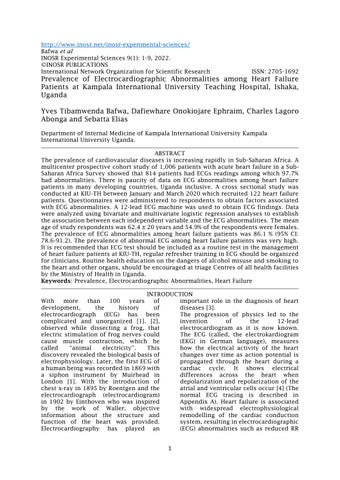
Normal ECG 13.9%
(95% CI: 8.78 21.40)


Abnormal ECG 86.1%
(95% CI: 78.60 91.21)

Figure 1. Prevalenceof ECG Abnormalities amongStudyRespondents
The results show that the prevalence of ECG abnormalities among heart failure patientswas86.1%(95%CI78.60 91.21). This shows that the majority 105 (86.1%)
ofHFpatientsthatreceivedhealthcareat KIU TH during the study period had abnormalECGs.
DISCUSSION
The prevalence of ECG abnormalities in this study was 86.1%. This is higher compared to [10] in Switzerland who found that 34.3% of study respondents had ECG abnormalities. The current prevalenceisalso higherthan the 36%of ECG abnormalities reported by [11] in United States and with the prevalence reported by [9] in Uganda who got 37.1% of ECG abnormalities. The disparities with the three studies above could be because [10,11,12,13,14,15] community based studies among populations without preexisting heart failure, while Buenzaconductedhisstudyamongheart failure patients but the proportion reported was that of arrhythmias only, leavingoutother ECGabnormalities. In India, [6] from their study reported a prevalence of 92% of ECG abnormalities among patients with either acute or chronic heart failure. [12] in their Sub Sahara survey reported a prevalence of electrocardiographic abnormalities of 97.7%amongacuteheartfailurepatients. Also, higher prevalence was reported by [13,14,15,16] in Nigeria from a cross sectional study which involved both patients of acute and chronic HF. They reported a prevalence of 98.2% of ECG abnormalities. A study conducted in Kumasi (Ghana) showed that the
prevalence of ECG abnormalities among HF patients was 93% [14]. These prevalencesarehigherthanthatreported in the current study. The discrepancy could be because the current study involved both patients with acute heart failure and those with chronic heart failure compared to the studyconducted by [12] that covered only patients with acuteHF.[13]and[6]studiesinvolvedall HF patients (acute and chronic) but the difference with the current study is that theyconductedtheirsinheartclinicsand respondents had been followed up for heartfailure foralong period.
The prevalence of ECG abnormalities amongHFpatientsinthisstudyissimilar tothestudyconductedbyGoudaetal.in 2016 in Canada which showed a prevalenceof82.5%ofECGabnormalities among heart failure patients. This similarity could be explained because Gouda et al. used the first available ECG in the admission of the HF patients, the same way like in this study. Subtle ECG changesmayoccurasapatientimproves or deteriorates and this may alter the relationship between an ECG and outcomes. A last ECG before death may also identify ECG changes unrelated to the initial presentation in a patient such
as terminal arrhythmias or while advanced medical therapyis instituted.
CONCLUSION
The findings of this study showed that the prevalence of ECG abnormalities among heart failure patients was very high at KIU TH and ECG abnormalities
were most common among heart failure patients in NYHA functional classes III andIV.
REFERENCES
1. Yang, X. L., Liu, G. Z., Tong, Y. H., Yan, H., Xu, Z., Chen, Q and Tan, S. H. (2015). The history, hotspots, and trends of electrocardiogram. Journal of geriatric cardiology: JGC, 12(4), 448.
2. Galvani, L and Aldini, G. (1953). Commentary on the effect of electricity on muscular motion. Cambridge,MA: Licht; 1953.
3. AlGhatrif, M. and Lindsay, J. (2012). A brief review: History to understand fundamentals of electrocardiography. Journal of community hospital internal medicineperspectives, 2(1),14383.
4.Dupre,A.,Vincent,S.andLaizzo,P. A. (2005). Basic ECG theory, recordings, and interpretation: In Handbook of cardiac anatomy, physiology, and devices. Humana Press, 191 201.
5. Nikolaidou, T., Ghosh, J. M. and Clark, A. L. (2016). Outcomes related to first degree atrioventricular block and therapeutic implications in patients with heart failure. JACC: Clinical Electrophysiology, 2(2), 181 192.
6. Vinit, A. T. and Jayesh, V. T. (2016). Prevalence of electrocardiographic abnormalities in heart failure patients attending Gujarat Adani institute of medical science, Kutch, Gujarat, India: a retrospective study. International Journalof Advancesin Medicine.
7. Oso, W. Y., and Onen , D. (2009). Writing research proposal and report:Ahandbookforbeginning Researchers.RevisedEdition,166.
8. Daniel,W.W.(2009).Biostatistics: A foundation for analysis in the healthsciences. 9th Ed.,JohnWiley andsons, 956.
9. Beunza, D. and Stark, D. (2004). Tools of the trade: The socio
technology of arbitrage in a Wall Street trading room. Industrial andCorporateChange,13(2),369 400.
10.Roos Hesselink, J , Baris, L , Johnson, M, De Backer, J , Otto, C, Marelli, A , Jondeau, G, Budts, W, Grewal, J , Sliwa, K, Parsonage,W ,Maggioni,A P ,van Hagen, I , Vahanian, A , Tavazzi, L , Elkayam, U , Boersma, E and Hall, R (2019). Pregnancy outcomes in women with cardiovascular disease: evolving trends over 10 years in the ESC RegistryofPregnancyandCardiac disease (ROPAC). Eur Heart J., 40(47):3848 3855. doi: 10.1093/eurheartj/ehz136. PMID: 30907409.
11.Auer, R., Bauer, D. C., Marques Vidal, P., Butler, J., Min, L. J., Cornuz, J and Rodondi, N. (2012). Association of major and minor ECG abnormalities with coronary heart disease events. Journal of theAmericanmedicalassociation, 307(14), 1497 1505.
12.Dzudie, A., Milo, O., Edwards, C., Cotter, G., Davison, B. A., Damasceno, A. and Sani, M. U. (2014). Prognostic significance of ECG abnormalities for mortality risk in acute heart failure: insight from the Sub Saharan Africa Survey of Heart Failure (THESUS HF). Journal of cardiac failure, 20(1), 45 52.
13.Karaye, K.M. and Sani, M. U. (2008) Factors Associated with Poor Prognosis among Patients Admitted with Heart Failure in a Nigerian Tertiary Medical Centre: A Cross Sectional Study. BMC CardiovascularDisorders, 8, 16.
14.Owusu, I. K., Boakye, Y. A., and Appiah, L. T. (2014). Electrocardiographic abnormalities in heart failure patients at a teaching hospital in
Kumasi. Ghana. Journal of cardiovascular diseases and diagnostic,2(2), 1 3.
15.Yves Tibamwenda Bafwa, Dafiewhare Onokiojare Ephraim, Charles Lagoro Abonga and Sebatta Elias (2022).Factors AssociatedwithElectrocardiograp hic Abnormalities among Heart Failure Patients at Kampala International University Teaching Hospital, Ishaka, Uganda. IAA Journal of Biological Sciences 9(1):166 175
16.Yves Tibamwenda Bafwa, Dafiewhare Onokiojare Ephraim, Charles Lagoro Abonga and Sebatta Elias (2022). Electrocardiographic Pattern among Heart Failure Patients at Kampala International University Teaching Hospital, Ishaka, Uganda IAA Journal of Biological Sciences 9(1):159 165 2022.